
Abstract
Oral squamous cell carcinoma (OSCC) is a lethal malignancy. It is reportedly demonstrated that long non-coding RNA (lncRNA) participates in the development of OSCC. The purpose of this study was to clarify the function and possible molecular mechanisms of lncRNA MCM3AP antisense RNA 1 (lncRNA MCM3AP-AS1) in OSCC. Quantitative real-time PCR (qRT-PCR) was adopted to investigate MCM3AP-AS1 expressions in OSCC tissues and cells. The proliferation, migration and invasion of HN-6 and SCC-9 cells were probed by cell counting kit-8 and Transwell assays, respectively. Dual luciferase reporter gene assay, Pearson's correlation analysis, qRT-PCR and western blot were used to detect the binding relationship among miR-204-5 p, MCM3AP-AS1 and forkheadbox C1 (FOXC1). MCM3AP-AS1 expression was elevated in OSCC tissues and cell lines. Overexpression of MCM3AP-AS1 facilitated the proliferation, migration and invasion of OSCC cells, while the knockdown of MCM3AP-AS1 suppressed these malignant phenotypes. Besides, MCM3AP-AS1 impeded miR-204-5 p by binding with it. MCM3AP-AS1 could also upregulate the expression of FOXC1 via repressing miR-204-5 p. MCM3AP-AS1 promotes the progression of OSCC cells by adsorbing miR-204-5 p and upregulating FOXC1 expressions.
Keywords
Significance of this study
What is already known about this subject?
miR-204-5 p acts as a tumor suppressor in oral squamous cell carcinoma (OSCC).
Forkheadbox C1 (FOXC1) is an oncogene in OSCC.
What are the new findings?
MCM3AP-AS1 expression was upregulated in OSCC.
Overexpression of MCM3AP-AS1 enhanced the proliferation, migration and invasion of OSCC cells.
MCM3AP-AS1 could upregulate the expression of FOXC1 via repressing miR-204-5 p.
How might these results change the focus of research or clinical practice?
MCM3AP-AS1 might be used as a novel target for clinical treatment of OSCC.
Introduction
In each year, oral squamous cell carcinoma (OSCC) gives rise to approximately 145 000 deaths worldwide, accounting for 2.1% of all cancer-related deaths.1 2 The 5-year overall survival rate of OSCC is only 40%-50% due to relatively low treatment responsiveness, drug resistance and late diagnosis.3 Therefore, it is urgent to explore the mechanism of OSCC progression for novel therapy strategies.
Long non-coding RNA (lncRNA), RNA molecule with over 200 nucleotides in length, takes up 68% of non-coding RNA (ncRNA) molecules.4 In recent years, lncRNA MCM3AP antisense RNA 1 (lncRNA MCM3AP-AS1) is reported to regulate the progression of cancers including gastric cancer, prostate cancer and cervical cancer.5–7 However, at present, the expression, function and underlying mechanism of MCM3AP-AS1 in OSCC await more research. MicroRNAs (miRNAs) is a kind of short ncRNA molecules with about 22 nucleotides in length, which is closely related to tumorigenesis.8 9 MiR-204-5 p expression is downregulated in a variety of tumors including OSCC, and it inhibits tumor cell proliferation and metastasis.10–13
In recent years, many forkhead box members have been identified as regulators in tumorigenesis.14 Forkheadbox C1 (FOXC1) has been shown to be upregulated in human cancers, and it can function as potential clinical biomarker and therapy target for the diagnosis or treatment of human cancers.15–17 This work aimed at probing into the expression, clinical significance and mechanisms of MCM3AP-AS1 in OSCC. Herein, we proved that the MCM3AP-AS1 modulated the progression of OSCC via miR-204-5 p/FOXC1 axis.
Materials and methods
Clinical data and ethical statements
This work was endorsed by the Ethics Committee of the First Affiliated Hospital of Nanchang University, and the patients involved in the research signed written informed consent forms. OSCC specimens (n=61) and matched normal adjacent tissue specimens were collected from 61 patients who had received surgery from October 2016 to July 2018, none of whom received radiotherapy or chemotherapy prior to surgery. All tissue specimens were frozen immediately after removal and then stored in liquid nitrogen until RNA extraction.
Cell line and cell culture
The American Type Culture Collection (Manassas, Virginia, USA) was the provider of normal human oral keratinocyte (NHOK) cell line and OSCC lines (including SCC-9, HN-6, Tca8113, ACC-3, and HSC-2 cells). Cells were cultivated in a PRMI-1640 medium (Gibco, Carlsbad, California, USA) containing 10% fetal bovine serum (FBS; Hyclone, Logan, Utah, USA), 100 U/mL penicillin and 100 µg/mL streptomycin (Hyclone) in an incubator in 5% CO2 at 37°C. The culture solution was replaced by fresh medium at the intervals of every 3 to 4 days. Subculture was carried out with 0.25% trypsin (Hyclone).
Cell transfection
OSCC cells were inoculated in a 24-well plate (2×10⁵ cells/well) and then cultured. Transfection was performed when cells reached 70% confluence with Lipofectamine 2000 (Invitrogen, Carlsbad, California, USA) according to the manufacturer's instructions. Short hairpin RNA (shRNA), overexpression plasmids, miRNA mimics and miRNA inhibitors were designed and synthesized by GenePharma (Shanghai, China).
Quantitative real-time PCR (qRT-PCR)
Cells or fresh tissues were lysed with TRIzol reagent (Invitrogen), and the total RNA was extracted. After that, the integrity of RNA was confirmed by agarose gel electrophoresis, and the concentration was measured by ultraviolet spectrophotometer. Reverse transcription was conducted with PrimeScript RT kit (TaKaRa, Dalian, China) in compliance with the instructions, and then PCR was performed using SYBR Premix Ex Taq II (TaKaRa). The specific experimental method could be found through the kit instructions. U6 and β-actin were the reference gene for the detection of miR-204-5 p, and MCM3AP-AS1 and FOXC1 mRNA, respectively. The expressions of the target genes were calculated using 2−ΔΔCt method. MCM3AP-AS1 upstream primer: 5′-GCTGCTAATGGCAACACTGA-3′, downstream primer: 5′-AGGTGCTGTCTGGTGTAGAGAT-3′; miR-204-5 p upstream primer: 5′-GCCAGATCTGGAAGAAGATGGTGGTTAGT-3′, downstream primer: 5′-GGCGAATTCACAGTCACCGTCAGTAGCA-3′; FOXC1 upstream primer: 5′-CAGAACAGCATCCGCCACA-3′, downstream primer: 5′-TGTTGTAGGAGTCCGGGTC-3′; U6 upstream primer: 5′-CTCGCTTCGGCAGCACA-3′, downstream primer: 5′-AACGCTTCACGAATTTGCGT-3′; β-actin upstream primer: 5′-GGACCTGACTGACTACCTC-3′, downstream primer: 5′-TACT-CCTGCTTGCTGAT-3′.
Cell counting kit-8 (CCK-8) assay
Cell growth was examined employing the cell proliferation reagent CCK-8 solution (Dojindo, Kyushu, Japan). Cells were inoculated in a 96-well plate (Corning Costar, Corning, New York, USA) at 1.0×10³/well, and after the cells were cultured for 24 hours, 48 hours and 96 hours, 10 µL CCK-8 solution was loaded into each well, respectively. After 1 hour, the cell viability was monitored by measuring the absorbance of cells at 450 nm with a microplate reader.
Transwell assay
The transfected cells were harvested and the cell concentration was modulated to 1×10⁵ cells/mL with serum-free medium. Two hundred microliters of cell suspension from each group was dripped into the upper chamber of the Transwell chamber (BD Biosciences, San Jose, California, USA), and 600 µL medium with 10% FBS was loaded into the lower chamber. After 24 hours of culture in the incubator, the chamber was removed, and the cells in the upper chamber were carefully wiped away before the migrated cells were fixed in 95% ethanol for 15 min. After being stained with 0.1% crystal violet staining solution for 20 min, the Transwell chambers were washed and dried. The number of cells in five randomly selected visual fields was observed under microscope. During the invasion experiment, the Transwell chamber was coated with Matrigel, and the other steps followed the migration assay.
Luciferase reporter gene assay
FOXC1 3′UTR (untranslated region)-wild type (WT) (or MCM3AP-AS1-WT) and FOXC1-mutant (MUT) (or MCM3AP-AS1-MUT) reporter plasmids were constructed. HEK293T cells were inoculated on a 12-well cell plate at 1×10⁵ cells/well. Afterward, the reporter plasmids mentioned above and miR-204-5 p mimics or negative control were cotransfected into HEK293T cells. After 48 hours of culture, the luciferase activity of each group was detected with dual luciferase reporter gene detection kit (Promega, Madison, Wisconsin, USA), and the ratio of firefly fluorescence intensity to Renilla fluorescence intensity was adopted to quantify the luciferase activity of each group.
Rna Immunoprecipitation (Rip) Assay
EZ-Magna RIP RNA-binding protein immunoprecipitation kit (Millipore, Bedford, Massachusetts, USA) was employed for RIP analysis according to the manufacturer's protocol. OSCC cells were lysed in lysis buffer containing protease and RNase inhibitors. The protein extracts were incubated at 4°C for 2 hours with RIP buffer with magnetic beads conjugated with human anti-argonaute 2 (Ago2) antibody or control IgG (Millipore). Free protein was removed with proteinase K so that co-immunoprecipitated RNA was extracted. qRT-PCR analysis was then conducted to determine the enrichment of the targets.
Western blot
Forty-eight hours after transfection, RIPA lysis buffer (Beyotime Biotechnology, Shanghai, China) was dripped into the cells to extract the total protein, and its concentration was detected in compliance with the instructions of the BCA protein quantitative kit (Beyotime Biotechnology). The protein samples were mixed with loading buffer and denatured in boiling water for 5 min. After denaturation, the protein was sampled at 20 μg/well, and the protein samples were then separated by 10% sodium dodecyl sulfate-polyacrylamide gel electrophoresis. Subsequently, the protein was transferred to the polyvinylidene difluoride membrane (Millipore). After the incubation with the blocking solution for 2 hours at room temperature, the primary antibodies including anti-β-actin antibody (1:2000, ab179467; Abcam, Shanghai, China) and anti-FOXC1 antibody (1:1000, ab227977; Abcam) were loaded and the membrane was incubated overnight at 4°C. After rinsing the membrane with tris buffered saline tween (TBST), the secondary antibody was loaded anterior to the incubation of membrane at room temperature for 1 hour. After the membrane was washed by TBST again, the bands were developed by electrochemiluminescence kit (Millipore).
Statistical analysis
Data analysis was conducted using SPSS V.20.0 (SPSS, Chicago, Illinois, USA). The measurement data were expressed as x±s, and the t-test was adopted in statistical analysis. Differences of p<0.05 were regarded statistically significant.
Result
Mcm3Ap-As1 Expression Was Elevated in Oscc Tissues and Cell Lines
To explore the expression pattern of MCM3AP-AS1 in OSCC, we extracted total RNA from 61 OSCC tissues and adjacent tissues for qRT-PCR analysis. As shown, the expression of MCM3AP-AS1 in OSCC tissues was significantly higher than that in control normal tissues (figure 1A), and the expression of MCM3AP-AS1 in OSCC cell lines was obviously higher than in NHOK cell line (figure 1B). These results signified that MCM3AP-AS1 could play a role in promoting cancer in OSCC progression.
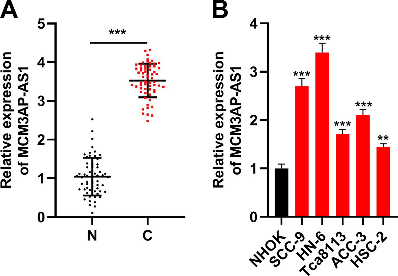
MCM3AP-AS1 was underexpressed in oral squamous cell carcinoma (OSCC). (A) Quantitative real-time PCR (qRT-PCR) was used to detect the expression of MCM3AP-AS1 in 61 pairs of OSCC tissues and normal tissues adjacent to cancer. (B) qRT-PCR was used to detect the expression of MCM3AP-AS1 in OSCC cell lines SCC-9, HN-6, Tca8113, ACC-3 and HSC-2 and normal cell line normal human oral keratinocyte (NHOK).
Mcm3Ap-As1 Gene Knockdown Impeded Proliferation, Migration and Invasion of Hn-6 and Scc-9 Cells
To investigate the biological function of MCM3AP-AS1 in OSCC cells, a specific shRNA targeting MCM3AP-AS1 was synthesized, and transfected into HN-6 and SCC-9 cells. qRT-PCR displayed that the expression of MCM3AP-AS1 in OSCC cells was dramatically decreased as expected (figure 2A). The proliferation, migration and invasion of HN-6 and SCC-9 cells were probed by CCK-8 and Transwell assays, respectively, and the experimental results of which demonstrated that downregulation of MCM3AP-AS1 remarkably blocked the proliferation of HN-6 and SCC-9 cells (figure 2B); the migration and invasion of OSCC cells transfected with MCM3AP-AS1 shRNA were markedly lower than those in the control group (figure 2C, D). These results connoted that MCM3AP-AS1 regulated the proliferation, migration and invasion of OSCC cells.
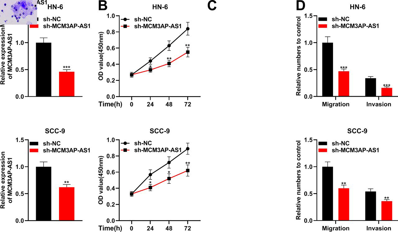
Knockdown of MCM3AP-AS1 inhibited the proliferation, migration and invasion of oral squamous cell carcinoma (OSCC) cells. (A) MCM3AP-AS1 siRNA (small interfering RNA) was transfected into HN-6 and SCC-9 cells to make MCM3AP-AS1 knocked down. The transfection efficiency was then detected by quantitative real-time PCR. (B) Cell counting kit-8 assay was used to detect the proliferation of HN-6 and SCC-9 cells. (C and D) Transwell assay was used to detect HN-6 and SCC-9 cell migration and invasion.
Mcm3Ap-As1 Directly Regulated Mir-204-5P Expressions in Oscc Cells
To delve into downstream miRNAs of MCM3AP-AS1, we carried out bioinformatic analysis with StarBase and noticed that MCM3AP-AS1 contained binding site for miR-204-5 p (figure 3A). We established luciferase reporter plasmids of MCM3AP-AS1-WT and mutant MCM3AP-AS1-MUT containing miR-204-5 p binding sites for dual luciferase reporting analysis. It was observed that the activity of WT luciferase reporter plasmid was decreased by miR-204-5 p, while that of MUT luciferase reporter plasmid was not changed (figure 3B). Additionally, Pearson's analysis confirmed a negative correlation between MCM3AP-AS1 and miR-204-5 p in OSCC samples (R2=0.5694; figure 3C). RIP experiments confirmed that MCM3AP-AS1 and miR-204-5 p were enriched in Ago2-containing microribonucleoproteins compared with that in IgG (figure 3D). Additionally, transfection of MCM3AP-AS1 shRNA markedly increased miR-204-5 p expression in OSCC cells (figure 3E). These results displayed that MCM3AP-AS1 could directly target miR-204-5 p in OSCC and negatively regulate its expressions.
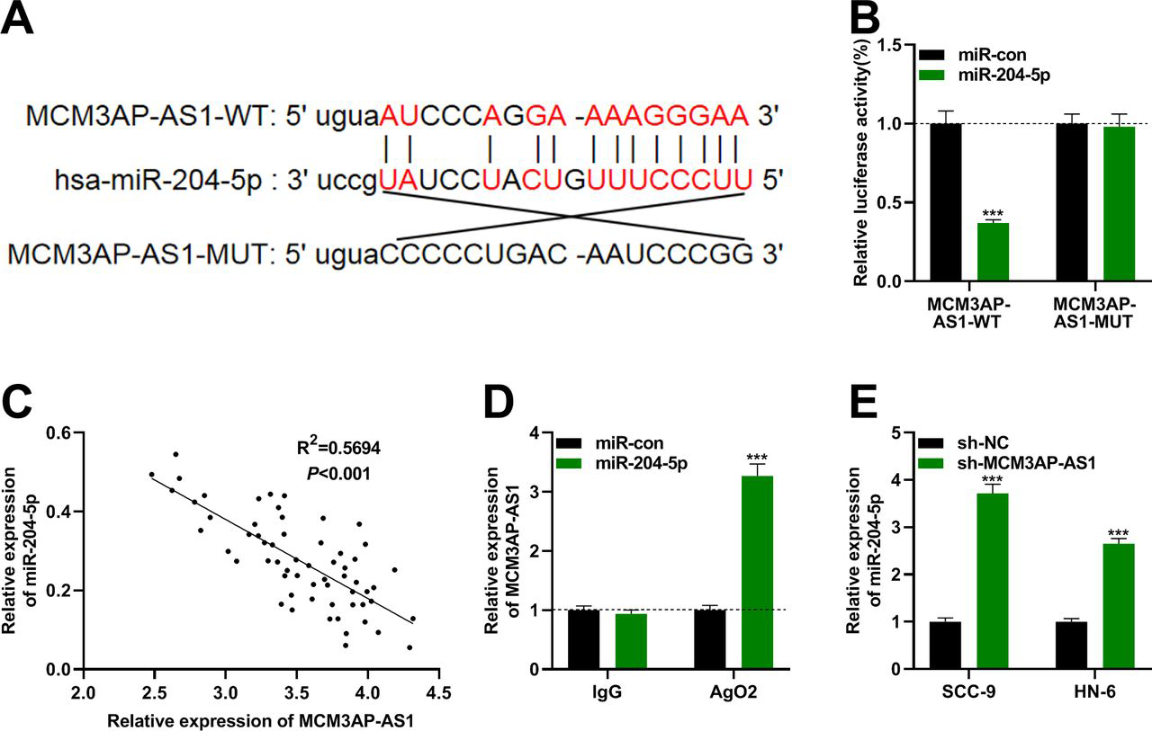
MCM3AP-AS1 adsorbed miR-204-5 p. (A) MCM3AP-AS1-WT and MCM3AP-AS1-MUT luciferase reporter vectors with miR-204-5 p binding site were constructed. (B) MCM3AP-AS1-WT or MCM3AP-AS1-MUT luciferase vector and miR-con/miR-204-5 p mimics were cotransfected into 293 T cells. The luciferase activity of the luciferase reporter was then determined. (C) Pearson's correlation analysis showed that there was a negative correlation between the expressions of MCM3AP-AS1 and miR-204-5 p in oral squamous cell carcinoma tissues. (D) RNA immunoprecipitation experiments confirmed that MCM3AP-AS1 and miR-204-5 p were enriched in argonaute 2 (Ago2)-containing microribonucleoproteins. (E) Quantitative real-time PCR was used to detect the expression of miR-204-5 p in HN-6 and SCC-9 cells after MCM3AP-AS1 was overexpressed.
Foxc1 Was the Target of Mir-204-5P in Oscc
Next, bioinformatic analysis was adopted to predict the possible targets of miR-204-5 p, and TargetScan database suggested that FOXC1 was one of the potential downstream target of miR-204-5 p (figure 4A). Dual luciferase reporter assay implied that miR-204-5 p mimics reduced the luciferase activity of FOXC1-WT reporter plasmids, but could not affect that of FOXC1-MUT reporter plasmids, suggesting the existence of the predicted binding site (figure 4B). Following this, Pearson's analysis confirmed that FOXC1 expression was positively interrelated with MCM3AP-AS1 expressions in OSCC tissues (figure 4C). Moreover, miR-204-5 p mimics significantly reduced FOXC1 mRNA and protein expressions in SCC-9 cells; miR-204-5 p inhibitors remarkably increased FOXC1 mRNA and protein expressions in HN-6 cells (figure 4D, E). These data signified that FOXC1 was the direct target of miR-204-5 p and was probably positively modulated by MCM3AP-AS1.
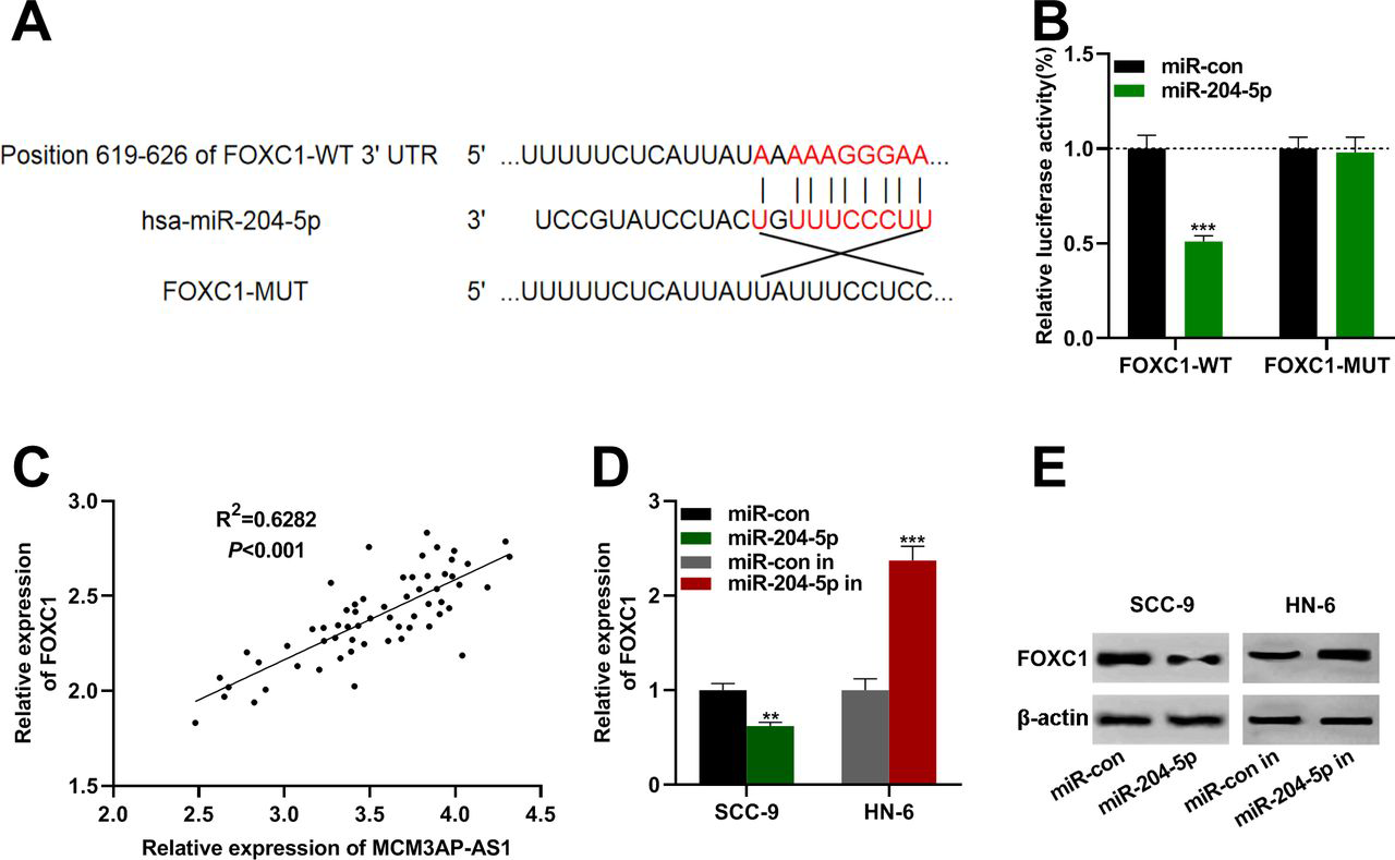
Forkheadbox C1 (FOXC1) was a functional target of miR-204-5 p. (A) Bioinformatics analysis showed that miR-204-5 p and the 3′UTR (untranslated region) of FOXC1 had potential binding sites. (B) Dual luciferase activity assay demonstrated that miR-204-5 p negatively regulated the luciferase activity of FOXC1 3'UTR-wild type (FOXC1-WT), but had no significant effect on the luciferase activity of FOXC1-mutant (FOXC1-MUT). (C) Person's correlation analysis indicated that there was a positive correlation between FOXC1 and MCM3AP-AS1 expressions in NPC tissues. (D) miR-204-5 p mimics were transfected into HN-6 and SCC-9 cells, and quantitative real-time PCR (qRT-PCR) was used to detect the expression of FOXC1 mRNA in both cells. (E) Western blot was used to determine the expression of FOXC1 protein after oral squamous cell carcinoma cells were transfected with miR-204-5 p mimics or inhibitors.
Mcm3Ap-As1 Regulated Mir-204-5P/Foxc1 Axis to Promote the Growth and Metastasis of Oscc Cells
To further pinpoint whether MCM3AP-AS1 promotes the OSCC process by modulating the miR-204-5 p/FOXC1 axis, SCC-9 cells were randomly divided into three groups: MCM3AP-AS1 overexpression group, MCM3AP-AS1 overexpression+miR-204-5 p mimics group and control group. As expected, overexpression of MCM3AP-AS1 greatly enhanced FOXC1 expressions on both mRNA and protein expression levels, while miR-204-5 p mimics reversed the elevation of FOXC1 induced by MCM3AP-AS1 (figure 5A, B). The proliferation, migration and invasion of the cells were subsequently verified by CCK-8 assay and Transwell assay, respectively. The data denoted that overexpression of MCM3AP-AS1 remarkably enhanced the proliferation, migration and invasion of OSCC cells, and miR-204-5 p mimics attenuated the effect of MCM3AP-AS1 on these processes (figure 5C, D). These data demonstrated that MCM3AP-AS1 could promote the growth and metastasis of OSCC cells by impeding miR-204-5 p and facilitating the expression of FOXC1 in OSCC.
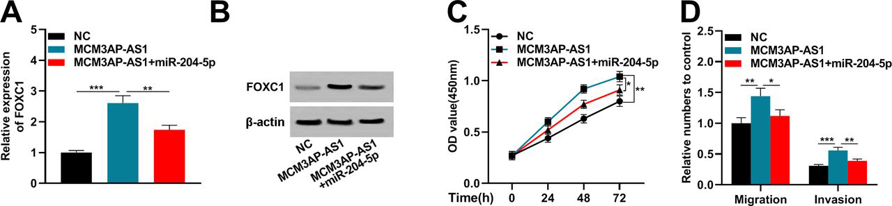
MCM3AP-AS1 promoted the progression of oral squamous cell carcinoma (OSCC) cells by inhibiting miR-204-5p and upregulating forkheadbox C1 (FOXC1). (A) MCM3AP-AS1 overexpression plasmid and miR-204-5 p mimics were transfected into SCC-9 cells, and quantitative real-time (qRT-PCR) was used to detect the FOXC1 mRNA expressions in the cells. (B) The expression of FOXC1 protein was detected by western blot after transfection. (C) Cell proliferation was determined by cell counting kit-8 (CCK-8) assay after transfection. (D) OSCC cell migration and invasion were detected by Transwell assay.
Discussion
OSCC occupies more than 90% of all oral tumors and 38% of head and neck tumors, with over 500 000 new cases diagnosed every year worldwide.18 Risk factors for OSCC include smoking, alcohol consumption, viral infection, betel nut chewing, immune deficiency, radiation, diet, genetic susceptibility and so on.19 The tumorigenesis of OSCC is a complex pathological process. Accumulating studies display that ncRNA plays crucial roles in this process.20 21
LncRNA cannot be translated to proteins, so in the past, it was regarded as the ‘junk RNA’ in the genome. However, existing studies manifest that lncRNA is a key participant in regulating gene expressions, including chromatin remodeling, transcription and post-transcriptional modification, so lncRNAs become a hot research issue as a new class of regulatory molecules.22 Imbalanced lncRNAs are closely relevant to the progression of tumors. For instance, lncRNA UCA1 expression is upregulated in OSCC and facilitates cancer cell proliferation23; lncRNA PDIA3P is overexpressed in OSCC and reduces the survival rate of patients with OSCC24; lncRNA ANRIL is overexpressed during the carcinogenesis of OSCC and is associated with cancer development and metastasis, and ANRIL knockdown inhibits the proliferation of tumor cells by restraining the expressions of MRP1 and ABCC2.25 Studies on MCM3AP-AS1 in cancer depict that overexpression of MCM3AP-AS1 enhances the growth and metastasis of gastric cancer, prostate cancer, cervical cancer, lung cancer, pancreatic cancer, hepatocellular carcinoma, papillary thyroid carcinoma and gliobastoma.5–7 26–30 In this work, for the first time, it was demonstrated that MCM3AP-AS1 was highly expressed in OSCC tissues and cells. It is also found that MCM3AP-AS1 knockdown impeded the proliferation, migration and invasion of HN-6 and SCC-9 cells, while overexpression of MCM3AP-AS1 stimulated the above malignant biological behaviors of OSCC cells. Hence, MCM3AP-AS1 was considered to be a pro-cancer lncRNA in OSCC.
FOXC1 is originally identified as a key prognostic indicator of basal-like breast cancer, and it has recently been proven to function in diverse cancer types.15 Studies manifest that FOXC1 is highly expressed in OSCC and functions as an oncogene, which may be a crucial therapeutic target and predictive biomarker for OSCC.31 Moreover, as an inhibitory miRNA involved in malignant transformation of tumors, the expression of miR-204-5 p is reduced in most OSCC tissues and cancer cell lines, and overexpression of miR-204-5 p blocks OSCC cell proliferation and metastasis.32 In the present work, we further confirmed that miR-204-5 p had a negative regulatory effect on FOXC1 in OSCC, which is consistent with the report in laryngeal squamous cell carcinoma.33
Recently, the mechanism of competitive endogenous RNAs (ceRNAs) has attracted a lot of attention in cancer research. LncRNA can affect post-transcriptional regulation by competitively binding with miRNA via miRNA response element, thus exerting an impact on gene expression.34 So far, certain lncRNAs have been validated as functional ceRNA in OSCC. For example, lncRNA MALAT1 can regulate STAT3 expression by sponging miR-125b in OSCC35; lncRNA FTH1P3 promotes the OSCC progression by functioning as a molecular sponge for miR-224-5 p36; H19 expedites cell proliferation and invasion via decoying miR-138 and releasing EZH2 mRNA in OSCC.37 In this work, we were curious about whether MCM3AP-AS1 functioned as a ceRNA for miR-204-5 p and FOXC1 to mediate the progression of OSCC. Through the experiments, we proved that FOXC1 was a downstream target of miR-204-5 p, and further research proved that MCM3AP-AS1 could upregulate the expression of FOXC1 via suppressing miR-204-5 p and promoting the proliferation, migration and invasion of OSCC cells, which identified that MCM3AP-AS1 was a ceRNA in OSCC.
In summary, we prove that MCM3AP-AS1 is highly expressed in OSCC, and MCM3AP-AS1 regulates the malignant biological behaviors of OSCC cells including proliferation, migration and invasion. Mechanistically, MCM3AP-AS1 decreases the expression of miR-204-5 p and upregulates the expression of FOXC1 as a molecular sponge. Therefore, we make a conclusion that MCM3AP-AS1 is a promising target for OSCC treatments. However, this study is limited to in vitro experiments and this conclusion still needs to be further confirmed by animal studies in the future.
Footnotes
Contributors
Conceived and designed the experiments: HL, JJ; performed the experiments: HL; statistical analysis: JJ; wrote the paper: HL, JJ. All authors read and approved the final manuscript.
Funding
The authors have not declared a specific grant for this research from any funding agency in the public, commercial or not-for-profit sectors.
Competing interests
None declared.
Patient consent for publication
Not required.
Provenance and peer review
Not commissioned; externally peer reviewed.
Data availability statement
Data are available on reasonable request. The data used to support the findings of this study are available from the corresponding author on request.