
Abstract
Immune thrombocytopenic purpura (ITP) is a disorder caused by autoimmune antibodies which target glycoprotein IIb/IIIa complex or other platelet membrane antigens leading to platelet destruction. These platelets are then cleared by the spleen resulting in thrombocytopenia. Immune thrombocytopenic purpura affects about 1 to 6.4 cases in 100 000 children making it one of the most common causes of symptomatic thrombocytopenia in the pediatric population. It is rare that children or adolescents present with serious bleeding due to ITP. Common presentations include petechiae, bleeding gums, or bruising. Bleeding requiring hospitalization or transfusions is unusual and only occurs in approximately 5% of children. Even more uncommon is the presentation of severe bleeding complications requiring surgery for resolution. We present a case of a 17-year-old girl with acute ITP complicated by intraperitoneal hemorrhage and refractory thrombocytopenia due to ovarian cyst requiring oophorectomy.
Although immune thrombocytopenic purpura (ITP) manifests as 2 different clinical syndromes, both will present with isolated thrombocytopenia despite normal bone marrow. In children, acute ITP typically follows a viral illness. This form of ITP usually resolves spontaneously without treatment. Patients present with clinical manifestations of thrombocytopenia including bruising, petechiae, mucosal bleeding, or menorrhagia. These symptoms are common if the platelet count is lower than 20 000. Bleeding risks increase when platelet counts decrease to 10 000 or less. 1
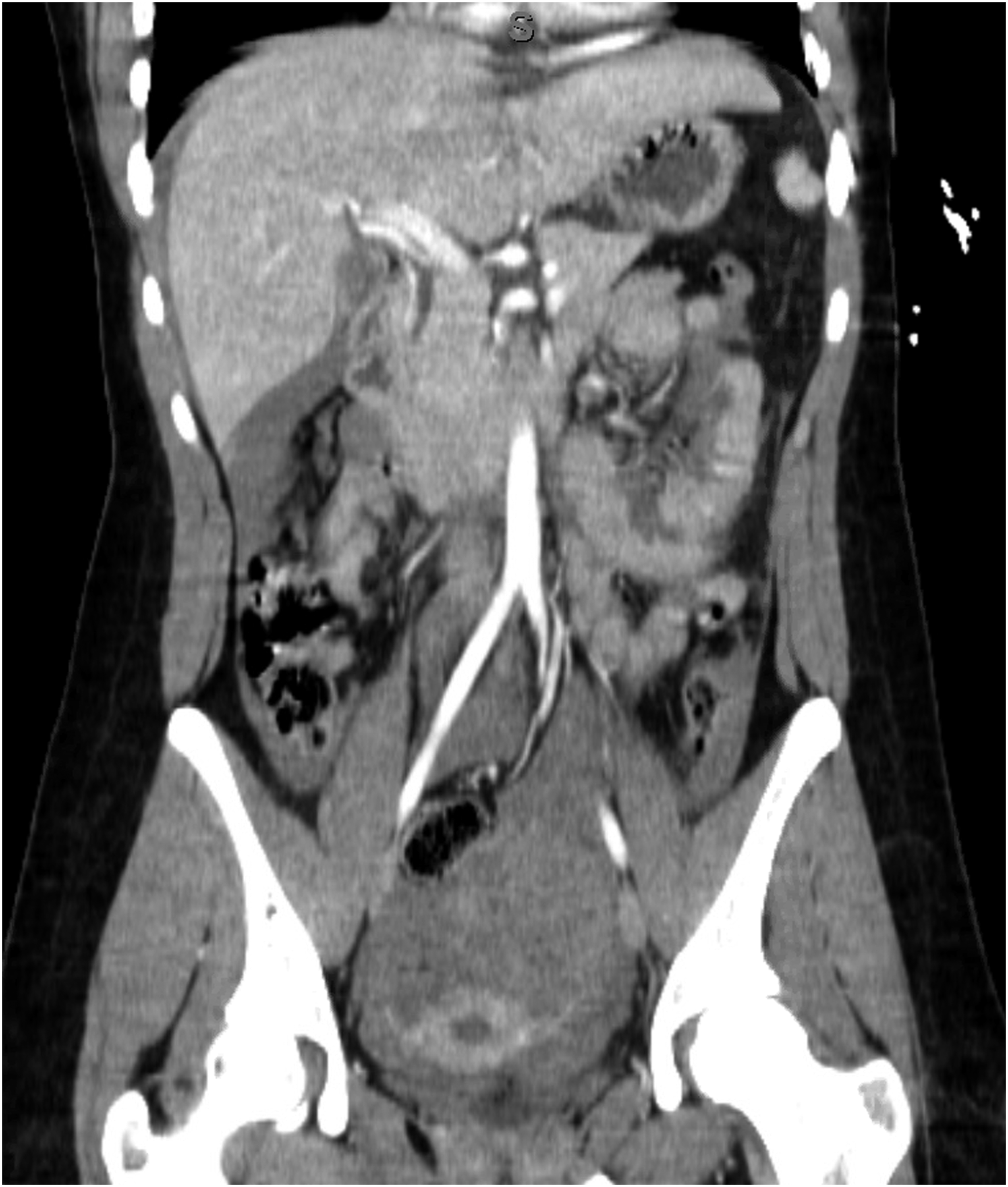
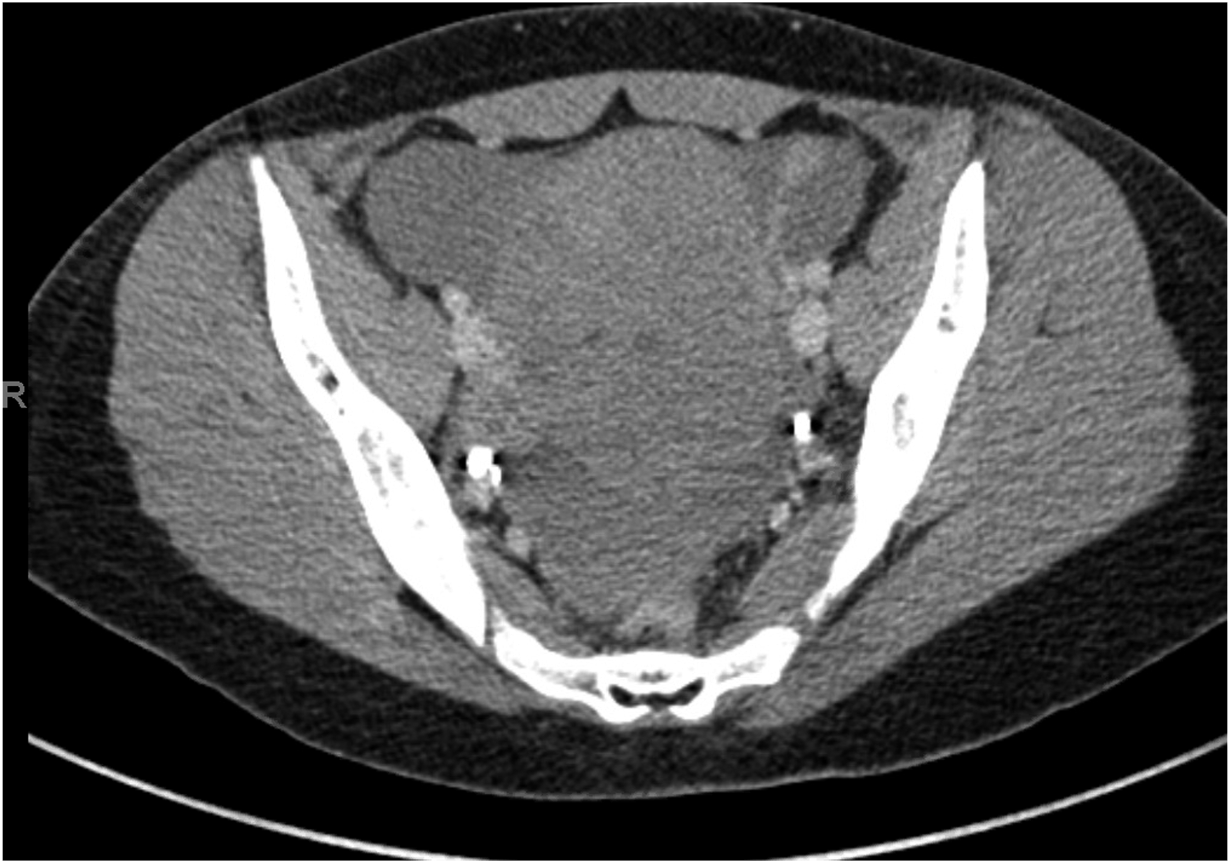
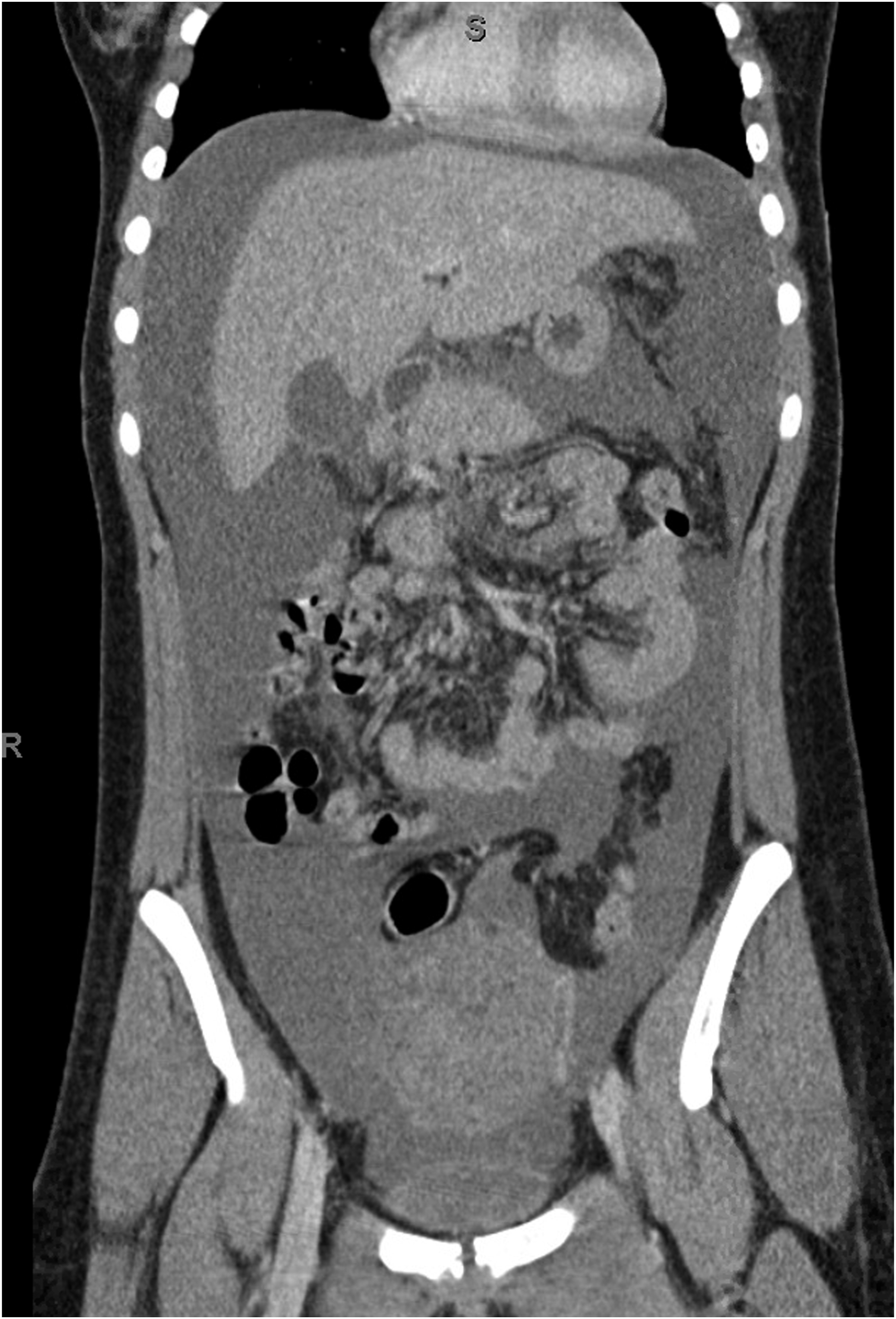
A 17-year-old girl with no significant past medical history presented to the pediatric emergency department with complaints of syncope, abdominal pain, and petechiae of her bilateral lower extremities and lips. Additionally, she had epigastric and right-sided abdominal pain associated with nausea and hematemesis. Laboratory values revealed a white blood cell count of 23.4, hemoglobin of 10.5, and a platelet count of <3000. Computed tomography (CT) of the abdomen and pelvis revealed a large heterogeneous mass in the mid pelvis measuring 5.8 × 5.6 × 6.8 cm as well as large volume hemoperitoneum (Figures 1 and 2). Patient was admitted and intravenous immunoglobulin (IVIG) was initiated. Platelet count remained <3000, and laboratory values for ANA, DAT, C3, and C4 were negative, increasing the suspicion for primary ITP. Despite a 1 g/kg dose of IVIG and 60 mg intravenous steroids every 6 hours, there was no improvement in her thrombocytopenia. On hospital day three, due to increasing pain and continued episodes of syncope, a repeat CT head and abdomen/pelvis were obtained. CT head imaging did not show any acute abnormality, but imaging of the abdomen/pelvis showed significant worsening of hemoperitoneum and mass on left ovary now measuring 9.6 × 7.7 × 8.9 cm (Figures 3 and 4). At this point, the poor response to IVIG and steroids was thought to be secondary to a consumptive process caused by her ovarian mass. The patient’s hemoglobin had decreased to 6.3, and she was transfused red blood cells as well as platelets. It was determined the patient had a major bleeding event categorized by a drop in hemoglobin >3 and a transfusion requirement of greater than 2 units of blood. The patient continued to have syncopal episodes, increasing abdominal pain, and tachycardia. Due to continued transfusion requirements despite optimal medical management, the patient was taken to the operating room. She underwent exploratory laparotomy through a lower midline incision and left oophorectomy. A large hemorrhagic, ruptured mass was noted on the left ovary as well as large volume hemoperitoneum. The patient was also transfused 2 units of red blood cells and 2 units of platelets intraoperatively. After removal of the ruptured hemorrhagic ovary and evacuation of over 2 L of hemoperitoneum, the abdomen was inspected and found to be hemostatic. Commonly, surgical intervention for ITP involves a splenectomy for thrombocytopenia unresponsive to medical management. Due to the patient’s acute presentation with ITP, and no signs of chronic thrombocytopenia as well a known source of hemorrhage, splenectomy was not performed. We suspected the consumptive process of platelets would cease with removal of the ovary. If hemorrhage was unable to be controlled, or if thrombocytopenia continued, splenectomy would be considered. Avoidance of splenectomy spared our young patient from a new set of risks associated with asplenia. Postoperatively, thrombocytopenia improved and transfusion requirements ceased. The patient only required one platelet transfusion in the immediate postoperative period and remained stable with a platelet count >30 000 for the remainder of her hospital course. Final pathology reported fragments of ovarian tissue with cystic follicles and extensive hemorrhage consistent with torsion. Patient was seen for routine follow-up and was doing remarkably well, with platelet count at 213 000 and no further transfusion requirement. Axial computed tomography images of pelvis at presentation. Coronal computed tomography images of abdomen and pelvis at presentation. Repeat computed tomography scan axial images of pelvis. Repeat computed tomography scan coronal images of abdomen and pelvis.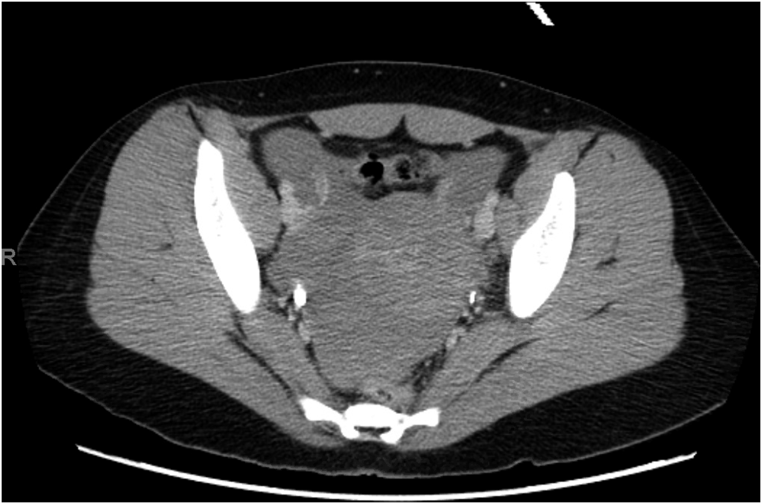
The risk of serious bleeding in children diagnosed with acute onset ITP is approximately 3%, and the risk of life threatening bleeding is less than 1%. 2 When bleeding occurs, combination medical therapy is the first-line treatment. Treatment includes high dose steroids, IVIG, and platelet transfusions. The role of surgery in ITP is typically limited to splenectomy which is reserved for chronic ITP with severe thrombocytopenia refractory to medical management or those with bleeding complications. We present a rare case of necessary surgical intervention for intraperitoneal hemorrhage due to ovarian torsion with acute onset ITP. Few cases have been reported linking ITP and hemoperitoneum secondary to ovarian follicle rupture, ovarian cyst rupture, or ovarian torsion.3,4 In each of these cases, bleeding was controlled with IVIG, platelet transfusions, and high dose methylprednisolone. Despite maximal medical management, our patient continued to require transfusions. Complete resolution occurred status post oophorectomy. This case highlights the consideration of surgical intervention despite thrombocytopenia in patients who do not respond to medical management.
Although clinically significant bleeding in adolescents with ITP is exceedingly rare, the consideration of ovarian pathology must be considered in menstruating women.
Footnotes
Declaration of Conflicting Interests
The author(s) declared no potential conflicts of interest with respect to the research, authorship, and/or publication of this article.
Funding
The author(s) received no financial support for the research, authorship, and/or publication of this article.