
Abstract
Prostate adenocarcinoma most commonly metastasizes to bone and lymph nodes; isolated soft-tissue presacral metastasis without a known primary is exceedingly rare and poorly characterized. We report a 69-year-old man presenting with hematuria and an incidental 4-cm presacral mass. MRI demonstrated a solid, heterogeneously enhancing lesion within the right mesorectal compartment without intraluminal involvement, most consistent with a primary neurogenic tumor. Serum PSA was not obtained preoperatively, as symptoms were attributed to chronic suprapubic catheter irritation and imaging did not suggest a prostatic origin. Preoperative biopsy was not pursued given the lesion’s deep posterior location at the S4 level—proximity to sacral nerve roots precluded safe percutaneous access, and transrectal biopsy is contraindicated. The mass was resected via a posterior transsacral (Kraske) approach for presumed primary presacral neoplasm. Histopathology revealed metastatic prostatic adenocarcinoma with positive PSA and prostatic acid phosphatase immunostaining, identifying an occult primary tumor. This case illustrates an atypical diagnostic scenario in which resection was undertaken for a presumed primary lesion; the metastatic diagnosis was an unexpected pathologic finding that would have substantially altered management had it been established preoperatively. This case underscores the importance of including serum PSA in the standard workup of solid presacral masses in older male patients and demonstrates that the Kraske approach can provide adequate exposure for selected posteriorly situated extrarectal masses when diagnosis remains uncertain and resection is clinically indicated.
Prostate adenocarcinoma demonstrates a well-established metastatic pattern involving the axial skeleton, regional lymph nodes, liver, and lungs. Isolated soft-tissue metastasis to the presacral space without concurrent skeletal involvement, and in the absence of a known primary malignancy, is exceptionally uncommon; to our knowledge, no prior cases of this specific presentation have been reported in the literature. Presacral masses encompass a wide spectrum of congenital, neurogenic, mesenchymal, and metastatic lesions, and radiographic characteristics are frequently nonspecific. The most common primary tumors of the presacral space include neurogenic tumors (schwannoma, neurofibroma), developmental cysts (tailgut cyst, dermoid), and chordoma; metastatic deposits are comparatively rare and typically arise in the setting of known advanced pelvic malignancy with concurrent osseous involvement. On MRI, schwannomas classically appear as well-circumscribed, T2-hyperintense masses with heterogeneous enhancement, often with cystic degeneration, arising in proximity to sacral foramina. These features overlap substantially with other presacral lesions, making preoperative diagnosis challenging even with advanced imaging.1,2 Surgical excision is often required both for diagnosis and definitive management when imaging alone cannot establish the nature of a presacral mass. 3
A 69-year-old man with diabetes, hypertension, recurrent urinary tract infections, and chronic hematuria from a longstanding suprapubic catheter presented for evaluation of worsening bleeding. He had no prior history of prostate cancer and no known malignancy. Contrast-enhanced computed tomography incidentally revealed a right-sided presacral mass. Magnetic resonance imaging (MRI) demonstrated a 4.1 × 3.8 cm solid lesion within the right mesorectal compartment, abutting but not invading the rectal wall, with heterogeneous T2 signal and avid contrast enhancement (Figure 1). The lesion was well-circumscribed, did not involve the sacral cortex, and arose in proximity to the sacral neural foramina at the S4 level. Colonoscopy excluded intraluminal pathology. Based on these imaging characteristics—a well-defined, T2-heterogeneous, enhancing extrarectal mass in proximity to sacral foramina in a patient without a known primary malignancy—the leading preoperative diagnosis was a neurogenic tumor, most likely schwannoma, with additional considerations including tailgut cyst and gastrointestinal stromal tumor. Metastatic prostate cancer was considered unlikely given the absence of a known primary, the solitary soft-tissue presentation without bone involvement, and the lack of classic radiographic features of prostate metastasis such as sclerotic or lytic sacral lesions. Serum PSA was not obtained preoperatively; this decision reflected both the absence of imaging features suggesting a prostatic primary and the clinical context in which hematuria and lower urinary tract symptoms were attributed to chronic suprapubic catheter-related irritation rather than urologic pathology. In retrospect, this represents a significant oversight, as PSA testing is inexpensive, non-invasive, and—had it been obtained—might have prompted targeted prostate imaging and fundamentally altered the diagnostic pathway. Preoperative tissue sampling was considered but not pursued: the lesion’s location immediately anterior to the sacrum at the S4 level, in close proximity to the sacral nerve plexus, made standard percutaneous biopsy prohibitively high-risk for neural injury; a transgluteal approach, used in selected presacral lesions accessible lateral to the sacrum, was evaluated but was not anatomically feasible given the lesion’s medial position and relationship to adjacent sacral nerve roots; transrectal biopsy is generally contraindicated for presacral lesions due to risk of tumor seeding, fistula formation, and abscess
4
; and endoscopic ultrasound-guided biopsy, though available at our institution, was not pursued, as the clinical and imaging picture strongly favored a primary neurogenic tumor and the decision was made to proceed directly to resection under that diagnostic presumption—a context in which tissue sampling would not have altered the operative plan. Given the presumed primary presacral neoplasm, the technical barriers to safe tissue sampling, and the need for both definitive diagnosis and management, operative intervention was planned. T1-weighted and T2-weighted MRI of the presacral mass from proximal to distal (left to right). Arrows indicate the boundaries of the well-circumscribed right mesorectal lesion abutting the rectum, demonstrating heterogeneous T2 signal and avid contrast enhancement without intraluminal rectal extension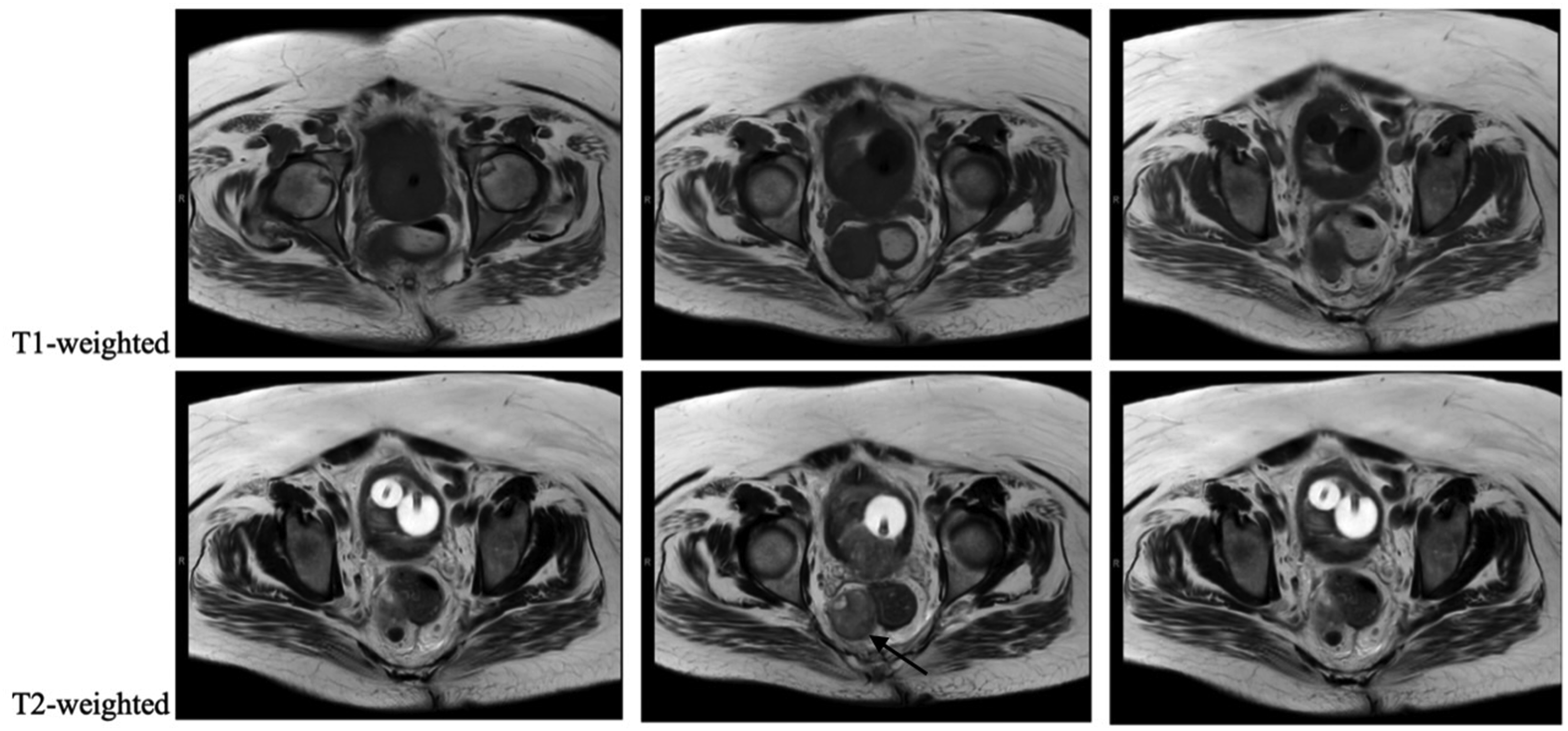
Digital rectal examination confirmed a firm extraluminal mass to the right of midline. Given the radiographic picture most consistent with a primary neurogenic tumor and the lesion’s position in the deep posterior presacral space at the S4 level with extrarectal extension, operative resection was undertaken for both definitive diagnosis and management. A posterior transsacral (Kraske) approach was selected, as this provided direct access to the presacral space with optimal visualization, avoided the need for rectal mobilization, and was anatomically best suited to the lesion’s inferior posterior position—which would have been inaccessible via a transanal or purely transperineal route. The decision to proceed with resection rather than awaiting preoperative biopsy was driven by the presumed primary presacral neoplasm, the technical barriers to safe tissue sampling at this anatomic location, and the recognition that complete surgical excision is the definitive treatment for neurogenic presacral tumors irrespective of benign or malignant designation.
The mass was successfully resected. It was situated adjacent to the puborectalis muscle and extended superiorly toward S4 without rectal invasion. Pathologic evaluation revealed metastatic prostatic adenocarcinoma. Immunohistochemical staining was positive for prostate-specific antigen and prostatic acid phosphatase, confirming prostatic origin. Subsequent prostate-specific antigen testing and staging studies identified an occult primary prostate carcinoma. The patient recovered uneventfully and was referred for systemic oncologic therapy.
This case is best understood as a diagnostic dilemma rather than a surgical paradigm. The operative decision was driven not by histologic confirmation but by a coherent preoperative presumption of primary presacral neoplasm: the lesion was solitary, well-circumscribed, and T2-heterogeneous, arising adjacent to sacral foramina in a patient without a known primary malignancy—features that closely mirror the MRI profile of sacral schwannoma.1,2 In contrast, presacral metastasis from prostate cancer most often arises in the setting of known advanced disease with concurrent osseous involvement; isolated soft-tissue presacral metastasis in the absence of a known primary is exceedingly rare, and our review of the literature identified no prior reports of this specific presentation.
The most preventable gap in this workup was the omission of serum PSA. PSA is inexpensive, non-invasive, and routinely available; an elevated result would have prompted targeted prostate imaging and potentially redirected the entire diagnostic pathway before operative planning began. Its absence reflected anchoring on catheter-related irritation as the cause of hematuria—a plausible attribution in isolation that illustrates how anchoring bias can inappropriately narrow the differential when competing explanations exist for urinary symptoms. We propose that serum PSA be considered a standard component of the preoperative evaluation of solid presacral masses in all male patients, irrespective of imaging characteristics.
Preoperative tissue sampling was not pursued for the anatomic and clinical reasons detailed in the case presentation. As the operative plan would not have changed under the presumptive diagnosis of a primary neurogenic tumor, proceeding to resection without prior biopsy was consistent with published practice for potentially resectable presacral lesions where imaging supports a primary neoplasm.3,4 In retrospect, the principal lesson is not that biopsy routes were insufficient, but that PSA should have been obtained before committing to resection—a simple test that might have reframed the differential and altered the clinical course entirely.
We acknowledge that surgical excision of metastatic prostate cancer is not standard oncologic practice, and this case must not be interpreted as endorsing surgical metastasectomy. Critically, had the diagnosis been established preoperatively, the management pathway would have differed substantially: systemic therapy and metastasis-directed therapy (MDT), not operative resection, represent the established standard of care. Prospective randomized trials including STOMP, ORIOLE, and EXTEND have demonstrated that SBRT-based MDT delays androgen deprivation therapy and improves progression-free survival in oligorecurrent prostate cancer, with meta-analytic data confirming favorable local control and acceptable toxicity.5-8 The resection in this case was performed under a preoperative diagnosis of primary presacral neoplasm, with the metastatic histology an unexpected finding that fundamentally changed its oncologic context. With respect to operative approach, the Kraske approach provides direct access to the presacral space and is anatomically well suited for posteriorly situated extrarectal masses at or below S4 that are inaccessible via transanal or transperineal routes—a technical advantage independent of tumor histology. The indication for resection, however, must be distinguished from the choice of approach: the Kraske approach is appropriate when resection is warranted for diagnostic uncertainty, symptomatic compression, or excision of a presumed primary presacral tumor, not as a strategy for known metastatic disease.
In conclusion, this case demonstrates that isolated soft-tissue presacral metastasis from an occult prostate primary can closely mimic primary neurogenic tumors on preoperative imaging, leading to operative intervention prior to tissue confirmation. Operative resection was undertaken under a preoperative diagnosis of a primary presacral neoplasm, and the metastatic histology was discovered incidentally on final pathology; management would have differed substantially had this diagnosis been established preoperatively, with systemic therapy and SBRT-based MDT representing the appropriate initial treatment strategy. Surgical excision of metastatic prostate cancer is not standard practice, and this case should not be interpreted as endorsing such an approach. The Kraske approach remains an appropriate operative strategy for selected posteriorly situated presacral lesions when resection is clinically indicated and the diagnosis is uncertain. Multidisciplinary evaluation—including routine serum PSA measurement, advanced cross-sectional imaging, and exhaustive assessment of available biopsy strategies—should be incorporated into the workup of solid presacral masses in older male patients to characterize pathology before operative intervention is undertaken.
Footnotes
Funding
The authors received no financial support for the research, authorship, and/or publication of this article.
Declaration of Conflicting Interests
The authors declared no potential conflicts of interest with respect to the research, authorship, and/or publication of this article.