
Abstract
Stroke remains a major cause of morbidity and mortality worldwide. Despite preventive measures, effective management strategies are needed to reduce the morbidity and mortality associated with this devastating condition. While the management of hemorrhagic stroke is mostly limited to supportive care, reperfusion strategies in ischemic stroke have been developed and continue to evolve. Conceptually, the pathophysiology of ischemic stroke is similar to that of acute myocardial infarction and the objective of management is similar (ie, to rapidly restore normal flow to reduce permanent damage). It is, therefore, not surprising that the management of acute ischemic stroke includes intravenous (IV) thrombolysis, the only Food and Drug Administration (FDA)-approved strategy at this point. In addition, there are a myriad of emerging endovascular interventional techniques. We review the current literature and discuss some of the technical aspects of endovascular therapy in the setting of acute ischemic stroke.
Introduction
Approximately 795 000 individuals in the United States suffer from a stroke annually. 1 Stroke remains the leading cause of disability in adults and is responsible for 150 000 deaths annually, making it the third leading cause of death in the United States (after heart disease and cancer). 2 Stroke may be hemorrhagic (15%) or, more commonly (85%), ischemic in etiology. 3 Ischemic stroke is usually the result of an embolic or a thrombotic mechanism. The extent of ischemic brain injury will depend on the time from the onset of symptoms to reperfusion, the presence of collateral circulation, and the size of the region of the brain surrounding the infarct area (the so-called necrotic core) in which the blood supply is significantly reduced and metabolism is maintained by way of collateral flow (the so-called penumbra). 4
The principle of ischemic stroke management is simple—to restore flow to the affected area as soon as possible (“time is brain”) without causing intracerebral hemorrhage. This objective, however, is complicated due to the heterogeneous nature of ischemic strokes etiologies and the lack of a widely accepted approach to achieving this objective.
Therefore, there is no single effective approach, rather the authors' recommended strategy is to individualize the approach based on the presentations and patient characteristics; taking into account the underlying mechanism of stroke, the clinical variables at presentation, timing of presentation, and with the intention to utilize all available options to achieve the objective (the so-called multimodal approach to reperfusion). 5
In this review, we discuss the current state of ischemic stroke reperfusion strategies with emphasis on endovascular therapy.
Intravenous Thrombolytic Therapy
The use of intravenous (IV) thrombolytic therapy in the management of acute ischemic stroke is the only Food and Drug Administration (FDA)-approved therapy for patients with no contraindication to IV thrombolytic therapy (Table 1 , criteria for thrombolytic therapy 6 ) and who present within 3 hours from the onset of symptoms. The support for this therapy was derived from the landmark National Institute of Neurological Disorders and Stroke (NINDS) randomized trial of IV thrombolytic therapy in the management of acute ischemic stroke. 7 In this study, 524 patients were randomized within 3 hours after the onset of an ischemic stroke to receive either IV recombinant tissue plasminogen activator (rtPA) or a placebo. The rtPA was given IV at a dose of 0.9 mg/kg (maximum dose 90 mg) infused over 1 hour. At 24 hours, there was no significant difference in the percentage of patients with neurologic improvement between the group given rtPA and that given placebo; however, at 3 months follow-up, patients in the rtPA arm had less severe disability (odds ratio [OR] for a favorable outcome, 1.7; 95% confidence interval [CI], 1.2-2.6). Furthermore, patients in the rtPA arm had a full, or nearly full, recovery rate of 34% versus 21% in the placebo arm. Despite a higher rate of symptomatic intracerebral hemorrhage (6.4% with rtPA vs 0.6% with placebo; P < .001), there was no difference in mortality at 3 months (17% in the rtPA group and 21% in the placebo group, P = .30).
Characteristics of Patients With Ischemic Stroke Who Could Be Treated With Thrombolytic a
Abbreviations: INR, international normalized ratio; CT, computed tomography; aPTT, activated partial thromboplastin time.
a Adapted from Adams HP Jr et al. 10
Despite efficacy, fewer than 5% of patients with ischemic strokes receive IV thrombolytic therapy due to the short window of opportunity (3 hours, although therapy is most effective within 90 minutes) 7,8 and numerous contraindications. The recently published European Cooperative Acute Stroke Study (ECASS)—3 trials showed some marginal benefit in stroke treatment from 3 to 4.5 hours (more favorable outcome with rtPA than with placebo, 52.4% vs 45.2%; OR, 1.34; 95% CI, 1.02-1.76; P = .04). However, this benefit was in a more limited patient population (nondiabetics, under the age of 80, and no prior history of stroke) as compared to the under 3-hour NINDS trial patients. 8 The current guidelines for acute ischemic stroke give the use of IV rtPA a class I (level of evidence A), with emphasis on careful selection of patients which is crucial in achieving a favorable outcome with this therapy. 9 The guidelines state that IV rtPA (0.9 mg/kg; maximum dose 90 mg) is recommended for selected patients who may be treated within 3 hours of onset of ischemic stroke. 9 Imaging of the brain is recommended before initiating any specific therapy to treat acute ischemic stroke and in most cases computed tomography (CT) will provide the information to make decisions about emergency management.
Endovascular Therapy for Acute Ischemic Stroke
The major cerebral vessels—the intracranial internal carotid artery (ICA), the middle cerebral artery (MCA), the anterior cerebral artery (ACA), the intracranial vertebral artery (VA), the basilar artery (BA), and the posterior cerebral artery (PCA)—are most commonly involved in the pathogenesis of ischemic stroke and fortuitously the most amenable to endovascular intervention. These vessels are connected through the posterior communicating arteries and the anterior communicating artery, forming the circle of Willis, a major source of collateral blood supply.
Endovascular therapy for the management of acute ischemic stroke includes intra-arterial thrombolysis, mechanical thrombectomy, or balloon angioplasty with or without stent placement. Conceptually, these interventional techniques are closely related to techniques applied in the coronary and other peripheral vascular beds. However, interventions in the cerebrovascular bed are associated with specific and unique challenges. For example, the lack of adventitia and external elastic lamina in cerebral vessels make them more prone to perforation. 10 Furthermore, access to these vessels is complicated by extracranial carotid and vertebral arteries tortuosity and aortic arch abnormalities.
Despite these limitations, endovascular therapy when done in properly selected patients carries the advantages of more effective reperfusion, lower dose of thrombolytic therapy (the dose of intra-arterial thrombolytic therapy is about one third the dose of IV thrombolytic therapy) 11 may be safer in older patients (>80 years) 12 and can be of benefit up to 6 hours from the onset of symptoms 13 or even arguably longer in properly selected patients. 5,14
Intra-Arterial Thrombolysis
Intra-arterial thrombolysis involves direct catheterization of the intracerebral arteries with subsequent infusion of a thrombolytic agent directly into the clot. Treatment requires the patient to be at an experienced stroke center with immediate access to cerebral angiography and qualified interventionalists. The effectiveness of intra-arterial thrombolysis has been established in the Prolyse in Acute Cerebral Thromboembolism II (PROACT II) trial, which was the largest trial that addressed the use of intra-arterial thrombolysis in acute ischemic stroke. In this seminal trial, 180 patients with acute MCA occlusion were randomized to either 9 mg of intra-arterial recombinant prourokinase (r-proUK) infused directly into the MCA over 2 hours (with 2000 units of heparin given as an IV bolus and followed by IV heparin drip at a rate of 500 U/h for 4 hours) or heparin alone in the control group. Patients were enrolled if they presented within 6 hours (median 5.3 hours) of symptom onset. 13 Patients receiving r-proUK were more likely to have recanalization (66% vs 18%; P < .001) and to achieve functional independence at 90 days (40% vs 25%; P = .04; number needed to treat was 7). Mortality was similar in the 2 groups (25% vs 27%), although the r-proUK group had a significantly higher incidence of symptomatic intracranial hemorrhage within 24 hours (10.9% vs 3.1%; P = .06).
This trial supported the ongoing use of intra-arterial thrombolytic therapy in the management of acute ischemic stroke as have successful experiences from various centers. 15 It is important to point out that in the PROACT II, mechanical disruption of the clot was prohibited by design. Also of note is the relatively higher rate of intracranial hemorrhage observed in this trial when compared with IV thrombolysis trials. This may be explained by the greater baseline stroke severity in PROACT II when compared with other studies (median National Institute of Health Stroke Score [NIHSS] 16 was 17 in PROACT II, 14 in NINDS and 11 in ECASS-II). 7,8,13,17
A meta-analysis of 5 randomized control trials (including PROACT II) with 395 participants that compared intra-arterial thrombolysis versus control (mostly IV heparin) for the management of acute stroke secondary to a large artery occlusion (mostly MCA strokes) confirmed the effectiveness of intra-arterial thrombolytic therapy in achieving a better outcome (modified Rankin scale score 18 of 0 to 2 in 43% versus 28% in the control group (OR = 2.05) and a modified Rankin scale score of 0 to 1 in 31% versus 18% in the control group (OR = 2.14), with a high rate of partial or complete vessel recanalization (65% vs 18%, OR = 6.4), and with no increase in mortality (21% vs 24%, OR = 0.8, 95% CI 0.5-1.4), despite higher risk of symptomatic intracerebral hemorrhage (9% vs 2%, OR = 2.9). 15
A class I (level of evidence B) indication is given, in the current acute stroke management, to intrarterial thrombolysis in patients who have a major ischemic stroke, presenting within 6 hours and who are not candidates for IV thrombolysis, and a class IIa (level of evidence C) for patients who have contraindications to IV thrombolysis use. The guidelines also emphasize that such therapy requires a well-intergrated stroke program with experienced interventionalists. 9
Technique
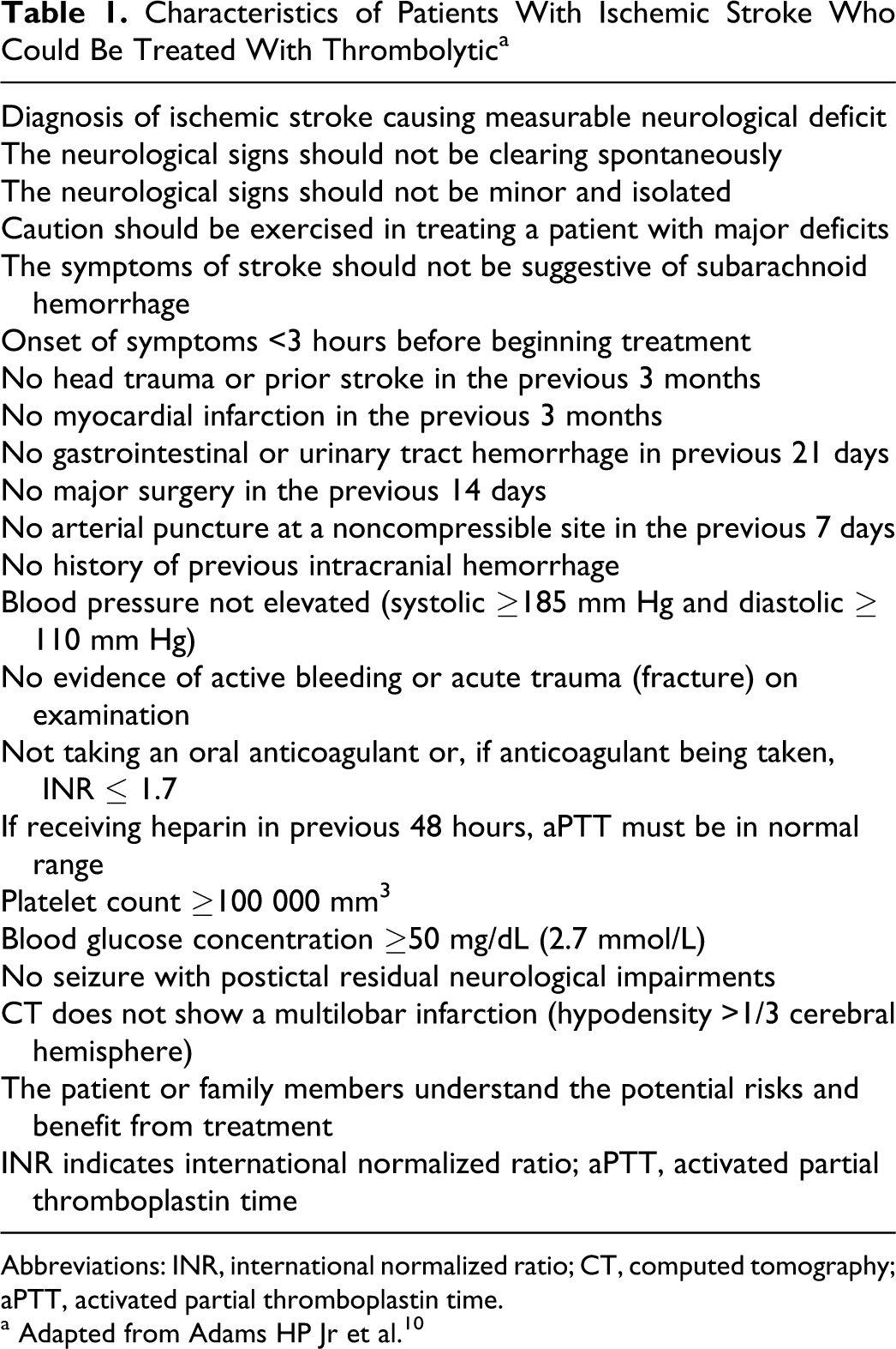
After access is obtained, usually through the common femoral artery, the symptomatic artery should be quickly cannulated (6F guide catheter over an 0.035-inch hydrophilic wire) and angiography obtained by injecting into the common carotid artery if the ischemia is in the ICA territory, or the VA (preferably, the left since it is easier to cannulate) in vertebro-basilar ischemia. An arch aortogram should not be performed as it consumes valuable time and should only be considered if the canulation of the great vessels is difficult. Once a cervical angiogram is obtained, cerebral digital substraction angiography is performed (cranial AP view, Figure 1
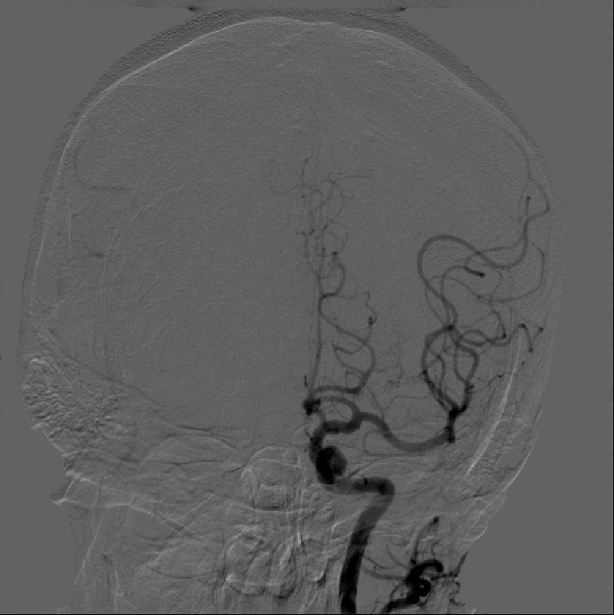
and true lateral, Figure 2
). If the ICA is patent in the neck, it should be cannulated and injected selectively. The field of view should be large enough to see the entire cerebral vascular tree (ie, entire skull), and filming should be continued until the end of the venous phase. The following angiographic findings need special attention: Obvious vessel cutoff sign of one of the major vessels or branches. Early venous shunting and delayed arterial filling and emptying.
Anteroposterior view of the cerebral circulation.
Lateral view of the cerebral circulation.
Collateral blood supply and retrograde filling of the distal MCA, ACA, PCA, or cerebellar cortical branches via pial collaterals (the presence of collaterals is a positive prognostic sign for clinical recovery and probably decreased risk of intracranial hemorrhage). Radiologic contraindications to thrombolysis such as AV malformations and large aneurysms thrombosed.
After screening angiography of the culprit artery is completed, the contralateral vessel may be cannulated to evaluate collateral blood supply from the anterior communicating and posterior communicating arteries and the pial collaterals from the PCA to the MCA or ACA. Ideally, however, the operator already gathered this information, including the arch configuratrion, from the CT angiogram obviating the need for this step. While it takes slightly longer to perform a CTA in addition to the screening head CT, it typically will take less time than it would take to perform selective angiography of the contralateral ICA and a VA to search for collaterals.
When the occlusion is identified, the 6–F guide catheter should be seated securely in the mid-distal ICA or the distal V2 segemnt of the VA. If there is severe tortuosity or difficulty maintaining guide access in the desired vessel through a short femoral sheath, a long sheath placed into the distal common carotid artery or subclavian artery will allow for better support. Intra-arterial or IV heparin bolus of 2000 U is given immediately after sheath insertion, followed by 500 U/h of IV heparin drip (PROACT II protocol).
Once the access is secured, a 0.014-inch soft-tipped hydrophilic wire is advanced through a microcatheter (or small balloon angioplasty catheter if an atherosclerotic occlusion is suspected) across the occluded segment. Care must be taken in advancing the guidewire and familiarity with the natural course of the occluded segment, and the location of the perforator branches is essential to avoid complications due to inadvertent anterior choroidal, lenticulostriate, posterior communicating, and thalamoperforator artery perforation. Once the guidewire is across, the microcatheter is advanced into the clot. At this point, the infusion of thrombolytic agents commences.
There is no concensus for the best location of the microcatheter (in the clot or proximal to it), the infusion rate of the lytic agent, the duration of infusion, the use of adjunctive pharmacologic agents (such as glycoprotein IIb/IIIa inhibitor or heparin) and mechnical inerventions (such as clot distruption, angioplasty, and stenting) and the best thrombolytic agent to achieve reperfusion.
In addition to procedural technical complications, intracrainial bleeding remains the most dreaded complication. Older age (>80), high blood pressure ([BP] >185/110 mm Hg), high serum glucose, duration of ischemia (>4 hours) in unselected patients, large clot burden necessitating multiple devices passes, evidence of infarction on CT or magnetic resonance imaging (MRI), elevated international radomized ration (INR), and thrombocytopenia are factors that are associated with higher risk of bleeding and poorer prognosis. 19
Mechanical Clot Disruption and Extraction
While IV and intrarterial fibrinlytic therapy can be effective in treating patients with ischemic stroke, these modalities are limited by contraindications for the use of fibrinlytic agents, the low recanalization rate with large vessel occlusion, 20 the inability to effectively dissolve platelet-rich clots, 21 and more importantly the ineffectiveness in preventing abrupt vessel closure following intial success (reported to be up to 34% following IV thrombolytic therapy and to be up to 17% following intra-arterial thrombolytic therapy 22,23 ).
Mechanical clot disruption has been used and reported to be effective in conjunction with intra-arterial thrombolysis or as a stand-alone therapy. It is usually effective in cases of small clot burden and a fresh clot. 24,25 Most operators will combine these techniques with intra-arterial thrombolytic therapy to increase the permability of the agent into the thrombus. A number of techniques have been utilized such as microcatheter or microwire clot maceration, photoacoustic laser catheter use, angioplasty, and a variety of embolectomy and thrombectomy devices. 25–27
In the nonrandomized safety and efficacy of mechanical embolectomy in acute ischemic stroke (MERCI) study, 28 the MERCI Mechanical clot Retreiver device (Concentric Medical, Mountain View, California) was utilized in 141 patients who were ineligible for IV rtPA, and who could be treated within 3 to 8 hours, and who had angiographic demonstration of thrombotic occlusion in the intracranial ICA, MCA, BA, or VA. The MERCI device is a corkscrew-like coil that is deployed distal to the thrombus, then retracted to snare the thrombus, back through a guide catheter that is balloon tipped (the balloon is inflated to obstruct antegrade flow, limiting distal emobolization and improving thrombectomy efficacy). The median baseline NIHSS score was 19 (higher than PROACT II). The study group was compared with the placebo group from the PROACT II trial. There was no evidence that the treated patients had improved outcome at 90 days compared to the PROACT II historical control. However, patients in the MERCI trial had a higher rate of recanalization Thrombolysis in Myocardial Infarction (TIMI 2 or 3 flow) than the historical control group (46% vs 18%). The overall mortality was higher in the MERCI trial than PROACT II (44% vs 27%), which may be due in part to the differences in patients enrolled in the 2 tirals. The MERCI study included older patients with more severe strokes with larger vessel occlusions (increased thrombus burden) that included BA and internal carotid occlusions. The rate of intracranial hemorrhage was reported at 8%. The rates of procedural complications (embolization, dissection, subarachnoid hemorrhage, perforation, and groin hemorrhage) were reported at 13%.
Another thrombectomy device, the Penumbra system (Penumbra, Inc, Alameda, California) has been used with limited reported data. In a study of 27 consecutive patients, mechanical thrombectomy was performed alone or in combination with IA thrombolysis or intracranial stent placement (n = 4). The primary end point was the achievement of TIMI 2 or 3 flow in the target vessel and the secondary end points were improvement of >4 points on the NIHSS score at discharge, improvement in the overall mortality at 3 months, modified Rankin score, the rate of symptomatic intracranial hemorrhage, and procedure-related adverse events. The mean NIHSS score was 14 ± 7. Recanalization to TIMI 2 or 3 was achieved in 25 patients (93%). None of the patients developed symptomatic intracranial hemorrhage. At hospital discharge, 15 patients (56%) had an NIHSS improvement of ≥4 and 13 patients (48%) had a modified Rankin score of ≤2, at 3 months. There was a significant correlation between complete vessel recanalization and favorable outcome. The all-cause mortality 29 at 3 months was 11%. The use of penumbra device was further tested in the prospective, multicenter, single-arm Penumbra Pivotal Stroke Trial of 125 patients with ischemic strokes (NIHSS ≥8), who presented within 8 hours and had angiographic occlusion (TIMI 0-1) of the culprit artery. Recanalization (achieving TIMI 2-3) was achieved in 81.6%. However, only 25% of patients recovered to a modified Rankin scale 30 of ≤2, with a 3-month mortality rate of 32.8% and symptomatic intracranial hemorrhage of 11.2%.
Another method of clot disruption and removal has been reported, mostly through case reports and case series, with the use of snare devices (Gooseneck or Lasso). This technique mandates the removal of the guidewire prior to snaring, and if snaring is unsuccessful, it may be difficult to redirect guidewire into the culprit vessel. However, its efficacy appears to be comparable with the other available embolectomy devices.
In current guidelines, the use of the MERCI device is given a class IIb recommendation (level of evidence B) and emphasizes that more studies are warranted to further define the role of this device and other available mechanical devices. 9
Intracranial Angioplasty and Stent Placement
Angioplasty with or without stent placement has been used effecitvely by experienced operators as first-line therapy in patients with acute ischemic stroke in the context of stenotic atherosclerotic lesions in the intracrainial vessels. These lesions could precipitate an acute ischemic event, as thrombotic occlusion (and distal embolization) develops over a stenotic segment. Mechnical disruption of the in situ clot will not resolve the underlying mechanism (as decreased turbulent flow through the stenotic segment will lead to platelet aggregation and thrombus formation), causing a high rate of abrupt reocclusion. Therefore, to ensure the restoration of normal flow, angioplasty is perfomed with an undersized balloon and may be all that is needed to restore normal flow, 31–34 and then stent is placed if warranted or intended. 5,35
Two strategies have been advocated; angioplasty with provisional stenting (if warranted by lesion irregularities, residual high grade lesion, or elastic recoil) versus primary stent placement (the intention to place a stent following angioplasty to reduce elastic recoil and possibly restenosis).
The drawbacks of angioplasty and stenting are dissection, perforation, and plaque shift into the perforating branches. 35,36 Also in, the long term, the need for dual antiplatelet therapy and the possiblity of instent restenosis may be associated with adverese outcomes.
Technical Aspect of Intracranial Angioplasty and Stenting
While the technical aspects of these proceudres are similar to that of coronary angioplasty and stent placement, there are uniqe aspects to the intracranial vascular bed that warrant special attention. The intracranial vessles are more prone to perforation and dissection. These vessels are also tortous, particularly the cavernous carotid artery segment, with some segments surrounded by bone (petrous carotid) or tough dura (vertebrals) making them inflexible. 37
Intracranial angioplasty and stenting in the management of acute stroke (as opposed to management of intracranial atherosclerosis to prevent stroke 38 ) is idealy used as part of multimodel approach in patients with an acute thrombotic occlsuion superimposed on atherosclerotic flow limiting lesion. 5 In this setting, much like plaque rupture in acute coronary syndrome, thrombosis in situ with or without distal embolizationon is the underlying mechanism of stroke and without an effective resolution of the lumen stenosis, recurrent thrombosis is likely following clot aspiration, maceration, or dissolution. 39
Technically, once access is achieved (as described above), 0.014 hydrophilic wire is advanced meticulously through the obstructive lesion. Balloon angioplasty is done (with a slightly undersized balloon) using low inflation pressure (2-5 atmospheres) for 30 to 60 seconds. Post angioplasty spasm and elastic recoil are commonly encountered. If angiography confirms the restoration of flow with no dissection, then some operators advocate to end the procedure at this point. However and drawing mostly from the coronary arena, stent placement has been advocated to complement the results of angioplasty by possibly reducing elastic recoil, improving flow in atherosclerotic segment and reducing restenosis.
Balloon Expandable or Self-Expanding Stents?
The initial experience with intracranial revascularization using stents involved the use of coronary balloon mounted stents (BMS) as a rescue procedure following failed pharmacologic and mechanical interventions. 35 The overall 35 recanalization rate varies from 63% to 79%. Stent placement was an independent predictor of recanalization (OR = 4.8, CI = 1.8-10.0; P < .001), in a retrospective analysis of 168 consecutive patients treated with endovascular therapy for acute ischemic stroke. 35
The advent of self-expanding stents (SES) specifically designed for intracranial revascularization has revolutionized cerebral vascular stenting. These devices have excellent profile (when compared to coronary BMS), are more likely to conform the vessel tortuousity, and are much more easily delivered. 35–40
Five intracranial SES are currently available; the Neuroform stent (Boston Scientific, Natick, Massachusetts), the Enterprise stent (Codman Neurovascular, Raynham, Massachusetts), The Leo Stent (Balt Extrusion, Montmorency, France), the Solitaire/Solo stent (ev3, Inc, Irvine, California), and the Wingspan stent (Boston Scientific). Of these, the first 3 were designed for the treatment of wide-necked cerebral aneurysms. The Solitaire/Solo stent was designed for both aneurysm and acute stroke treatment and represents a new generation of devices sometimes called “stent-retriever.” This device, and other in development, can act as retriever devices for clot extraction, as temporary scaffolding devices to permit perfusion and to facilitate thrombolysis, or as a permanent stent implant. The Wingspan stent was designed for elective treatment of atherosclerotic intracranial stenoses.
The reported experience in single-center case series was salutary, with a stent placement success rate of about 90%, and a rate of restoration of TIMI 2 to 3 nearing 80%. However and bearing in mind that these studies 41–44 were small, the mortality rates reported ranged between 30% and 40%, with intracerebral beelding reported in 11% to 40% and stent thrombosis to be about 0.5% to 11%. In these series, stenting was a bailout after other treatments had failed so the patients were more likley to have prolonged ischemia and more interventions and therefore were more likely to have poor outcomes and complications. Also in some series, stenting was reserved for patients with undelying atherosclerotic occlusion, while in others stenting was performed regardless of pathophysiology including a majority of cases with embolic occlusions, that is stenting of normal arteries.
In the Stent-Assisted Recanalization in acute Ischemic Stroke (SARIS), SES placement was used for 20 patients who did not improve after IV thrombolytic therapy or had a contraindication to that therapy. The mean time from stroke onset to intervention was 5 hours and 13 minutes. Total time from procedure onset to vessel recanalization was 45 minutes. The average presenting NIHSS score was 14. Stenting was performed in 19 (95%) of 20 enrolled patients successfully. TIMI 2 or 3 flow was achieved in all stented patients (100%). An improvement by ≥4 points in NIHSS score was reported in 65% of patients after treatment. In all, 1 patient had symptomatic intracerebral hemorrhage ([ICH] 5%) and 2 had asymptomatic ICH (10%). At 1 month follow-up, 12 (60%) of 20 patients had a modified Rankin Stroke score of ≤2 and 9 (45%) had score of ≤1. Mortality at 1 month was 25% (5 patients). None of the patients enrolled in this study died due to any cause related to stent placement; all deaths were due to the severity of the initial stroke and associated comorbidities. 45 The experience with SES in acute stroke mangement is promising; however, most studies are small. It is important to point out that currently available SES can only be used in large vessels, such as the ICA, proximal MCA (M1), BA, intracranial VA, proximal anterior (A1), and proximal posterior cerebral arteries (P1). The characteristics of the available stents vary as well, with regard to trackability, crossing profile, stent pore size, and radial strength. The latter 2 aspects may have important effects on the recanalization efficacy and acute closure. More important perhaps are the consequences of stent placement in mostly normal arteries. These include the need for dual antiplatelet therapy and even the intraprocedural infusion of glycoprotein (GP) IIb/IIIa receptor antagonists. All such pharmacological adjuncts will increase the risk of ICH to some degree, which will greatly increase the risk of mortality. Some patients who receive stents may develop malignant cerebral edema requiring decmpressive hemicraniectomy which may not be possible in the presence of potent antiplatelet agents. Many patients with large vessel occlusion will have atrial fibrillation as the underlying cause requiring anticoagulation, which in combination with dual antiplatelet agents is problematic in the setting of acute stroke. Acute vessel closure and in-stent stenosis are also major concerns. At present, the guidelines state that combinations of interventions to restore perfusion cannot be recommended outside the setting of clinical trials. Therefore, clincial trials with adequate numbers of patients are needed on the safety of this approach. It should be noted that the Stenting and Aggressive Medical Management for Preventing Recurrent stroke in Intracranial Stenosis (SAMMPRIS) study, the first prospective randomized multicenter trial to compare aggressive medical management alone versus aggressive medical management plus angioplasty combined with stenting in patients with symptomatic highgrade (70%-99%) stenosis of a major intracranial artery (intracranial carotid, MCA, intracranial VA, and BA), was recently stopped due to a higher risk of stroke and death in the stented group. 46
Conclusions
The potential for endovascular intervention to restore normal perfusion to the acutely ischemic brain has great appeal. However, currently available evidence does not provide conclusive support for the safety or efficacy of available interventional strategies. Intravenous rtPA (0.9 mg/kg; maximum dose 90 mg) is recommended for selected patients who may be treated within 3 hours of onset of ischemic stroke. If thrombolytics are used, physicians should be vigilant for bleeding complications. Intra-arterial thrombolysis is an option for the treatment of selected patients who have major stroke of <6 hours duration and who are not otherwise candidates for IV rtPA. Although the MERCI and Penumbra devices are reasonable interventions for extraction of intra-arterial thrombi in carefully selected patients, their utility in improving outcomes after stroke is unclear. These and newer mechanical clot removal devices need to be be studied in additional clinical trials to help define their role in the emergency management of stroke. Emergency angioplasty and or stenting appear promising and may have a role in management. As with the intra-arterial administration of thrombolytics, the use of these devices is limited to those stroke centers that have the resources and physician expertise to perform these procedures safely. In addition to reperfusion strategies, optimal medical therapy of the high-risk patients is of paramount importance to improve clinical outcomes. 47–51
Footnotes
The authors declared no potential conflicts of interest with respect to the research, authorship, and/or publication of this article.
The authors received no financial support for the research, authorship, and/or publication of this article.