
Abstract
We assessed the relation between platelet-to-lymphocyte ratio (PLR) on admission and contrast-induced nephropathy (CIN) in patients with non-ST-segment elevation acute coronary syndrome (NSTE-ACS). A total of 488 patients with NSTE-ACS who underwent urgent coronary angiography were enrolled. Levels of PLR and creatinine were measured before angiography and at 72 hours after angiography. Patients were divided into 2 groups, namely, the CIN group, 80 patients (16.3%; age 65.3 ± 12.5years; 66.7% men) and the non-CIN group, 408 patients (83.7%; age 61.2 ± 12.3 years; 72.5% men). Patients in the CIN group had significantly higher PLR than those in the non-CIN group (152.9 ± 99.6 vs 120.4 ± 66.1, P < .001). In logistic regression analysis, PLR (odds ratio [OR] 1.004, 95% confidence interval [CI] 1.001-1.007, P = .02), diabetes mellitus (OR 1.75, 95% CI 1.02-2.98, P = .03), and ST-segment depression on admission electrocardiogram (OR 1.68, 95% CI 1.00-2.81, P = .04) were independent predictors of CIN. The PLR was an independent predictor of CIN after angiography in patients with NSTE-ACS.
Introduction
Contrast-induced nephropathy (CIN) is an important complication of invasive cardiovascular (CV) procedures that require the administration of contrast media and is associated with prolonged hospitalization and increased short- and long-term morbidity and mortality. 1 The risk of developing CIN varies depending on the patient population evaluated, definition criteria, and preventive strategies. 2 The incidence of CIN is 3-fold higher in patients with acute coronary syndrome (ACS), and patients with diabetes mellitus or baseline renal impairment have a risk of almost 50% for developing CIN. 3 –5 Therefore, it is crucial to identify patients with ACS who may be at risk of CIN. Neutrophil gelatinase-associated lipocalcin level, cystatin C level, and the neutrophil-to-lymphocyte ratio (NLR) have been used to predict CIN. 6 –9
The platelet-to-lymphocyte ratio (PLR) has been proposed as anti-inflammatory marker and predictor for major adverse CV outcomes in coronary artery disease (CAD). High PLR has emerged as a significant independent predictor of no reflow in patients with ST-segment elevation myocardial infarction (STEMI) and long-term survival in patients with non-ST-segment elevation myocardial infarction (NSTEMI). 10,11 However, no study has investigated the relationship between PLR and CIN in non-ST-segment elevation ACS (NSTE-ACS).
Because of the potential role of inflammation in the development of CIN, we investigated the role of PLR in predicting CIN in patients with NSTE-ACS undergoing early invasive strategy.
Methods
Patients
A total of 488 consecutive patients with NSTE-ACS who underwent urgent angiography at our institution between January 2011 and January 2014 were enrolled. Exclusion criteria were high-risk features warranting emergency coronary angiography (within 2 hours), acute renal failure or end-stage renal failure requiring dialysis, known malignant or chronic inflammatory disease, recipient of transplanted organs, contrast dye exposure within the last 10 days, and chronic treatment with steroid or nonsteroidal anti-inflammatory drugs. According to these criteria, 26 patients were excluded (acute infection: 10 patients, history of malignancy: 5 patients, urgent coronary bypass surgery: 6 patients, and chronic dialysis: 5 patients). The remaining 488 patients (mean age: 61.8 ± 12.4 years; 71.4% men) formed our study population. The study was planned and conducted according to the ethical standards detailed in the Declaration of Helsinki and approved by ethical committee of Istanbul University.
Definitions and Diagnoses
At presentation, diagnosis of NSTE-ACS was based on the presence of chest pain, electrocardiogram (ECG) findings, and troponin T levels. Prolonged (>20 minutes) angina pain at rest, new-onset (de novo) angina (class II or III of the Classification of the Canadian Cardiovascular Society), recent destabilization of previously stable angina with Canadian Cardiovascular Society Class III angina characteristics (crescendo angina), or postmyocardial infarction (post-MI) angina and absence of persistent ST-segment elevation were criteria for diagnosis of NSTE-ACS. Patients with normal troponin T levels were considered to have unstable angina pectoris, and those with high troponin T levels were considered to have NSTE-ACS. 12
Venous blood samples were drawn immediately after hospital admission. The white blood cell (WBC), platelet, and lymphocyte count were determined. Cardiac enzymes (creatine kinase [CK], CK-MB, and troponin T), glucose, creatinine, and lipid levels were recorded in all patients. Serum creatinine level measurement was repeated at 72 hours after contrast medium administration. The estimated glomerular filtration rate (GFR) was calculated by applying the Levey-modified Modification of Diet in Renal Disease formula: (186.3 × serum creatinine−1.154) × (age−0.203) × (0.742 if female). 13 The PLR was calculated as the ratio of the platelet count to the lymphocyte count.
The 6-month Global Registry of Acute Cardiac Events (GRACE) risk score was calculated for each patient by assigning the appropriate number of points for each of the 9 prognostic variables entered into the calculation: age, history of heart failure, history of acute MI, heart rate and systolic blood pressure at admission, ST-segment depression, serum creatinine at admission, elevated myocardial necrosis markers or enzymes, and lack of percutaneous coronary revascularization during admission. 14
All patients were assessed with transthoracic echocardiography (Vivid-E9; GE Vingmed Ultrasound AS, Horten, Norway) within 24 hours after admission. Left ventricular ejection fraction (LVEF) was calculated using the Simpson method.
On admission, all patients were administered aspirin, clopidogrel (loading dose of 300 mg followed by 75 mg/d), enoxaparin, and a statin if they were not already on a statin. Percutaneous coronary intervention (PCI) was performed immediately after diagnostic angiography when appropriate. After coronary angiography, all patients continued to take aspirin (100 mg/d orally) indefinitely and clopidogrel (75 mg/d) for at least 12 months. In accordance with our standard protocol, all patients received intravenous hydration with isotonic saline (1 mL/kg/h, 0.9% sodium chloride for 12 hours both before and after the procedure). Hydration rate was reduced to 0.5 mL/kg/h in both groups for patients with LVEF <40%.
Study End Point
The primary end point was CIN, defined as an increase in serum creatinine level by ≥0.5 mg/dL or ≥25% over the baseline value within 72 hours after contrast agent administration. 15
Statistical Analysis
All analyses were performed using SPSS for Windows version 21.0 (IBM Corporation). Continuous variables are presented as mean ± standard deviation and categorical variables are presented as percentages. Continuous variables were checked for normal distribution using the Kolmogorov-Smirnov test, and those that did not satisfy the criteria were log transformed to obtain a normal distribution. Comparisons of parametric values between the 2 groups were performed by means of independent-samples Student t tests. Categorical variables were compared by the chi-square test. Receiver–operating characteristic (ROC) analyses were used to detect the cutoff value of PLR in the prediction of CIN. To determine the independent predictors of CIN, parameters that were found to be significant in the univariate analysis were evaluated by stepwise forward logistic regression analysis. Two-sided P values <.05 were considered significant.
Results
There were 80 (16.3%) patients in the CIN group (mean age 65.3 ± 12.5; 66.7% men), and 408 patients in the non-CIN group (mean age 61.2 ± 12.3; 72.5% men). Baseline clinical characteristics of all study patients are listed in Table 1. The patients in the CIN group were significantly older, with a higher prevalence of diabetes mellitus, hypertension, chronic kidney disease (CKD), previous MI, ST depression on ECG, and higher heart rate on admission and lower LVEF than those in the non-CIN group. No significant differences in the frequency of smoking history, hyperlipidemia, and proportion of male sex were observed between the groups. Laboratory findings are summarized in Table 2. In the CIN (+) group, serum glucose (143 ± 57 vs 128 ± 57, P = .04), baseline creatinine (1.29 ± 1.36 vs 0.99 ± 0.47, P = .001), GRACE score (155.5 ± 43.3 vs 140.0 ± 37.0, P = .01), and platelet count (258 ± 86 vs 238 ± 69, P = .02) were higher, whereas hemoglobin (12.4 ± 1.9 vs 13.2 ± 1.9, P < .0001) and baseline GFR (76 ± 30 vs 82 ± 24, P = .03) were lower compared with the CIN (−) group. The PLR was significantly higher in the CIN group than in the non-CIN group (152.9 ± 99.6 vs 120.4 ± 66.1, P < .001; Table 2).
Baseline Characteristics of the Study Population.
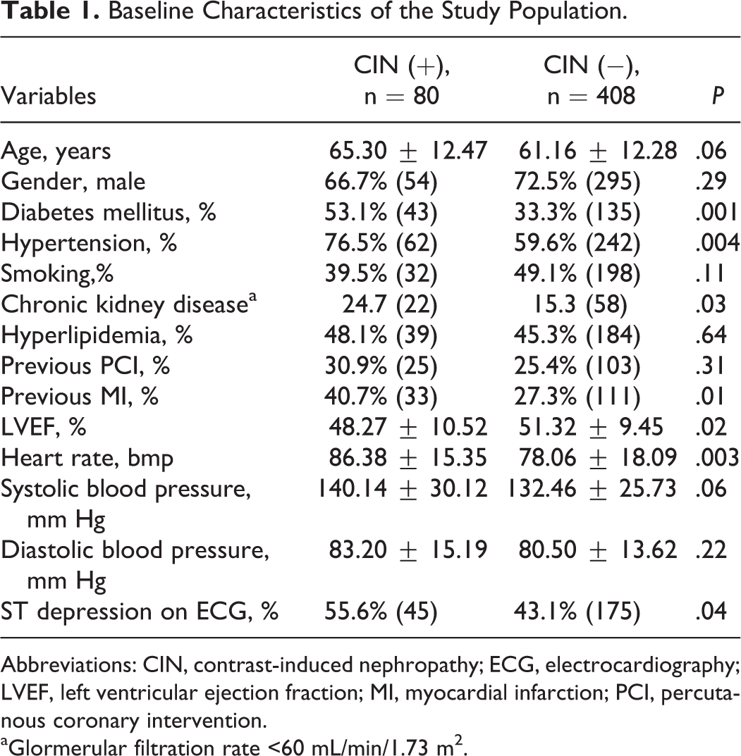
Abbreviations: CIN, contrast-induced nephropathy; ECG, electrocardiography; LVEF, left ventricular ejection fraction; MI, myocardial infarction; PCI, percutanous coronary intervention.
aGlormerular filtration rate <60 mL/min/1.73 m2.
Laboratory Measurements of Study Groups.
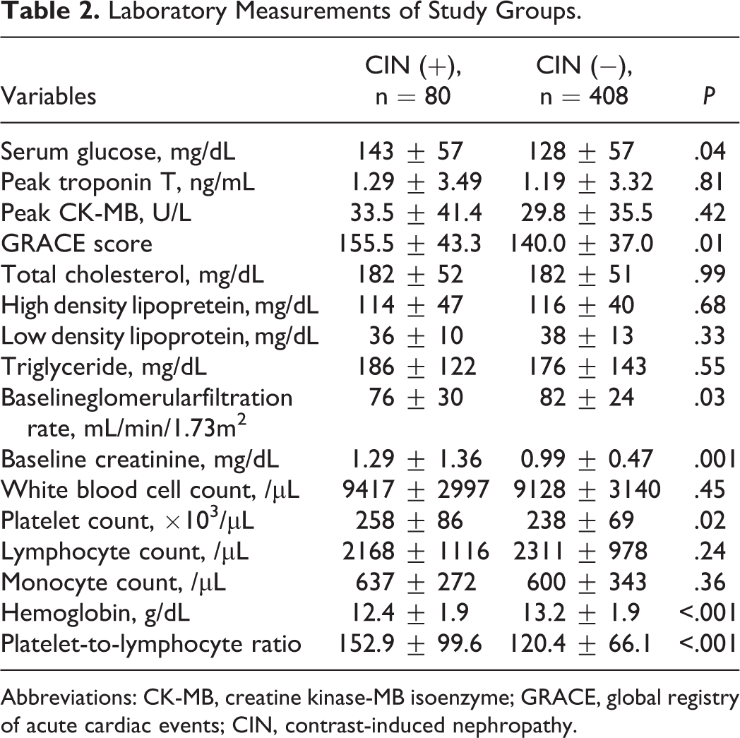
Abbreviations: CK-MB, creatine kinase-MB isoenzyme; GRACE, global registry of acute cardiac events; CIN, contrast-induced nephropathy.
The angiographic and procedural characteristics of the patients are presented in Table 3. After undergoing angiography, PCI was performed on 31 (38.8%) patients, 20 (25.0%) patients went on to coronary artery bypass grafting (CABG), and 29 (36.3%) were treated medically in the CIN (+) group (P = .03). In the CIN (−) group, PCI was performed for 219 (53.6%) patients, 65 (15.9%) patients underwent CABG, and 124 (30.3%) were treated medically (P = .03). Amount of contrast media (229.6 ± 162.7 vs 210.9 ± 126.5 mL, P = .25) and frequency of multivessel disease (71.6% vs 57.3%, P = .06) were higher in the CIN than non-CIN group, but these differences were not statistically significant.
Angiographic and Procedural Characteristics of Study Groups.
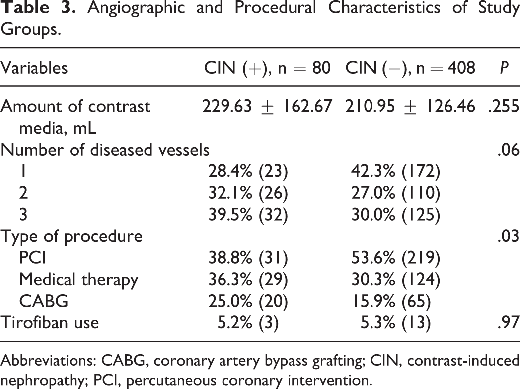
Abbreviations: CABG, coronary artery bypass grafting; CIN, contrast-induced nephropathy; PCI, percutaneous coronary intervention.
Univariate and multivariate logistic regression analyses of the association between the CIN and multiple parameters are listed in Table 4. In multivariate analyses, PLR (odds ratio [OR] 1.004, 95% confidence interval [CI] 1.001-1.007, P = .02), diabetes mellitus (OR 1.75, 95% CI 1.02-2.98, P = .03), and ST-segment depression on admission ECG (OR 1.68, 95% CI 1.00-2.81, P = .04) were independent predictors of CIN. The Hosmer-Lemeshow test showed that the model fit the data well (P = .67).
Independent Predictors of Contrast-Induced Nephropathy.
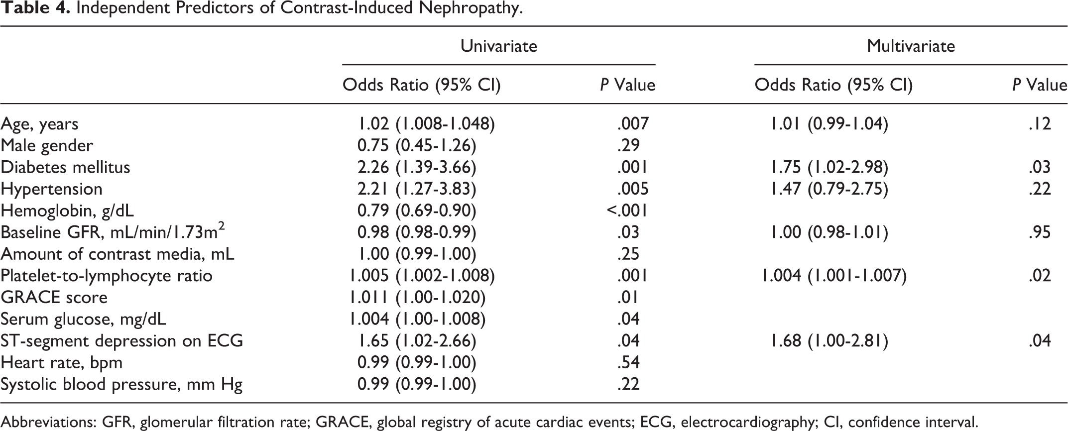
Abbreviations: GFR, glomerular filtration rate; GRACE, global registry of acute cardiac events; ECG, electrocardiography; CI, confidence interval.
The area under the ROC curve for PLR was 0.68, and a PLR of >112 predicted CIN with sensitivity of 68% and specificity of 58% (0.684, 95% CI 0.641-0.725, P < .001; Figure 1).
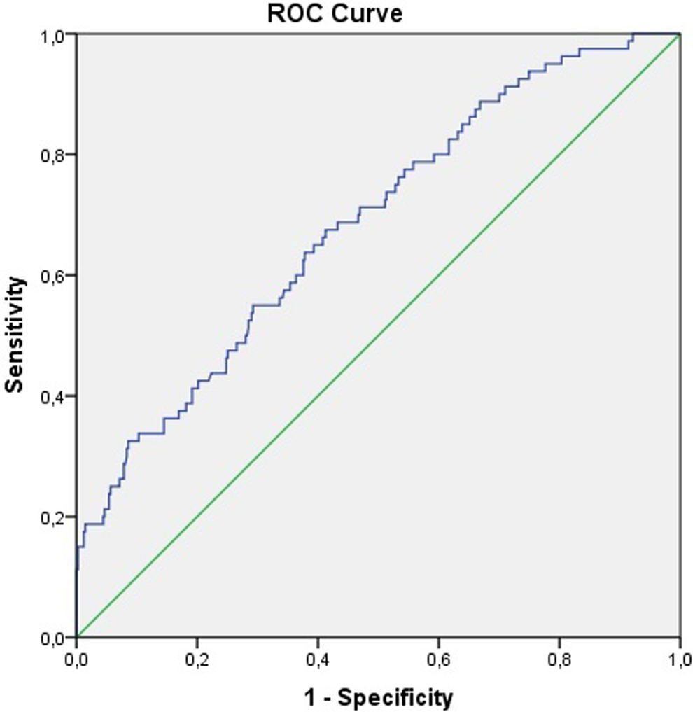
Receiver–operating characteristics curve analysis of platelet-to-lymphocyte ratio and contrast-induced nephropathy.
Discussion
Our study findings showed that PLR, a readily accessible parameter, is an independent risk factor for CIN in patients with NSTE-ACS. In our study, prevalence of CIN was 16.3% in the whole study population, 24.7% in the population with CKD, and 24.2% in the diabetic population. Although the pathogenesis of CIN is not completely understood, multiple mechanisms may be involved, and inflammation is known to play a key role. Experimental studies have shown a significant relationship between inflammation and acute renal injury. Inflammatory cells, such as macrophages, natural killer cells, lymphocytes, and particularly neutrophils, infiltrate damaged tissue leading to further renal destruction. 16 Following experimental studies, clinical investigations also reported the role of inflammation in the initiation and extension of CIN using C-reactive protein levels. 17 Furthermore, favorable effects of statin treatment on prevention of CIN have been demonstrated in many studies, and the underlying mechanism of this protection is linked to the anti-inflammatory effects of statins. 18,19 In our study, all patients were given statin therapy on admission according to current guidelines. 12
Decreased GFR, DM, hypovolemia, and the amount of contrast media are known risk factors for the development of CIN. 20 Furthermore, older age, hypertension, dyslipidemia, metabolic syndrome, and heart failure are also associated with CIN. 20 In our study, DM, ST-segment depression on admission, hypertension, CKD, and previous MI were more frequent in the CIN (+) group, but in multivariate analysis, apart from PLR, only DM and ST depression on admission were predictors of CIN. Although the association of DM and CIN is well established in previous studies, 3 –5 the effect of ST depression has not been investigated in this issue and this is another novelty of our study. Another interesting finding of our study was that PCI in CIN (+) group was less frequent than in CIN (−) group. High prevalence of multivessel disease in CIN (+) group may explain this finding.
Complete blood count is an easy, readily available, and routine examination that provides information about red blood cells and WBCs, platelets, the count and dimensions of subgroups of cells including red cell distribution width and platelet distribution width, and parameters such as the PLR and NLR. White blood cell count and subtypes are recognized as inflammatory markers in CV diseases. 21 Kaya et al investigated the role of NLR in predicting CIN among patients with STEMI. 9 They found that NLR was higher in the CIN group and an independent predictor of CIN along with advanced age, diabetes mellitus, low baseline GFR, and high amount of contrast media.
Platelet-to-lymphocyte ratio has been investigated as a new inflammatory marker and predictor of major adverse events for malignancies and CV diseases. 22,23 High PLR emerged as a significant independent predictor of no reflow inpatients with STEMI and of long-term survival in patients with NSTEMI. 10,11
Balta et al 24 compared NLR with PLR among patients with end-stage renal disease (ESRD) for predicting inflammatory status. They concluded that PLR was positively correlated with NLR, interleukin 6, and tumor necrosis factor α levels. Additionally, patients with ESRD having higher PLR had higher levels of inflammation. Finally, PLR was found to be superior to NLR in terms of predicting inflammation in ESRD. Both lymphopenia and high platelet count have been shown to be associated with adverse events in a wide range of populations withCAD. 25 –27 The advantage of PLR is that it reflects both hyperactive coagulation and inflammatory pathways, and as both of them are the underlying mechanisms of CIN, PLR may be a valuable marker for CIN.
As mentioned previously, recent studies have demonstrated the importance of PLR for short- and long-term CV events among different CAD subgroups, but to our knowledge, no study has demonstrated the importance of PLR for the prediction of CIN in patients with NSTE-ACS.
Our study has several limitations. It was an observational, single-center study with a relatively small number of patients. Additionally, we did not compare PLR with other inflammatory markers (eg, fibrinogen or myeloperoxidase) because they were not routinely measured in our study population.
We conclude that PLR, a readily available parameter, is an independent risk factor for CIN in this particular group of patients with NSTE-ACS. The PLR may help identify high-risk candidates who require intensive hydration prior to PCI and careful monitoring of renal function.
Footnotes
Authors’ Note
All of the authors were included in all the following processes: (1) substantial contributions to conception and design, or acquisition of data, or analysis and interpretation of data; (2) drafting the article or revising it critically for important intellectual content; and (3) final approval of the version to be published.
Declaration of Conflicting Interests
The author(s) declared no potential conflicts of interest with respect to the research, authorship, and/or publication of this article.
Funding
The author(s) received no financial support for the research, authorship, and/or publication of this article.