
Abstract
We present our single-center results on ultrasound-assisted thrombolysis (USAT) in patients with pulmonary embolism (PE) at intermediate high risk (IHR) and high risk (HR). Our study consisted of 75 patients with PE who underwent USAT (60 at IHR and 15 at HR). The median time delay from symptoms to USAT was 5 days. Ultrasound-assisted thrombolysis resulted in improvements in tricuspid annular plane systolic excursion; pulmonary artery (PA) systolic and mean pressures; Qanadli score; right to left ventricle diameter ratio and right to left atrial diameter ratio; and diameters of main, right, and left PA regardless of the baseline risk status (P < .0001 for all). Death was documented in 4 patients, and major and minor bleeding were noted in 2 and 5 of the patients, respectively. No PE-related event was noted during postdischarge follow-up period of median 310 days. Our study revealed that USAT facilitates the resolution of PA thrombotic burden, recovery of pulmonary hemodynamics, and right heart functions with acceptable rates of procedure-related complications in patients with PE, irrespective of the IHR or HR status.
Introduction
Although various catheter-directed treatment (CDT) technologies including thrombus fragmentation, suction or rheolytic embolectomy, conventional catheter-directed thrombolysis, and pharmacomechanical thrombolysis have been reported in patients with acute pulmonary embolism (PE), none of these was tested in randomized clinical trials. 1 –3
A novel ultrasound-assisted thrombolysis (USAT) technology, EkoSonic Endovascular System (EKOS Corporation, Bothell, Washington) was reported to facilitate thrombolysis with a reduced bleeding risk in PE series. 4 –9 This system was tested in the ULTrasound Accelerated ThrombolysIs of PulMonAry Embolism (ULTIMA) randomized clinical trial and a prospective, single-arm, multicenter trial of ultrasound-facilitated, low-dose fibrinolysis for acute massive and submassive PE. 10,11 In the ULTIMA trial, a standardized USAT regimen was reported to be superior to heparin alone in reversing right ventricular (RV) dilatation at 24 hours, without an increased bleeding risk in patients at intermediate risk. 10 Furthermore, A Prospective, Single-arm, Multi-center Trial of EkoSonic® Endovascular System and Activase for Treatment of Acute Pulmonary Embolism (SEATTLE II) trial showed that USAT provides a significant improvement in right ventricle to left ventricle (RV/LV) ratio, pulmonary artery (PA) pressure, and thrombotic burden within 2 days, irrespective of the submassive or massive PE at presentation. 11 However, despite the improvements in CDT systems, absence of the convincing robust data other than ULTIMA study seems to preclude the current guidelines recommendations for more liberal use of USAT/CDT systems in intermediate-high risk (IHR) and high-risk (HR) PE. 12 Nonetheless, there is a paradoxical situation for CDT in terms of the source of the evidence and address of risk group in guideline recommendations. Although the majority of the convincing evidences for CDT have been derived from patients at IHR or formerly submassive PE subset, indications are directed to patients at HR in whom the data for efficacy of CDT remain inconclusive.
In our single-center study based on retrospective analysis of patients with acute PE at IHR or HR treated by USAT system, we evaluated the acute changes in RV systolic function as assessed by echocardiography, PA pressures, PA diameters and PA thrombotic burden, right atrial to left atrial diameter (RA/LA) ratio, RV/LV ratio as assessed by computed tomography (CT), bleeding events, and PE-related or all-cause mortality.
Methods
This single-center study comprised 75 patients referred to our tertiary cardiovascular center with a diagnosis of acute PE and underwent USAT treatment from October 2012 to July 2015. The diagnosis of acute PE and definition of HR and IHR subgroups were based on the criteria as recommended by European Society of Cardiology/European Respiratory Society (ESC/ERS) 2014 PE Guidelines. 12 Inclusion criteria were acute symptomatic PE confirmed by contrast-enhanced CT with embolus located in at least 1 main or proximal lower lobe PA. Lower extremity venous Doppler ultrasound (US) and echocardiographic data were also available in all of patients.
Clinical end points
Echocardiographic, invasive, and CT measures were used as surrogates of efficacy. Safety outcomes included minor and major bleeding, recurrent venous thromboembolism, serious adverse events, and death (all cause and cardiovascular), up to 30 days after USAT treatment. Major bleeding was defined as overt bleeding associated with a fall in the hemoglobin level of at least 2.0 g/dL or with transfusion of 2 units of packed red blood cells or involvement of a critical site. Clinically overt bleeding not fulfilling the criteria of major bleeding was classified as a minor bleeding complication.
Echocardiographic assessment
Short-axis view was used to evaluate the presence of D-shaped interventricular septum, and the apical four-chamber view was used for estimation of PA systolic pressure (PASP) from tricuspid regurgitation jet, M-Mode measurement of tricuspid annular plane systolic excursion (TAPSE), and tissue velocity of tricuspid annular longitudinal systolic motion (St). 12 –14
Contrast-enhanced chest CT
Images were acquired before and after the USAT procedure on a 64-slice helical CT scanner with angiographic contrast material (Omnipaque 350; Toshiba Aquilion 64; Toshiba Medical Systems Corp, Tokyo, Japan) and the stored images recorded at the time of diagnosis and following the USAT were retrospectively evaluated. The main, right, and left proximal PA diameters; RV/LV diameter ratio; RA/LA diameter ratio; and PA thrombotic occlusion score were measured from the CT images.
Quantification of PA thrombotic obstruction
For evaluation of PA thrombotic obstructive burden, a score proposed by Qanadli et al (Qanadli score [QS]) was used, and PA occlusion was quantified as the product of N × D, where N was the value of the clot site, and D was the degree of obstruction. 15 Nonobstructive thrombus located in a lobar or main PA received score points equal to the number of arising segmental PA branches (maximum 10 points per lung). A segmental PA containing nonobstructive thrombus without any thrombus in its proximity received 1 score point, and score points were multiplied by 2 in case of occlusive clot (range 0-40 points, maximum 20 points per lung).
Right Heart Catheterization, Pulmonary Angiography, and USAT Procedures
Pulmonary artery systolic pressure, PA diastolic pressure (PADP), and PA mean pressure (PAMP) were measured and recorded prior to PA angiograms. EkoSonic (EKOS, Bothell, Washington) Endovascular Device consisting of the Intelligent Drug Delivery Catheter (IDDC) and the MicroSonic Device (MSD) containing multiple small US trancducers distributed over the treatment zone was used. A 0.035-inch hydrophilic guidewire was used to cross the thrombotic segment of target PA, and after a safe position within a large segmental PA, the multipurpose catheter was exchanged with the IDDC of the USAT system. Following the removal of guidewire, an MSD was inserted and advanced throughout the IDDC and connected to the EkoSonic control unit. Recombinant tissue-type plasminogen activator (tPA) was used as thrombolytic agent, and a continuous infusion of tPA according to the selected dose and duration and saline coolant at 35 mL/h per catheter were initiated.
Statistics
Sample size calculation was performed using previous trials. 8–9 We calculated sample size for all primary surrogate markers (PASP, PAMP, PADP obtained by cardiac catheterization; PASP, TAPSE, and St obtained by echocardiography; and RV/LV ratio obtained by CT) and the larger sample size was chosen. Finally, we determined that a minimum of 49 patients were needed. Descriptive statistics were reported as mean ± standard deviation for normally distributed continuous data, median (interquartile range) for nonnormally distributed data, and percentages for categorical data. The distribution of continuous data was assessed by Kolmogorov-Smirnov test. Student t test or Mann-Whitney U test was used for between-group comparisons. The outcomes (PAMP, CT obstruction scores, and RV/LV ratio) before and after USAT were assessed using paired t test or Wilcoxon matched-pair signed rank test. A 2-sided P value < .05 was considered significant. All statistical analyses were performed using SPSS Statistics version 20.0 (IBM SPSS Statistics for Mac; IBM Corp, New York).
Results
Baseline Clinical Characteristics and Treatment Regimen
The baseline clinical characteristics and laboratory data were summarized in Table 1. The median time delay from symptom onset to admission was 5 (3-10) days. In 22 (30.6%) patients, USAT treatment was started after the failure of the intravenous infusion of tPA. The placement of the system catheters was technically successful in all patients. The mean hospital stay was 5.1 ± 2.5 days.
Baseline Clinical, Laboratory, and Treatment Characteristics of 75 Patients.
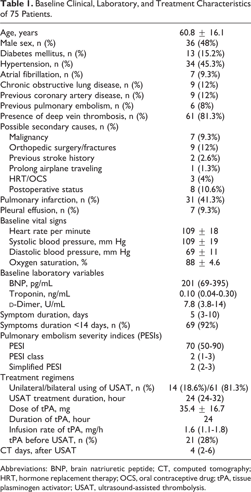
Abbreviations: BNP, brain natriuretic peptide; CT, computed tomography; HRT, hormone replacement therapy; OCS, oral contraceptive drug; tPA, tissue plasminogen activator; USAT, ultrasound-assisted thrombolysis.
Primary Analysis of Surrogate Markers for Efficacy Outcomes
Echocardiographic assessment
Measures obtained at baseline assessment and after USAT were given in Table 2. Compared with baseline measures, the estimated PASP from tricuspid regurgitation jet was significantly decreased (55.8 ± 12.6 mm Hg vs 37.3 ± 8 mm Hg, P < .001) and the TAPSE (18.1 ± 3.9 mm vs 21.3 ± 4.1 mm; P < .001) and St (13.1 ± 2.8 vs 14.1 ± 2.5; P = .043) were increased after USAT treatment (Table 2; Figure 1).
Echocardiographic, CT, and Invasive Hemodynamic Measures Before and After USAT.
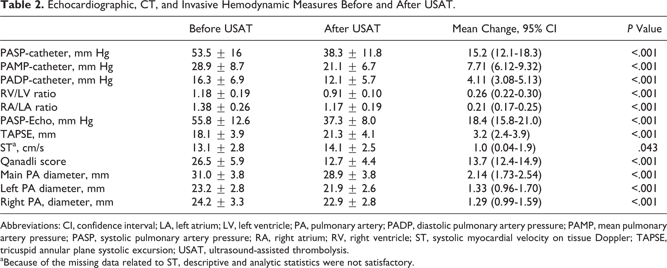
Abbreviations: CI, confidence interval; LA, left atrium; LV, left ventricle; PA, pulmonary artery; PADP, diastolic pulmonary artery pressure; PAMP, mean pulmonary artery pressure; PASP, systolic pulmonary artery pressure; RA, right atrium; RV, right ventricle; ST, systolic myocardial velocity on tissue Doppler; TAPSE, tricuspid annular plane systolic excursion; USAT, ultrasound-assisted thrombolysis.
aBecause of the missing data related to ST, descriptive and analytic statistics were not satisfactory.
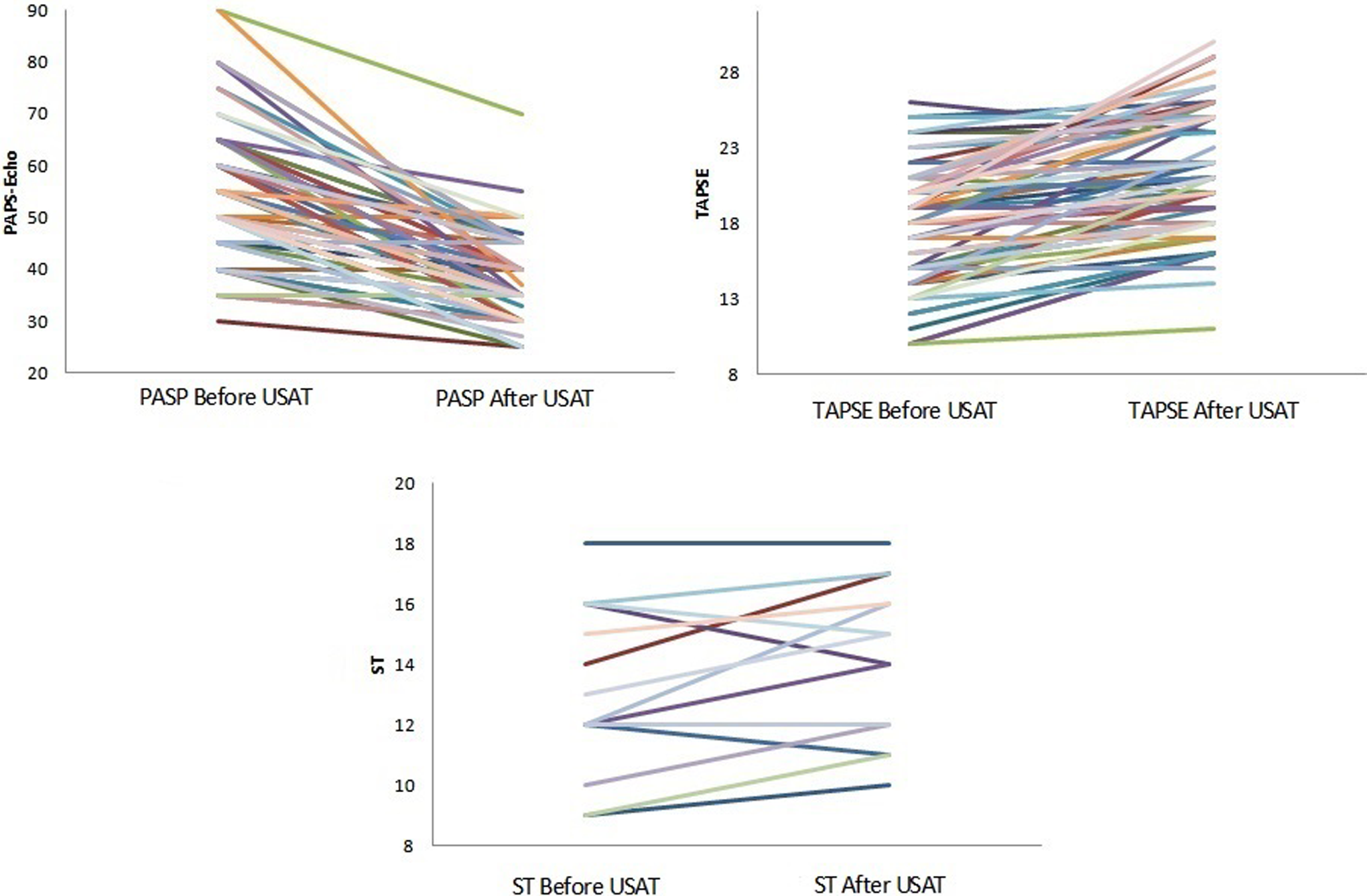
Echocardiographic measures (systolic pulmonary artery pressure [PASP], tricuspid annular plane systolic excursion [TAPSE], and tissue velocity of tricuspid annular longitudinal systolic motion [St]) before and after ultrasound-assisted thrombolysis (USAT) treatment.
Baseline and final CT assessment
Baseline and postprocedural CT assessment were performed in all patients. The CT data were summarized in Table 2. In the subgroup of the 22 patients who underwent intravenous tPA prior to USAT treatment, only CT images obtained after termination of intravenous tPA infusion were used as baseline CT data. Baseline assessment showed pulmonary infarction distal to the obliterated PA territories in 31 (43.7%) patients, and pleural effusion were noted in 7 (10%) patients (Table 2). These findings did not deteriorate after USAT as assessed by final CT assessment. The mean change (95% confidence interval [CI]) in main PA truncus, left, and right PA diameters were 2.14 (1.73-2.54), 1.33 (0.96-1.70), and 1.29 (0.99-1.59) mm, respectively. Both RV/LV diameter ratio (1.18 ± 0.19 vs 0.91 ± 0.10; P < .001) and RA/LA diameter ratio (1.38 ± 0.26 vs 1.17 ± 0.19; P < .001) were significantly improved after USAT treatment. The mean reduction in RV/LV diameter ratio and RA/LA diameter ratio were 0.26 (0.22-0.30) and 0.21 (0.17-0.25), respectively. Moreover, USAT resulted in a significant mean reduction (95% CI) in QS (26.5 ± 5.9 vs 12.7 ± 4.4; P < .001). The mean reduction (95% CI) in QS was 13.7 (12.4-14.9; Figure 2).
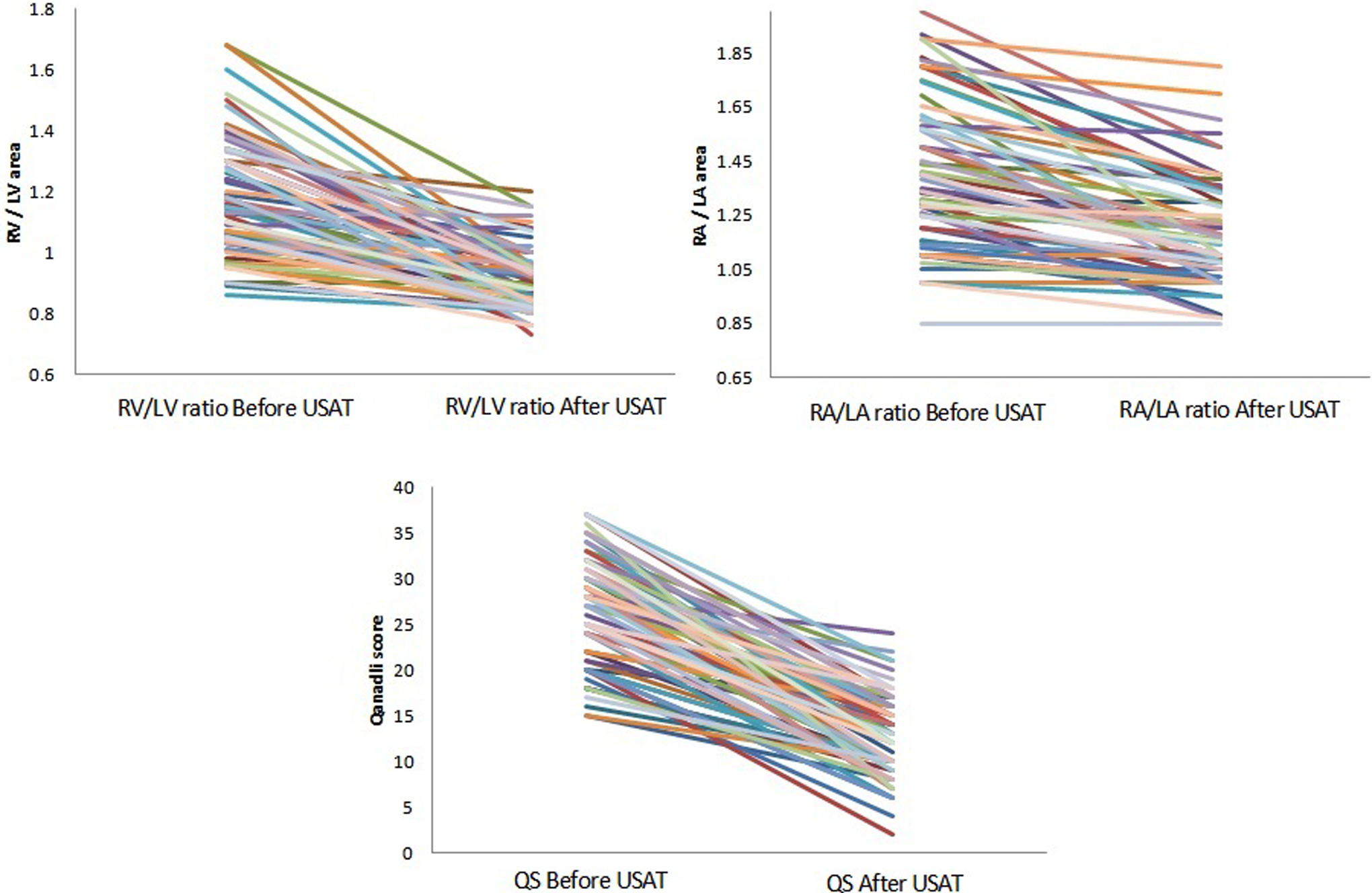
Computed tomography measures (RV/LV diameter ratio, RA/LA diameter ratio, and obstruction score) before and after ultrasound-assisted thrombolysis (USAT) treatment.
Invasive hemodynamic measures
Baseline and postprocedural invasive measures of PASP, PADP, and PAMP were given in Table 3. The PASP (53.5 ± 16 mm Hg vs 38.3 ± 11.8 mm Hg; P < .001), PADP (16.3 ± 6.9 mm Hg vs 12.1 ± 5.7 mm Hg; P < .001), and PAMP (28.9 ± 8.7 mm Hg vs 21.1 ± 6.7 mm Hg; P < .001) were significantly reduced after USAT treatment. The mean reduction (95% CI) in PASP, PADP, and PAMP were 15.2 (12.1-18.3), 4.11 (3.08-5.13), and 7.71 (6.12-9.32) mm Hg, respectively (Figure 3).
Comparison of Change in Absolute Values Between High-Risk and Intermediate-High-Risk Patients.
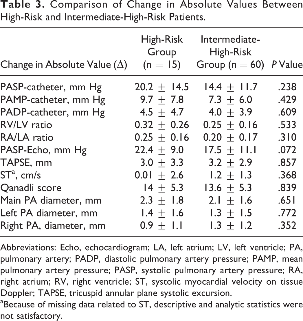
Abbreviations: Echo, echocardiogram; LA, left atrium; LV, left ventricle; PA, pulmonary artery; PADP, diastolic pulmonary artery pressure; PAMP, mean pulmonary artery pressure; PASP, systolic pulmonary artery pressure; RA, right atrium; RV, right ventricle; ST, systolic myocardial velocity on tissue Doppler; TAPSE, tricuspid annular plane systolic excursion.
aBecause of missing data related to ST, descriptive and analytic statistics were not satisfactory.
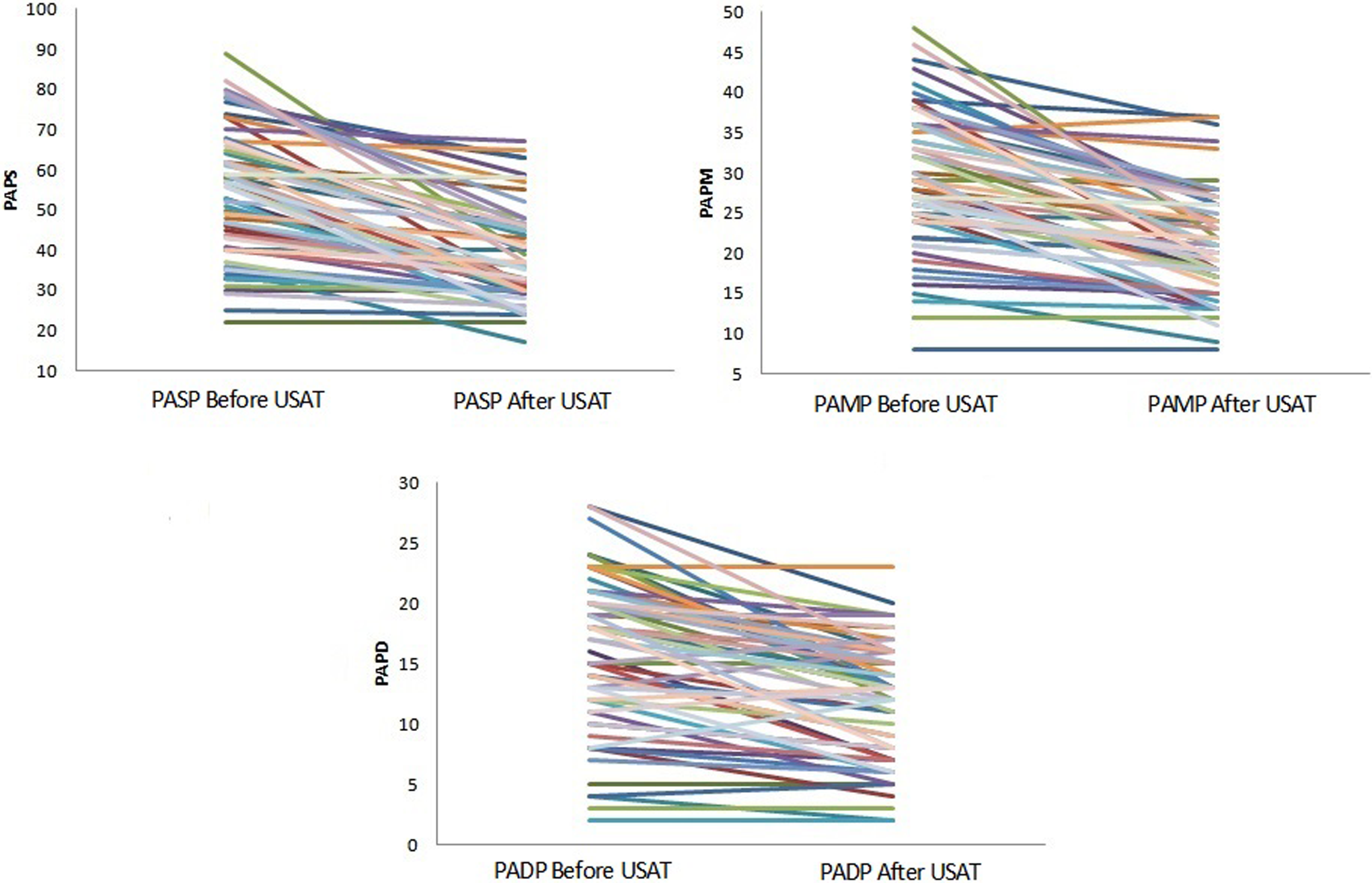
Invasive hemodynamic measures (systolic pulmonary artery pressure [PASP], mean pulmonary artery pressure [PAMP], and diastolic pulmonary artery pressure [PADP]) before and after ultrasound-assisted thrombolysis (USAT) treatment.
The comparison of the HR and IHR groups
The comparison of the HR and IHR groups demonstrated that absolute change in echocardiographic measures of PASP, TAPSE, and St; invasive measures of PA pressures; and CT measures of PA diameters, QS, RV/LV diameter ratio, and RA/LA diameter ratio were comparable between groups (P > .05; Table 3).
Primary Analysis of Safety Outcomes
Minor bleeding episodes at puncture sites and major bleeding were noted in 2 and 5 patients, respectively. In 1 patient with post-coronary artery by-pass surgery (CABG) shock due to bilateral massive PE, tPA treatment needed to be terminated at 6 mg of the drug infusion because of the neck hematoma originating from previous jugular venous puncture, but bilateral USAT treatment was maintained for 24 hours only with coolant and heparin infusion. This treatment regimen improved clinical status and PA thrombus burden, and patient was discharged from hospital at 11th day of the treatment. Death was documented in 4 patients. In 2 patients, death was due to sudden deterioration of the RV function and hemodynamic status following an initial improvement in the clinical findings under USAT treatment. In a third patient, death was due to intracranial bleeding developing at the 12th hour of the treatment. In the last patient (91-year-old female), mortality was not primarily due to treatment failure or complication but was associated with a nosocomial pneumonia documented after satisfactory response in hemodynamic measures and clinical status.
Heparin-induced thrombocytopenia occurred in 4 patients, and 3 patients were treated with fondaparinux, and 1 was treated with rivaroxaban. No episodes of minor or major bleeding, recurrent venous thromboembolism, or death related to PE were documented during the follow-up period of median 310 days (minimum 68-maximum 983 days).
Discussion
This study is based on the retrospective evaluation of the patients with PE at IHR and HR who underwent percutaneous treatment with USAT system that combines a novel technology of ultrasonic facilitation with the low-dose and slow infusion tPA treatment. This treatment modality provided statistically significant benefits in terms of the resolution in PA thrombus burden, PA pressure overload, and recovery of the RV and RA functions with acceptable complication rates regardless of the HR or IHR status at baseline stratification.
The need for ideal risk-adjusted PE treatment strategies that combines the efficacy with safety issues remained unmet for at least 4 decades. Despite the clear instructions of PE guidelines recommending the intravenous thrombolytics in patients at HR, avoidance from this treatment is reported in more than two-thirds of the patients with PE even in the presence of the hemodynamic instability and/or RV dysfunction. 12,16 –21 The systemic use of thrombolytics carries a high bleeding risk of up to 20%, and especially large tPA dosage up to 100 mg over 2 hours is associated with a 3% to 5% risk of intracranial hemorrhage. 12,16,19 –23 Therefore, current treatment strategies in PE should aim to facilitate the resolution of PA obstruction, right-sided cardiac dysfunction, and hemodynamic instability, and to prevent evolution to chronic thromboembolic pulmonary hypertension without an increased bleeding risk compared with those observed with current lytic or anticoagulant therapies.
The efficacy of thrombolytics depends on the penetrance of the agent throughout the fibrin strands to access plasminogen receptor sites embedded into the clot. 4,5,8,23 In the meta-analysis by Kuo et al on CDT modalities for massive PE, utilization of CDT was associated with a high clinical success of 86.5% with minor and major complication rates of 8% and 2.4%, respectively. This success rate was 91.2% in series in which the rate of systemic thrombolytic use >80% and 89.2% in a series in which the rate of intra-PA local thrombolytic use was >80%. 24 In the recently published initial results of prospective multicenter Pulmonary Embolism Response to Fragmentation, Embolectomy, and Catheter Thrombolysis registry, the success rates for CDT including fragmentation, embolectomy, and catheter thrombolysis techniques were 85.7% in patients with massive PE and 97.3% in patients with submassive PE. Moreover, CDT was associated with significant reduction in PAMP (mm Hg) and improvement in RV systolic strain in 89.1% of the patients without major complications. 25 Ultrasound-assisted thrombolysis technology integrates CDT with high-frequency and low-power acoustic streaming that facilitates disaggregation and separation of fibrin fibers and increases the penetration of the lytics into the clot and accelerates thrombolysis. 7,12,25,26 This system was documented to facilitate recovery of RV function and hemodynamic status with a lower dose of lytics and a lower risk of bleeding events in intermediate and HR PE compared with other CDT technologies. 1 –3,7,9,13,26–27 In the ULTIMA randomized clinical trial, USAT, but not heparin alone treatment, was associated with a significant reduction in RV/LV diameter ratio at 24 hours. Moreover, EKOS compared with heparin alone provided a greater benefit in RV/LV diameter ratio at 90 days, whereas bleeding outcomes were comparable between the 2 treatment strategies. 7 The SEATTLE II trial showed a significant improvement in RV/LV diameter ratio, PA pressures, modified Mastora score from baseline to 48 hours irrespective of the submassive or massive PE status at presentation. 11 In reference to these positive results, the Food and Drug Administration (FDA) approved this USAT system for the treatment of PE in May 2014.
The treatment regimen and results of USAT in our single-center study are comparable with those in previous studies, the ULTIMA randomized trial and the SEATTLE II multicenter registry. 10,11 This treatment modality resulted in significant improvements in QS, PASP, PADP, PAMP, PA diameters, RV/LV diameter ratio, RA/LA diameter ratio, TAPSE, and St, regardless of the baseline risk status at presentation. Mortality was documented in 4 of the 75 patients. Intracranial bleeding was noted in 1 patient and resulted in death. In 2 patients, initial improvement with USAT was complicated by severe RV failure and shock possibly due to reembolization or treatment failure. Other patient died from a hospital-acquired pneumonia despite the satisfactory hemodynamic and clinical response to USAT treatment. The minor and major bleeding rates were 6.6% and 2.6%, respectively. The median follow-up of 310 days revealed an event-free survival of 100%.
Current ESC/ERS PE guidelines recommend systemic thrombolysis as the treatment of choice for patients with HR PE (class I, level B) and surgical embolectomy in those in whom thrombolysis has failed to improve the hemodynamic status or that option was contraindicated, if surgical expertise and resources are available (class I, level C). As an alternative to surgery, CDT is recommended in HR PE if expertise with this method and the appropriate resources are available on-site (class IIa, level C). However, systemic thrombolysis is not routinely recommended as primary treatment for patients with IHR PE (class III, level B) but is recommended to be considered if clinical signs of hemodynamic decompensation appear (class IIa, level B). Surgical pulmonary embolectomy (class IIb, level C) or CDT(class IIb, level B) may be considered as alternative, “rescue” procedures for patients with IHR PE, in whom hemodynamic decompensation appears imminent and the estimated bleeding risk under systemic thrombolysis is high. 12 The general use of the CDT term seems to refer to a wide spectrum of technologies and not specifically address the potential therapeutic advantageous of the currently used USAT system that is the only FDA-approved percutaneous CDT 4 decades after the approval of the first CDT, the Greenfield suction embolectomy catheter. 27
Because of the lack of the prospective and randomized study design, and the lack of a heparin alone arm in IHR PE group, and the lack of a standard dose intravenous thrombolysis alone arm in HR group may be considered as important limitations. However, according to our knowledge, in reference to patient number of the single-center studies, our study is the largest in the published or presented series. In addition, we cannot comment about contrast-induced acute kidney injury because we do not have comprehensive data. Finally, USAT was not a first-line treatment in all patients at IHR and was initiated after the documented failure of the intravenous thrombolytic treatment in 20 (27%) patients. However, only CT images obtained after termination of intravenous thrombolytic treatment were used as baseline reference CT data for the assessment of the efficacy of USAT strategy in these patients.
Retrospective evaluation of our single-center data from patients with PE at IHR or HR who underwent low-dose tPA treatment with USAT system demonstrated significant improvements in PA thrombotic burden, PA diameters, hemodynamic measures, and RV and RA functions with acceptable complication rates regardless of the risk status at presentation.
Footnotes
Authors’ Note
All authors contributed to (1) substantial contributions to conception and design, or acquisition of data, or analysis and interpretation of data, (2) drafting the article or revising it critically for important intellectual content, and (3) final approval of the version to be published. All statistical analysis was performed by I. H. Tanboga.
Preliminary results of this study were presented at the Annual American College of Cardiology Congress in San Francisco (United States), 2015 and Annual EuroPCR Congress in Paris (France), 2015, and an abstract based on final results of this study was accepted as the best abstract at the Annual European Society of Cardiology Congress London (United Kingdom), 2015.
Declaration of Conflicting Interests
The author(s) declared no potential conflicts of interest with respect to the research, authorship, and/or publication of this article.
Funding
The author(s) received no financial support for the research, authorship, and/or publication of this article.