






Abstract
Objective:
To evaluate the role played by adenoids as a reservoir for infection in children assigned for adenoidectomy.
Methodology:
The study included 35 children with adenoid hypertrophy. All patients underwent clinical examination and adenoidectomy, adenotonsillectomy, or myringotomy with insertion of aeration tube according to indications. Surgical specimens were processed for conventional bacterial culture examination and to assay for biofilm formation. The obtained adherence values using spectrophotometer at 595 nm (OD595) was used to classify isolates according to its biofilm forming capacity.
Results:
We did adenotonsillectomy and myringotomy with insertion of aeration tube in 5 patients having adenotonsillitis with otitis media with effusion. We did adenotonsillectomy in 12 patients having adenotonsillitis and adenoidectomy in 18 patients having adenoid hypertrophy. Thirty-one surgical specimens showed bacterial growth on conventional media, while 4 specimens failed to give growth. The predominant organism was H influenzae then Staph aureus and Strept pneumoniae. Thirty-two specimens showed biofilm forming capacity (BFC) of variable extent, while others showed no BFC.
Conclusion:
Adenoids act as a bacterial reservoir secondary to bacterial biofilm formation so could induce chronicity and initiate development of complications. Determination of BFC using the proposed protocol is feasible, inexpensive, and available and spares the need for sophisticated instruments or approaches.
Introduction
A biofilm is an aggregate of cells of 1 or several species that grow and adhere to a live or inert surface and are embedded in an exo-polysaccharide (EPS) matrix of their own making that has an electrical charge and a 3-dimensional structural organization and complex function. Biofilm construction starts with the formation or accumulation of a layer of organic waste, which neutralizes the excess charge and free energy of a surface to enable bacteria adhere to it. Then, a cascade of changes is triggered that will activate a group of genes, which will determine the phenotype of the biofilm. Bacteria are joined to the surface and to one another by reversible electrostatic attraction to remain irreversibly joined. When the basic structure has formed, the biofilm starts a ripening process where the cells will grow and reproduce inside the microenvironment, which has been created and determined by the substances belonging to the EPS and by the neighboring cells and proximity to a water channel. The final stage is the sweeping and the dispersal of the bacteria from the biofilm, either by external forces or by a physical movement.1,2
Several pathogenic bacterial species that are found in the environment can form complex multicellular structures on surfaces known as biofilms. Pseudomonas aeruginosa, Vibrio cholera, and certain species of nontuberculous mycobacteria are examples of human pathogens that form biofilms in natural aquatic environments. The dynamics of biofilm formation facilitates the transmission of pathogens by providing a stable protective environment and acting as a nidus for the dissemination of large numbers of microorganisms, both as detached biofilm clumps and by the fluid-driven dispersal of biofilm clusters along surfaces. Biofilm formation conveys a selective advantage to certain pathogens by increasing their ability to persist under diverse environmental conditions. 3
According to the National Institute of Health of the USA, “more than 60% of all microbial infections are caused by biofilms.” This is enough to consider that common infections like those of the genitourinary tract, infections produced by catheters, middle ear infections in children, and the formation of dental plaque and gingivitis are caused by biofilms for this statement to seem more realistic. These biofilms are more common than previously thought, and almost all of the people have been in contact with them in the form of infections in the teeth or humid, slippery areas. 4
Adenoid hypertrophy and associated symptoms of children are a common condition in pediatrics and ENT practice that is responsible for a great amount of medical visits and 1 of the main reasons for antibiotic treatment and parental leave off work. Nasopharyngeal obstruction caused by adenoid hypertrophy may lead to several other diseases and symptoms, such as hyponasality, snoring, obstructive sleep apnea syndrome (OSAS), acute otitis media, otitis media with effusion (OME), middle ear atelectasia, cholesteatoma formation, slow feeding, acute sinusitis, abnormal facial development, and behavioral problems.5-7 The current prospective study aimed to evaluate the role played by adenoids as a reservoir for infection in children assigned for adenoidectomy.
Materials and Methods
The current study was conducted at Departments of Ototrhinolaryngology and Medical Bacteriology in Saudi-German Hospital in Jeddah, Saudi Arabia, in collaboration with Clinical and Chemical Pathology department, Cairo University hospital in Cairo, Egypt, from January 2013 until August 2014. After approval of the study protocol by the local Ethical Committee and obtaining written fully informed parents’ consent, all children assigned for adenoidectomy for adenoid hypertrophy, irrespective of being associated with complications or not, were enrolled in the study.
Preoperative examination included history taking and complete otorhinolaryngological examination that included otoscopic and audiologic examinations (tympanometry) to determine the presence of associated OME. Clinical and radiological assessment of adenoid volume was performed. Only patients with adenoid tissue >2 cm in diameter, which could be removed by a sharp adenotom device without any surface injury or disruption of biofilm layers, were enrolled in the study.
Adenoidectomy was conducted under general inhalation anesthesia supplemented by intravenous non-depolarizing muscle blockers, short-acting narcotics, dexamethasone, and ondansetron at doses appropriate for patient weight. Prophylactic antibiotic were administered. The transoral removal was gently performed at the basis of adenoids avoiding surgical injury of torus tubarius or the nasal septum. If tonsillectomy was indicated, tonsillar tissue was removed using monopolar electrocautery and conventional technique, and hemostasis was obtained with suction electrocautery. In cases of OME, myringotomy and suction of the middle ear fluid content and insertion of the grommet’s aeration tube were performed.
Each specimen (adenoid or tonsil) was washed in a sterile saline solution and was processed for conventional bacterial culture examination and analyzed by the RapID NH System (BioMerieux, Marcy-l’Etoile, France). The tests used in the RapID NH System are based on microbial degradation of specific substrates detected by various indicator systems. The reactions employed are a combination of conventional tests and single-substrate chromogenic tests.
To assay biofilm formation, strains were grown overnight in BRUCELLA+VITK+HEM (MAIM-Spain) at 37oC and 5% CO2. The culture was diluted 1:20 in fresh BRUCELLA+VITK+HEM, and 200 µl of this suspension was used to inoculate sterile 96 well polystyrene microtitre plates (CELLSTAR-greiner bio-one). After 24 hours at 37oC and 5% CO2, wells were washed with phosphate-buffered saline (PBS) solution, dried in an inverted position, and stained with 1% crystal violet for 15 minutes. The wells were rinsed again, and the crystal violet was solubilized in 200 µl of ethanol-acetone (80:20 vol/vol). The optical density at 595 nm was determined using a microplate reader (Multiskan EC, Labsystem, Budapest). The adherence values (OD595) obtained allowed the classification of isolates into 4 groups: non-biofilm forming (OD595 ≤ 1), weak biofilm forming (1 < OD595 ≤ 2), medium biofilm forming (2 < OD595 ≤ 3), and strong biofilm forming (OD595 > 3). 8
Statistical Analysis
Obtained data were presented as mean ± SD, ranges, numbers, and ratios. Results were analyzed using chi-square test (χ2 test). Statistical analysis was conducted using the SPSS (Version 15) for Windows statistical package. P value <.05 was considered statistically significant.
Results
The study included 35 patients with mean age of 6.4 ± 1.3 years (range, 4-9 years); 20 were males and 15 were females. All patients had adenoid hypertrophy with a mean largest diameter of adenoid of 2.7 ± 0.3 cm (range, 2.1-3.5 cm). Five patients had adenotonsillitis complicated by OME, 12 patients had adenotonsillitis, and 18 patients had adenoid hypertrophy.
Traditional culture of obtained specimens showed predominance of H influenzae that could be isolated in 17 specimens (48.6%). Interestingly, 4 of the 5 specimens (80%) obtained from patients with adenotonsillitis complicated by OME gave a positive culture for H influenzae, 5 specimens of those had adenotonsillitis (41.7%), and 8 specimens (44.4%) of those had adenoid hypertrophy also gave a positive culture for H influenzae. Six specimens (17.1%) gave growth for Streptococcus pneumoniae; 4 specimens obtained from patients had adenoid hypertrophy (22.2%), and 2 specimens were obtained from patients with adenotonsillitis (16.7%). Seven specimens (20%) gave growth for Staphylococcus aureus; 3 specimens obtained from patients had adenoid hypertrophy (16.7%) and 4 from patients had adenotonsillitis (33.3%). One specimen of those who had adenoid hypertrophy gave growth to Moraxella catarrhalis (Figures 1 and 2). Four specimens (11.4%) failed to give bacterial growth, for a negative culture rate of 11.4% (Table 1).
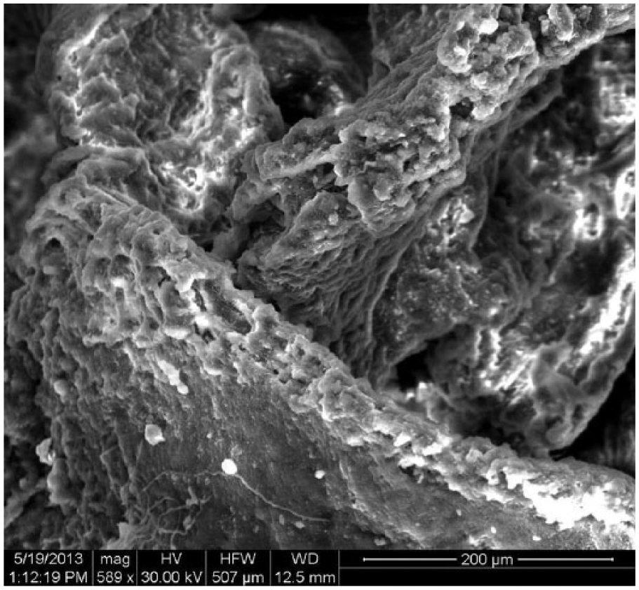
Scanning electron micrograph showing evidence of bacterial biofilms in samples of adenoid tissue of a child with adenotonsillitis. A mature biofilm containing thousands of bacteria.
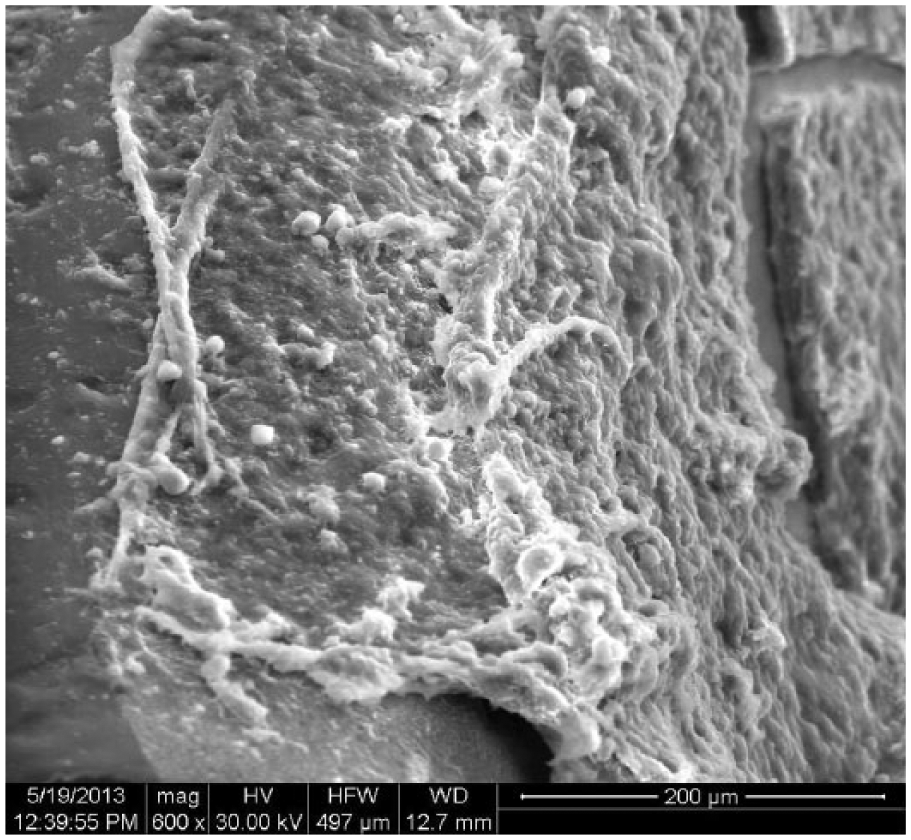
Scanning electron micrograph in samples of adenoid tissue of a child with adenotonsillitis. Showing biofilm sheets of exo-polysaccharide (EPS) and number of trapped cocci.
Distribution of Obtained Specimens According to the Result of Culture.a

Eight specimens (22.9%) had strong biofilm forming capacity (BFC); 4 specimens were obtained from patients who had adenoid hypertrophy, 2 specimens were obtained from patients who had adenotonsillitis, and the other 2 were obtained from patients who had adenotonsillitis complicated by OME. Seventeen specimens (48.6%) had medium BFC; 9 specimens were obtained from patients who had adenoid hypertrophy, 6 specimens were obtained from patients who had adenotonsillitis, and the other 2 were obtained from patients who had adenotonsillitis complicated by OME. Another 7 specimens (20%) had weak BFC; 3 specimens were obtained from patients who had adenoid hypertrophy, 3 specimens were obtained from patients who had adenotonsillitis, and the last 1 was obtained from patients who had adenotonsillitis complicated by OME. On the contrary, no biofilm formation was detected in 3 specimens (8.5%); 2 of them were obtained from patients who had adenoid hypertrophy, and the third was obtained from a patient who had adenotonsillitis (Table 2).
Distribution of Obtained Specimens According to Their Biofilm Forming Capacity (BFC).

The frequency of BFC of specimens was significantly higher with specimens obtained from cases of adenotonsillitis complicated by OME compared to the frequency in cases of adenoid hypertrophy (χ2 = 9.151, P = .009) or adenotonsillitis (χ2 = 6.363, P = .013), with nonsignificant (χ2 = 0.135, P > .05) higher frequency among those who had adenotonsillitis versus those who had adenoid hypertrophy (Figure 3).
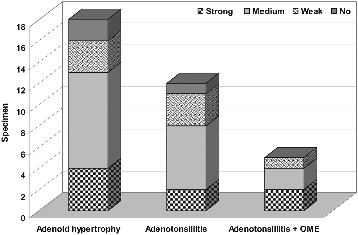
Distribution of obtained specimens according to their biofilm forming capacity and the clinical diagnosis.
Discussion
The current study included 18 patients who underwent adenoidectomy and 17 patients who underwent adenotonsillectomy; of which 5 were complicated by OME. Thirty-one surgical specimens showed bacterial growth on conventional media, while 4 specimens failed to give growth for a negative culture rate of 11.4%. The predominant organism was H influenzae detected in 17 cultures, while Staph aureus and Strept Pneumoniae were detected in 7 and 6 cultures, respectively. Only 1 adenoid specimen gave growth to Moraxella catarrhalis. Thirty-two specimens showed BFC of variable extent, while only 3 specimens showed no BFC for a rate of 91.4%. Eight specimens showed high BFC, 17 specimens showed moderate BFC, and 7 specimens showed low BFC.
These data point to a fact that the adenoid acts as a reservoir of bacteria in the form of biofilm, which provided a continuous flow of organisms leading to chronicity of inflammation with subsequent development of complications. In support of this assumption, there was a significantly higher frequency of specimens showing BFC in patients with adenotonsillitis complicated by chronic otitis media (COM) compared both to those who had adenotonsillitis alone or adenoid hypertrophy alone.
In line with the obtained data and supporting the provided assumption, Drago et al 9 evaluated production of biofilm, in vitro by means of spectrophotometry after growth in microplates and staining with crystal violet, of the isolates from intraoperative samples and found that 44.7% were either moderate or strong biofilm producers compared with 27% of isolates obtained at 6 months after surgery with decrease in biofilm production for H influenzae and S aureus and concluded that the decreased rate of isolation and ability to form biofilm in bacteria isolated subsequent to adenoidectomy and/or tonsillectomy suggests a role for biofilm in pathogenesis of recurrent and chronic pharyngeal diseases and rhinopharingitis.
Torretta et al 10 reported that nasopharyngeal biofilm-producing otopathogens (BPO) were more frequently isolated in patients with recurrent acute otitis media (RAOM) than in controls, and H influenzae (70.6%) was confirmed as the main pathogen in the RAOM group, and concluded that the presence of nasopharyngeal BPO is an important factor favoring RAOM; it is therefore useful to investigate biofilms even in children with non-severe RAOM without adenoid hypertrophy. Torretta et al 11 found biofilm-producing bacteria were significantly more frequently located near the ostium of the Eustachian tube (ET) and suggested that the adenoids are a reservoir for bacteria and so hypertrophic adenoids (particularly hypertrophy near the ostium of the ET) play a role in recurrent acute OM and/or COME.
Saafan et al 12 found adenoids removed from children with COM with effusion had a higher grade biofilm formation (74%) than that removed from children with adenoid hypertrophy without middle ear effusion (42%) and concluded that adenoids in COME may act as a reservoir of chronic infection rather than causing mechanical ET obstruction. Akyıldız et al 13 studied 19 patients, 10 with ear discharge history and 9 without discharge, and 9 patients who had undergone cochlear implantation as a control group and found that biofilm was detected in all patients with ear discharge history and in 2 of the patients without an ear discharge history, while all controls were free of biofilm formation, with significant difference between study and control groups and between study subgroups, and concluded that in the middle ear mucosa of patients with COM, biofilm formation is common, especially when ear discharge history is present.
Khosravi et al 14 studied swabs obtained from 45 patients with chronic rhinosinusitis, chronic tonsillitis, and COM and 15 asymptomatic control patients and detected H influenzae, Staph aureus, Strept pneumoniae, coagulase-negative staphylococci, and other Streptococcus species in both patient and control groups; biofilm was observed in approximately half of the smear prepared from swab samples obtained from patient group, and S aureus biofilm was most prevalent among nasal samples while H influenzae biofilm was more common among ear and throat samples and concluded that chronic otorhinolaryngologic diseases may be biofilm related. Wessman et al 15 examined 35 middle ear biopsies from patients with COM for the presence of mucosal biofilms and the bacteria within the biofilms and reported that Staphylococci were the most common bacteria in bacterial culture and bacterial biofilms were detected in 43% of middle ear biopsies and concluded that these findings may indicate that biofilms are a part of the pathogenesis in recurrent episodes of ear discharge in COM.
The obtained results allowed concluding that adenoids act as a bacterial reservoir secondary to bacterial biofilm formation so could induce chronicity and initiate development of complications. H influenzae and Staph aureus are the most common encountered organisms and showed moderate to high biofilm production capacity. Determination of biofilm production capacity using the proposed protocol is feasible, inexpensive, and available and spares the need for sophisticated instruments or approaches.
The information presented here has a clinical impact in the decision making of adenoidectomy operation in cases of recurrent AOM even if the adenoid is not large enough to completely close the nasopharyngeal airway.
Footnotes
Declaration of Conflicting Interests
The author(s) declared no potential conflicts of interest with respect to the research, authorship, and/or publication of this article.
Funding
The author(s) received no financial support for the research, authorship, and/or publication of this article.