






Abstract
Objective:
Extracapsular dissection (ECD) has become an accepted, less invasive alternative for the removal of select benign parotid lesions that may reduce complications. Minimal margin extracapsular dissection (MECD) with dissection on or closer to the tumor capsule may be a reasonable alternative to ECD. The objective of this study is to review the complications and safety of the MECD technique at a single institution.
Subjects and Methods:
Medical records for patients who underwent MECD for suspected benign parotid lesions were reviewed. Outcome measurements included intraoperative findings, complications, and recurrences.
Results:
Forty patients underwent a MECD for suspected benign parotid lesions. The average tumor size was 2.2 cm. Frozen section revealed low-intermediate grade mucoepidermoid carcinoma in 2 (5%) cases, requiring completion of a superficial parotidectomy at the same setting. There was 1 case of temporary facial nerve weakness and no cases of Frey syndrome. No tumor recurrences were observed within the follow-up period (average 3.5 years.)
Conclusion:
In the hands of an experienced surgeon, MECD may be a viable alternative to formal superficial parotidectomy. This study reports low rates of nerve weakness and Frey syndrome. Long-term follow-up is necessary to determine the ultimate risk of recurrence.
Introduction
The techniques available for surgical removal of benign parotid lesions have long been the subject of controversy. Pleomorphic adenoma is the most common benign parotid tumor, and these lesions frequently present in the lateral gland. 1 Historical approaches to benign parotid tumors involved removal through intracapsular and extracapsular enucleation, due to fear of injuring the facial nerve. 2 Extracapsular enucleation is interchangeable with “nodulectomy.” In many cases, the capsule would rupture or would be grossly left in situ after curettage of the internal tumor.3-5 This technique resulted in unacceptable recurrence rates—45% or higher.1,4-6 Interestingly, these recurrences appeared relatively rapidly, suggesting that “recurrence” was actually growth from primary tumor grossly left behind. 4
In the mid twentieth century, studies revealed that the pleomorphic adenoma capsule was frequently incomplete, 7 and microscopic extensions of the tumor (termed pseudopodia) protruded from the main lesion.6,8 The high recurrence rate observed with the earliest techniques were ascribed to these findings retrospectively.
Parotid surgery subsequently evolved in the following decades, mandating complete excision of the lesion and capsule, which significantly reduced the recurrence rate.8-10 Superficial (SP) and total parotidectomy (TP) techniques involve the identification and dissection of the main trunk and peripheral branches of the facial nerve. With partial superficial parotidectomy (PSP), only the portion of the gland involved with tumor was removed and only facial nerve branches involved in the dissection are delineated after identification of the main trunk.11,12
Recently, extracapsular dissection (ECD) has been advocated for certain superficial parotid lesions. This technique does not require identification of the main trunk of the facial nerve, and the surgeon aims to leave a cuff of 1 to 3 mm of normal parotid tissue around the lesion.16,17 Best practice guidelines published recently in Laryngoscope reported that ECD is effective and low risk for benign, superficial parotid tumors less than 4 cm in size when performed by an experienced surgeon. 18 Also, ECD has a reduced risk of postoperative Frey syndrome and facial nerve weakness when compared to SP and TP, most likely due to the decreased dissection of parotid tissue.19-21
With more limited resection, there is less resection of parotid tissue leaving a smaller cosmetic defect. Importantly, recurrence rates are similarly low for ECD and SP.13,22,23 A minimal margin extracapsular dissection (MECD) technique, equivalent to a meticulous nodulectomy, further reduces the amount of surrounding normal tissues dissected. Meticulous dissection along the capsule may allow for further reduction of the size of the surgical defect and potential complications. Long-term follow-up is necessary to determine the rate of recurrence. In this preliminary report, we review complications for patients who underwent such a procedure at a single institution.
Methods
Patient Selection and Data Collection
Internal review board approval was obtained for this study. A retrospective review of medical records was performed for patients who underwent MECD for suspected benign parotid lesions at a tertiary, academic medical center between 2008 and 2014. Tumors chosen for this procedure were primary tumors, superficial lesions, located in the lateral lobe or parotid tail, clinically benign, and 4 cm or less in size, or similar criteria for ECD. The determination of “clinically benign” was based on preoperative imaging results, history and physical examination (no facial nerve paresis, mobile lesion, painless), and fine needle aspiration (FNA) results. Preoperative FNA was performed in all cases. Outcomes such as facial nerve weakness, ear numbness, and gustatory sweating were obtained from the postoperative medical record.
Patients who underwent parotidectomy were identified from 4 surgeons’ operative case logs, and MECD technique was confirmed in the dictated operative note for each patient. Patient demographic, tumor characteristics (histology, size, presence of perineural invasion), and surgical technique data were collected and maintained in a secure, password-protected electronic database that was subsequently de-identified after collection. Data were recorded and analyzed using Microsoft Excel software version 14.1.0.
Surgical Technique
Details of the operative findings and procedure were reviewed from the surgeon’s dictated operative note. Residents participated in surgical procedures and perioperative care, and the amount of resident participation in the case depended on experience and surgeon judgment. Three separate surgeons performed the procedure in the following manner (Figure 1). Continuous electromyographic monitoring was not used for any of the cases. The size and level of the incision was tailored to the lesion. A skin flap over the parotid fascia was elevated anteriorly, just beyond the extent of the palpable mass. Blunt dissection in the capsular plane with meticulous hemostasis using bipolar cautery was performed. Both the bipolar cautery and high-frequency mechanical vibration devices were used to divide tissues. Any areas of concern for capsule loss or tumor extension were allowed a minimal cuff of normal tissue. If a nerve branch was encountered, the tumor capsule was carefully dissected free from the adjacent branch. If the lesion had concerning intraoperative features, such as infiltration into surrounding structures, frozen section was obtained. The procedure was converted to full superficial parotidectomy to achieve oncologic margins if there were malignant features on the frozen section pathology report.

Intraoperative photograph demonstrating the close-to-capsule nodulectomy technique. This patient had a benign epithelial cyst.
Results
Patients and Preoperative Workup
Forty patients underwent MECD for benign parotid lesions between May 2008 and July 2014. All patients presented with the primary complaint of facial or upper neck mass. The average age at the time of surgery was 57 years old. For preoperative imaging, computed tomography alone was used in 22 cases, magnetic resonance imaging alone in 12 cases, and both modalities in 1 case. No imaging was obtained in 5 cases.
Tumors and Intraoperative Findings
Details regarding pathology and operative findings are provided in Table 1. The average size of the excised lesions was 2.2 cm, ranging from 4 mm to 4 cm. A nerve branch was encountered in 5 cases (12.5%) during operative dissection and was dissected free of the tumor. There was intraoperative capsule rupture in 4 cases (10%), managed with thorough irrigation and meticulous removal of any tumor particles. The final pathology for the 4 ruptured cases were 2 pleomorphic adenoma, 1 basal cell adenoma, and 1 benign epithelial cyst. Frozen section was sent in 40% of cases. Pathology was upgraded to mucoepidermoid carcinoma on frozen section in 2 (5%) cases, requiring conversion to a traditional SP technique with facial nerve dissection and removal of additional parotid tissue.
Pathology and Operative Findings.
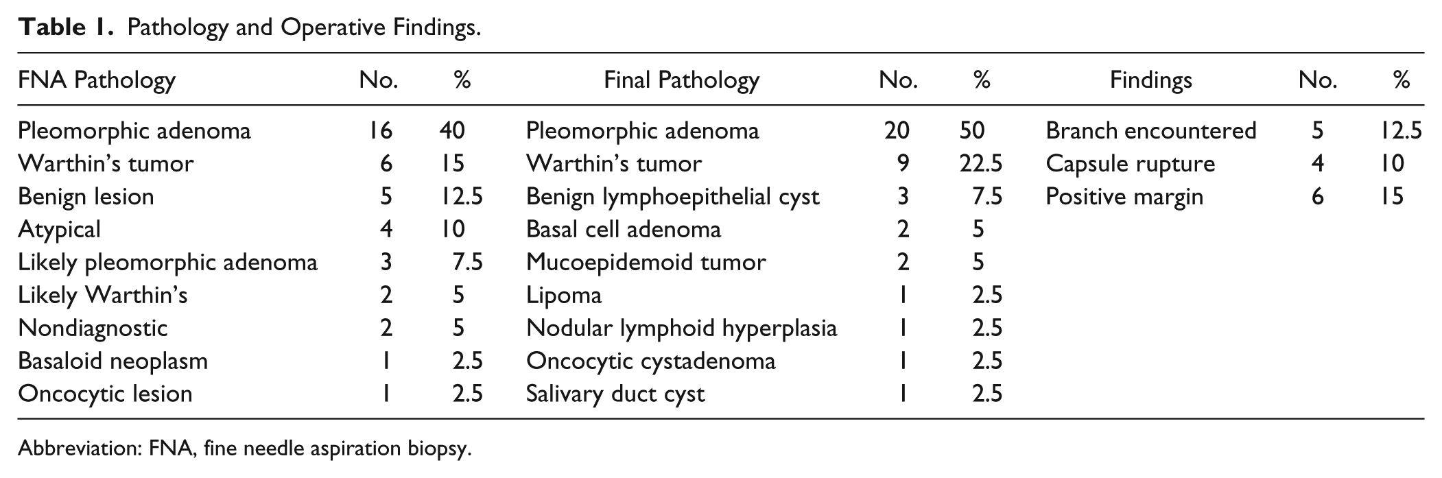
Abbreviation: FNA, fine needle aspiration biopsy.
Pathology
The FNA correlated with the final diagnosis in 90% of cases. Upon final analysis, there were 20 pleomorphic adenomas, 2 mucoepidermoid carcinomas (1 low-grade and 1 intermediate-grade), and various other benign lesions. Tumor was present at the margin of resection in 6 cases (15%)—all for benign lesions. The final margins for the mucoepidermoid cancer cases were negative.
Postoperative Complications
The most common postoperative concerns were temporary or prolonged earlobe numbness (27.5%) and scar aesthetic issues, including keloid formation and hyperpigmentation (17.5%). One patient developed temporary postoperative marginal mandibular branch weakness. A nerve branch was not encountered intraoperatively during that case. None of the patients reported gustatory sweating in the postoperative period. No tumor recurrences were observed within the follow-up period, with a mean of 3.5 years. The 2 patients with mucoepidermoid carcinoma did not receive adjuvant therapy and were without evidence of disease recurrence at follow-up, which was 22 months and 20 months, respectively.
Discussion
The most common benign tumor of the parotid gland, the pleomorphic adenoma, has microscopic extensions outside of the capsule and areas of this capsule and capsular disruption upon histopathologic analysis. 8 Tumor capsules vary in consistency and thickness,2,13 and there are frequently projections with narrow necks. 14 The currently favored techniques, as a consequence, resect normal parotid tissue around the tumor. Tumor-nerve interface occurs in the vast majority of parotid surgeries—approximately 60%,2,15,16 but tumor-nerve interface and tumor at the surgical margins do not appear to significantly increase the recurrence rates.2,15,17-19 Many studies suggest the critical factor is frank tumor spillage and/or capsular rupture, 18 although this also has been debated.20,21 We surmise that increased awareness of the potentially incomplete capsule, attention to tumor projections, and meticulous dissection may make MECD, similar to nodulectomy, a viable alternative to ECD.
Recently, there has been an increase in published literature advocating the advantages of limited dissection techniques, specifically, the extracapsular dissection. Albergotti et al 22 performed a meta-analysis of patients undergoing ECD or SP for pleomorphic adenoma of the parotid. Nine studies encompassing 1882 patients who underwent ECD or SP were included. The authors concluded that there was no difference in tumor recurrence between the techniques, but there was a significantly lower rate of transient facial nerve paresis with ECD versus SP (8% vs 20%). Frey syndrome was also significantly less common with ECD: 4.5% versus 26.1%. The recurrence rate for ECD was 1.5% and 2.4% for SP, but this was not statistically significant. 22 There appears to be an advantage regarding postoperative complications when reducing the amount of dissection of surrounding normal tissues.
The MECD further reduces the amount of normal tissue dissected compared to ECD, SP, and TP. This technique, in this current preliminary report, resulted in 0 patients with Frey syndrome and 1 patient with temporary marginal nerve weakness, lower than reported values for ECD and SP.10,18,22 Reported rates of transient and permanent facial nerve paresis with enucleation has been reported to be 11% and 3.5%, respectively, in an extensive review of the literature. 18 Factors that could explain the low rate of facial nerve weakness in the current report include decreased dissection of normal surrounding tissue and favorable tumor selection. Without an internal control comparing MECD to ECD or standard SP, it is difficult to draw definitive conclusions.
There are several potential complications from parotid surgery that are concerning to patients. Chief among them is facial nerve injury, especially when performed for benign lesions. Even with ECD, postoperative permanent facial nerve paralysis is 2%, and temporary weakness is approximately 8%. 3 Rates of facial nerve injury are significantly higher for SP and TP. Laccourreye, et al 10 reported a 64% rate of temporary paresis for SP. The present series using nodulectomy had 1 case of temporary marginal branch weakness (2%). This likely speaks to the minimal dissection of surrounding tissues involved with MECD. Outcomes of MECD as compared to ECD in the existing literature are briefly reviewed in Table 2.
Outcomes for the Current Study as Compared to Existing Literature.

Abbreviations: ECD, extracapsular dissection; MECD, minimal extracapsular dissection.
Frey syndrome is another concerning postoperative complication that presumably results from extensive dissection and aberrant reinnervation of cutaneous sweat glands. The incidence of Frey syndrome may be difficult to assess in retrospective review because without direct questioning, the patient may not notice any gustatory sweating, and very few patients require treatment. 9 Also, SP and TP result in significant rates of clinically apparent Frey syndrome—25% to 30% and higher.10,19 Hancock 19 reported no incidents of Frey syndrome after ECD. Our series using MECD had 0 cases of clinically apparent Frey syndrome.
Partial superficial parotidectomy and ECD, other limited dissection techniques, appear to be oncologically sound. McGurk et al 23 reported a series of 503 patients observed over a median of 12 years, ranging from 5 to 30 years’ follow-up. Oncologic outcomes were not compromised with ECD, and postoperative morbidities were reduced compared to more extensive techniques. In our series using the MECD technique, 2 patients were initially thought to have a benign mass, but frozen section revealed mucoepidermoid carcinoma. While a MECD was initially carried out in these 2 cases, the procedures were converted immediately to formal SP to resect an appropriate margin within the wound bed. No cases were upgraded on final pathology reports. While MECD is not appropriate for lesions discovered to be malignant, the surgeon could convert to SP to remove an appropriate margin, as was performed in our series.
Other complications are commonly observed clinically with more extensive parotid resection. These include cosmetic defect and facial asymmetry, salivary fistula, seroma formation, poor wound healing, and larger surgical scars. Numerous techniques have been described to fill the preauricular defect and potentially reduce the aberrant reinnervation attributed to the development of Frey syndrome. Techniques include autologous fat grafting, alloderm interposition, various muscle and fascia transpositions, and even free flaps.24,25-28 The MECD technique results in a minimal surgical defect, closely maintaining facial symmetry and minimizing surgical access scarring.
Tumor rupture is an important consideration in this study. With close-to-capsule dissection, tumor rupture is a certainly a risk of this procedure. In the present series (MECD), tumor capsular rupture occurred in 4 of 40 cases (10%). This appears to be slightly higher than published rates for ECD but much lower than previously published rates for enucleation/nodulectomy, techniques similar to MECD. Dell’Aversana Orabona et al 29 reported a 3.4% rate of capsular rupture with ECD and 1.8% capsular rupture with SP. Interestingly, Witt 18 reported a positive margin in 25% of cases and capsular rupture in 5% of ECD and SP procedures. Capsular rupture did not affect recurrence rates regardless of the technique used. A number of other authors report approximately a 7% to 8.5% rate of capsular rupture with ECD for pleomorphic adenoma, though it is not entirely clear whether margin status or capsular rupture actually affect recurrence rates at all.17,30,31
Capsular rupture rates have been reported to be much higher during what has been described in the literature as enucleation. Enluceation, as described by most authors, has also been termed local excision, minimal margin parotid surgery, and nodulectomy and is also akin to the MECD described in this report. Recurrence rates after enucleation vary widely in published literature, from 1% to 75%; however, many of these reports also have high tumor spillage rates associated with enucleation.18,32-36 A few authors have described treating limited pleomorphic adenoma tumors with nodulectomy followed by radiotherapy in the 1960s and early 1970s. These authors report an intraoperative capsular rupture rate of 21% and 49%.37,38 Comparatively, the rate of capsular rupture is lower (10%) in the present study. While the decreased rate of capsular rupture in our series of MECD patients as compared to reported rates with historical enucleation is significantly lower, the reason for this is not entirely clear. We postulate that highly meticulous dissection and minimizing traction on the lesion may decrease the rate of rupture.
The length of follow-up in this report should be carefully considered. Recurrent pleomorphic adenomas present in a multinodular fashion between 7 and 10 years after initial treatment.35,39-43 Recurrence of pleomorphic adenoma is troublesome and can result in malignant transformation, challenging surgical conditions, increased incidence of postoperative facial nerve paresis, and the need for adjuvant radiotherapy.40,42-46 The average follow-up in this heterogeneous group of patients was 3.5 years. While we are able to assess short-term postoperative complications, long-term follow-up of at least 10 years will be essential to determine the effect of this meticulous MECD technique on recurrence rates.
Additionally, there is significant patient selection bias. While basic criteria exist guiding patient selection, the decision to perform a MECD is essentially the experienced surgeon’s subjective consideration. Outcomes after parotid surgery are highly operator-dependent. Furthermore, the low rate of Frey syndrome (0%) reported here is based solely on patient reporting, and no objective testing, such as Minor starch-iodine testing, was performed. Subjective reporting is known to significantly underrepresent the rate of Frey syndrome, and symptoms may take up to a year or longer to develop. 47 Finally, this is a retrospective review of a small, single cohort of patients with heterogeneous pathologies and without an internal comparison group.
Conclusion
The benefits of minimally invasive surgery are evident in other areas of head and neck surgery, and the concepts are being examined in parotid disease as well. The MECD, when compared to SP or ECD, further reduces dissection of normal tissues, a technique that may correlate with fewer postoperative complications when performed by an experienced parotid surgeon. Avoidance of capsular rupture and tumor spillage is potentially a critical factor. Meticulous dissection and awareness of potential capsule irregularity may allow MECD to be a viable technique for certain benign parotid lesions. Long-term follow-up is absolutely essential to determine its oncologic soundness prior to routine implementation.
Footnotes
Declaration of Conflicting Interests
The author(s) declared no potential conflicts of interest with respect to the research, authorship, and/or publication of this article.
Funding
The author(s) received no financial support for the research, authorship, and/or publication of this article.