
Abstract
Objectives:
Metal hypersensitivity reaction to surgical implants is a well- known phenomenon that is associated with pain, swelling, inflammation, and decreased efficacy of the implant. We present a unique case of a patient with placement a metal Jackson tracheostomy tube that led to expeditious total subglottic stenosis.
Methods:
The patient was a 33-year old, severely atopic woman with history of asthma exacerbations requiring several intubations for acute respiratory failure with several subsequent tracheal dilations with steroid injections, and eventual tracheostomy placement with a metal Jackson tracheostomy tube that led to expeditious total subglottic stenosis.
Results:
Initial intervention included performing an airway evaluation, CO2 laser, and steroid injection of the area of complete subglottic stenosis. Follow up several months later revealed little improvement in level of tracheal narrowing proximal to the tracheostomy tube. Patient did not have shortness of breath but continued to be aphonic. Cricotracheal versus tracheal resection have been proposed but surgical morbidity was deemed too high due to patient’s obesity.
Conclusions:
Metal hypersensitivity reactions are well known phenomena as it relates to surgical implants in other surgical specialties but are seldom reported within the ear, nose and throat literature. Oftentimes, it takes astute observation to diagnose and establish a connection. Prompt recognition and treatment can be acquired from interdisciplinary collaboration with allergy.
Introduction
Delayed type hypersensitivity (DTH) reactions to surgical implants are a well- known phenomenon that is associated with swelling, inflammation, and decreased efficacy of the implant. 1 There is ubiquitous utilization of several of the culpable metals in surgical implants: nickel, cobalt, chromium. Several of the metal alloys that are employed in surgical implants such as stainless steel, titanium, Cobalt- chromium- molybdenum steel, incorporate a considerable proportion of the aforementioned metals. While these metals are often used due to their well- known flexibility, stability, and durability, at times, they are also immunogenic. 1
This type of allergy is initiated once the metal ions are released from the various alloys of the implant and act as potent haptens that trigger a cascade of inflammatory events. 2 Additional exposure to the same hapten would lead to activation of the hapten-specific T cells which produce signs of hypersensitivity at 48 to 72 hours within exposure. These allergies to surgical implants form the largest category of Medicare expenditures and imposes a huge financial and physical toll on the patient, the healthcare system, as well as the surgeon. 1
There is a plethora of research that details metal hypersensitivity reactions to various surgical implants. However, there is currently a dearth of literature that details an immunogenic reaction to a commonly used tracheostomy tube: the metal Jackson trach tube. This tracheostomy tube is often used for its cost-effectiveness, reusability, minimized infection risk, as well as for its thin wall which facilitates greater airflow. We report a case of a 33- year old African American female with severe atopy, asthma exacerbations requiring several intubations for acute respiratory failure with several subsequent tracheal dilations, and eventual tracheostomy placement with a metal Jackson tracheostomy tube that led to expeditious total subglottic stenosis within a brisk 2- week time frame.
Case Report
A 33- year old African American female with history of laryngopharyngeal reflux, severe asthma exacerbations requiring 4 separate intubations, subsequent subglottic stenosis refractory to several dilation procedures and steroid injections, underwent eventual tracheostomy with a 6-0 metal Jackson tracheostomy tube. Prior to the tracheostomy, the patient had undergone 3 tracheal 16 mm balloon dilations to 10 atmospheres with little sustained improvement in her subglottic stenosis.
It was noted that during her last airway evaluation with tracheal dilation, the area of subglottic stenosis elongated from 1.4 cm to approximately 6 cm with multiple shelves on different planes. Each recurrence of subglottic stenosis caused about 50% to 70% airway obstruction. She presented to clinic 2 weeks post-operatively with inability to voice with finger occlusion. Due to this, there was concern for suprastomal collapse or restenosis of the subglottis and the decision was made to take the patient back to the operating room for a repeat airway evaluation.
The airway evaluation revealed severe subglottic erythema, edema, and lymphoid hypertrophy. The suprastomal airway had ended in a blind pouch with discontinuity from the distal airway. Bronchoscopy revealed severe friability of irritation of the airway circumferentially in all areas where the metal Jackson tracheostomy tube was contacting the tracheal mucosa. We subsequently tied to reestablish airway continuity between the upper and distal airway by attempting to cannulate the proximal tracheal narrowing with a spinal needle under high-powered endoscopic visualization. Inability to do so confirmed that the proximal airway was in discontinuity with the distal airway secondary to scarring. The procedure at this point after confirming patency of the distal airway to maintain safety in the upper airway. The decision was ultimately made to exchange the tracheostomy tube to a 6-0 cuffless Shiley. Our clinical index of suspicion was high for an autoimmune or allergic etiology for the expeditious subglottic restenosis in a 2- week time frame.
As uncontrolled laryngopharyngeal reflux (LPR) can have extraesophageal and upper airway manifestations, it was imperative that this pathologic process be ruled out. The disease process has also been named as a likely cause of idiopathic subglottic stenosis.3,4 The patient underwent esophageal manometry with pH probe testing to rule out LPR. Manometry revealed ineffective esophageal motility that was not likely to contribute to her reflux symptoms as the pH study was not indicative of reflux. Her overall Demeester Composite Score was 5.0 (normal < 14.72) which rules out pathologic reflux.
The patient denied having any familial or personal history of any autoimmune disease. Basic autoimmune work up was initiated. All the markers were negative except Rheumatoid factor which was elevated at 40.0 IU/ml (Negative ref range 0-15.0 IU/ml), and Anti-DNA antibody, double-stranded which was indeterminate at 9 IU/ml (Negative ref range 1-4 IU/ml). Patient was subsequently seen by Rheumatology where further autoimmune studies were conducted. Ultimately, vasculitis panels, type II collagen antibodies and anti CCP antibodies were pan- negative.
The patient was evaluated by Allergists who performed patch testing- the gold standard for evaluating DTH reactions.5,6 The patch was placed and removed 2 days later. It revealed a robust reaction to Nickel (++ reaction associated with papules, possible pustules and erythema) that worsened from removal to final read. There weren’t any other clear positives that were appreciated. Due to the positive patch test to Nickel, the manufacturer for the metal Jackson tracheostomy tube was contacted and while it was hard to ascertain for certain which version was used, the stainless steel- alloy (item number 78-2031) was indicated as a definite, which may be comprised of 8.3% to 35% Nickel.
After an etiology was established for restenosis of her subglottic airway, repeat airway evaluation, steroid injection and CO2 laser of tracheal stenosis were performed. Airway evaluation was achieved with the Dedo laryngoscope- significant >90% tracheal stenosis was noted just below the cords. Shown in Figure 1. CO2 laser was used to ablate the tracheal scar tissue. It was noted that the tracheal tissue was still very collapsible. An unsuccessful attempt was made to pass a 5-0 endotracheal tube through the lumen. Measurements were taken and it was ascertained that the stenosis spanned approximately 10 mm. A Montgomery® T- Tube™ (Boston Medical Products, Inc., Shrewsbury, MA, USA) was attempted to be placed through the tracheal stoma however the tissue was too collapsible to be adequately placed. 0.5 cc triamcinolone acetonide (40 mg/mL) was injected into the tracheal scar tissue. Due to collapsible tracheal tissue and significant stenosis persisting after laser ablation, balloon dilatation, and endoscopic tracheoplasty were not performed and we elected to conclude the procedure. Shown in Figure 2.
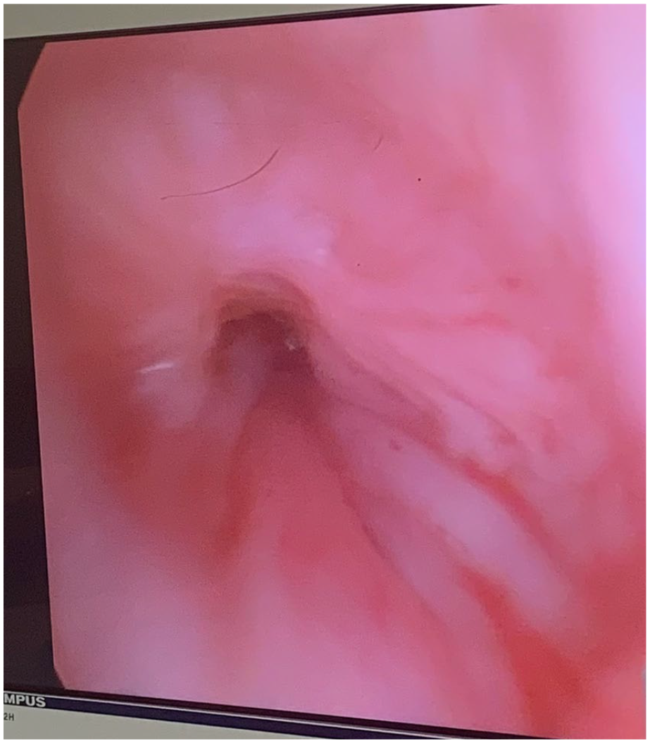
>90% subglottic stenosis following metal tracheostomy tube placement.
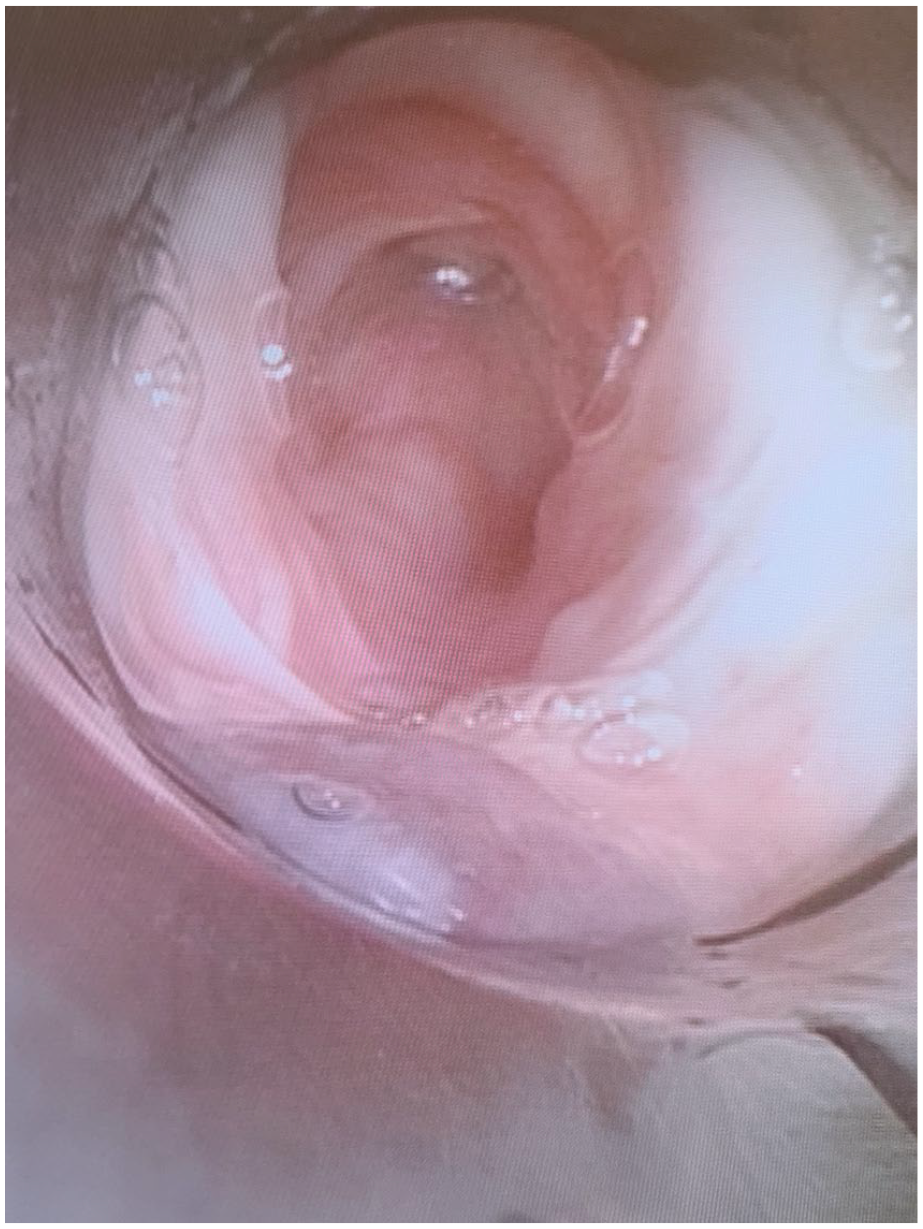
Modest improvement of subglottic stenosis following CO2 laser and steroid injection.
One month follow up in clinic revealed minimal improvement in the degree of subglottic stenosis. Subsequent follow up at 3 and 6 months later revealed refractory subglottic stenosis just proximal to the tracheostomy tube. Patient remained aphonic but denied any shortness of breath. She is to undergo a serial balloon dilation, CO2 laser, with steroid injection this month. A considerable discussion was held for potential future interventions including cricotracheal resection to alleviate the patient’s obstruction, however, the risk remains too great due to her morbid obesity. A healthy discussion was made concerning the need for continued weight loss to optimize her pre-operatively. In the interim, she remains stable with a 6-0 cuffless Shiley tracheostomy tube in place.
Discussion
A literature review was performed through PubMed/MEDLINE using the key terms subglottic, tracheal, stenosis, metal, allergy, tracheostomy tube, Jackson. Evidently, there is currently no literature out there that explains this phenomenon observed in this patient’s case of total subglottic stenosis due to a probable DTH reaction to the Nickel- imbued Jackson metal tracheostomy tube. Most orthopedic and dental implants, intracoronary stents, prosthetic valves, endovascular prostheses, and many gynecologic devices are derived from metal alloys. 7 The exact prevalence of metal hypersensitivity reactions to surgical implants is not well elucidated- current estimates range from 0% to 5%. 7 There is an abundant amount of literature out there for metal allergies secondary to orthopedic joint replacements due to the sheer volume of cases performed annually in the United States- >600 000 total knee arthroplasties 8 and ~421 000 total hip arthroplasties 9 performed annually. A study found that the prevalence of nickel hypersensitivity in a patch-tested population was approximately 18.5%. 10 Considering the severity of these severe metal reactions, it begs the question of whether routine pre-implant patch testing should be performed. An argument could be made to screen atopic patients in these types of situations, however, currently, there is a paucity of literature that can strongly argue for or against routine patch testing if the patient has no history of dermatitis or other concerning signs of previous metal allergies. Rosner and Fonacier 6 established an algorithm for evaluation of suspected metal implant hypersensitivity that is promising, albeit predicated on anecdotal evidence, and may help many medical professionals. However, ongoing research must be performed before strong recommendations may be made due to lack of scientific evidence and definitive guidelines.
This patient who had an unremarkable autoimmune work up with refractory stenosis despite several balloon tracheal dilations may have an underlying condition that potentiated the DTH reaction to the metal tracheostomy tube. It is still rather curious why the patient continued to have recurrence of subglottic stenosis every month that necessitated balloon dilations and steroid injections. Her propensity for restenosis was never elucidated but given the complete subglottic stenosis, and mucosal inflammation in all areas that were in contact with the metal tracheostomy tube, a metal hypersensitivity reaction was likely culpable in contributing to the complete stenosis. In conclusion, clinicians should consider nickel allergy in the differential diagnosis for patients with an unusual inflammatory pattern in patients with metal tracheostomy tubes. Through our interdisciplinary efforts with the allergy department, it was possible to make the astute observation that her expeditious airway narrowing was secondary to a metal hypersensitivity reaction that is not very well documented in the literature for tracheostomy tube placement.
Footnotes
Declaration of Conflicting Interests
The author(s) declared no potential conflicts of interest with respect to the research, authorship, and/or publication of this article.
Funding
The author(s) received no financial support for the research, authorship, and/or publication of this article.