
Abstract
Objectives:
The submental island flap is a dependable option for head and neck reconstruction. Venous drainage depends on the submental vein, which typically drains into the facial vein and, subsequently, the internal jugular vein. Variations in venous anatomy often involve drainage into the anterior jugular or external jugular venous systems. This study evaluates the likelihood of encountering submental venous anatomy variants and the accuracy of preoperative imaging in identifying them.
Methods:
Twenty-two patients who underwent a submental island flap procedure at the University of New Mexico Hospital from 2015 through 2023 with defined submental venous anatomy were analyzed. Three surgeons, blinded to intraoperative findings, predicted venous anatomy from preoperative imaging, with inter-rater reliability assessed using Fleiss Kappa.
Results:
Fifteen patients exhibited typical venous anatomy. Four patients’ submental venous vasculature showed drainage into the external jugular vein, and 3 into the anterior jugular vein. Imaging reviews showed accuracy rates of 72.23%, 90.91%, and 86.36%, respectively. Analysis of cases with CT scans yielded k = 0.46 (P < .001).
Conclusion:
The submental island flap is versatile and reliable but demonstrates common variant venous anatomy. Accurate imaging-based predictions can optimize surgical efficiency and outcomes.
Keywords
Introduction
First described by Martin et al 1 in 1993, the submental island flap is a reliable and versatile reconstructive option for many complex head and neck defects. Specific benefits include excellent cosmesis,2-5 conservation of function, 6 improved quality of life,7,8 and tissue bulk9,10 similar to recipient head and neck sites. Use of the submental island flap obviates the need for a second donor site, reduces operative length,11-14 and is associated with faster recovery rates and shorter hospital stays for patients.11,13-19 All of these benefits are especially advantageous for patients who are not good candidates for free tissue transfer. 20 Literature investigating the utility of pedicled flaps in orofacial reconstruction shows that submental island flaps have lower postoperative morbidity when compared to other pedicled flap techniques, including pectoralis major, deltoperctoral, latissimus dorsi, and trapezius flaps. 20 It has been noted that substantial variation exists in the venous anatomy of the submental island flap. The venous anatomy of the flap is generally described as follows: submental vein drains into the facial vein, into the common facial vein, and finally into the internal jugular vein 21 (Figure 1). However, variant anatomy is common and can substantially increase the risk of flap compromise during elevation, 22 along with potentially limiting the arc of flap rotation. 21 The most common variations of the venous anatomy involve drainage of the submental vessels ultimately into the external jugular venous (Figures 2 and 3) system or anterior jugular venous (Figures 4 and 5) system.23,24 To date, no articles have established the relative rates of the various venous patterns seen in the submental island flap. Based on our extensive reconstructive experience with this flap, we hypothesized that the rate of aberrant submental venous anatomy would be high. Additionally, we hypothesized that the venous anatomy of the submental island flap could be determined preoperatively by review of contrast enhanced cross-sectional imaging.
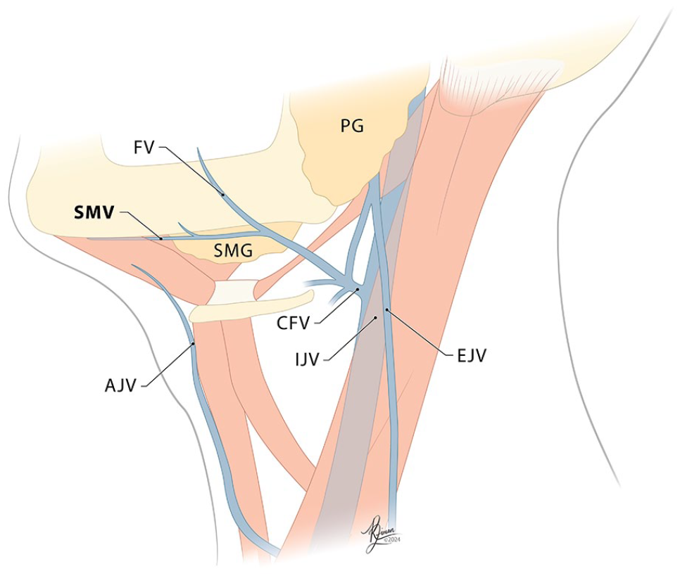
Figure of submental venous anatomy, Type 1—Submental vein branches off of the internal jugular vein.
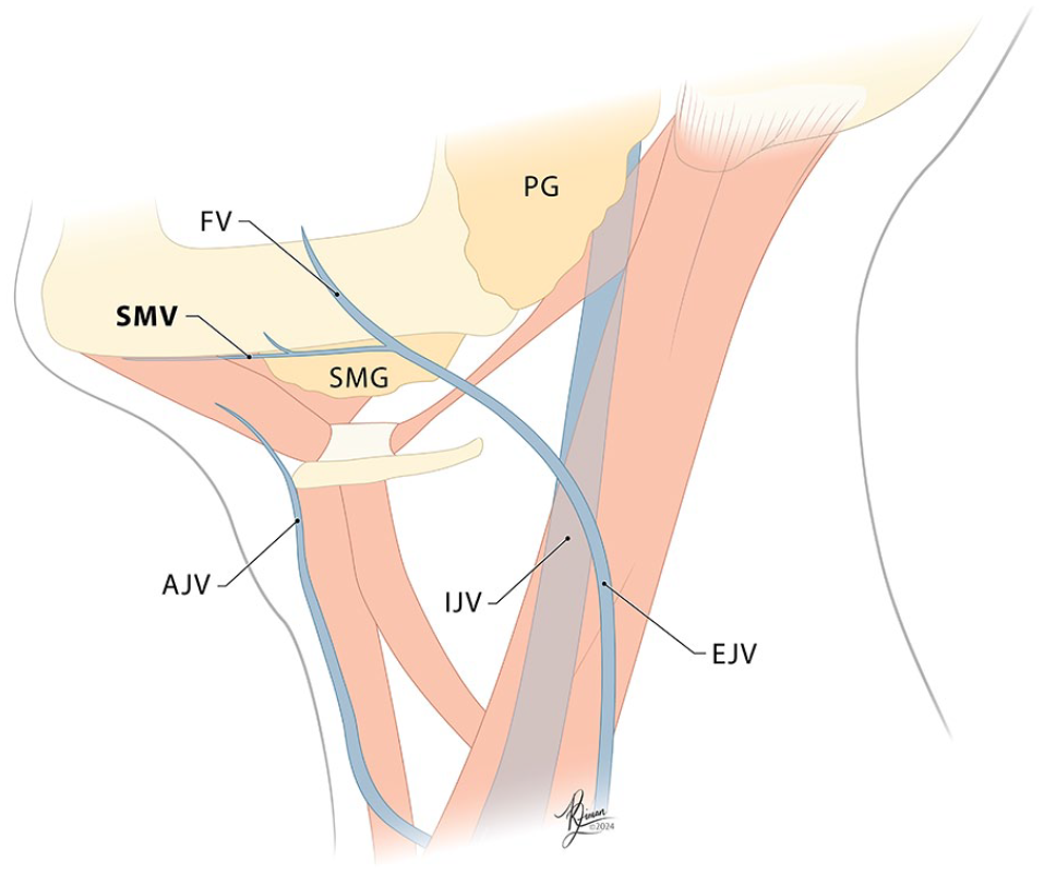
Figure of submental venous anatomy, Type 2—Submental vein branches off of the external jugular vein (Created by Roxanne Ziman, used with permission).
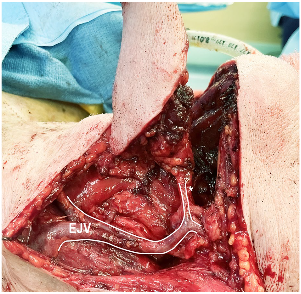
Intraoperative photo of submental venous anatomy, Type 2—Submental vein branches off of the external jugular vein.
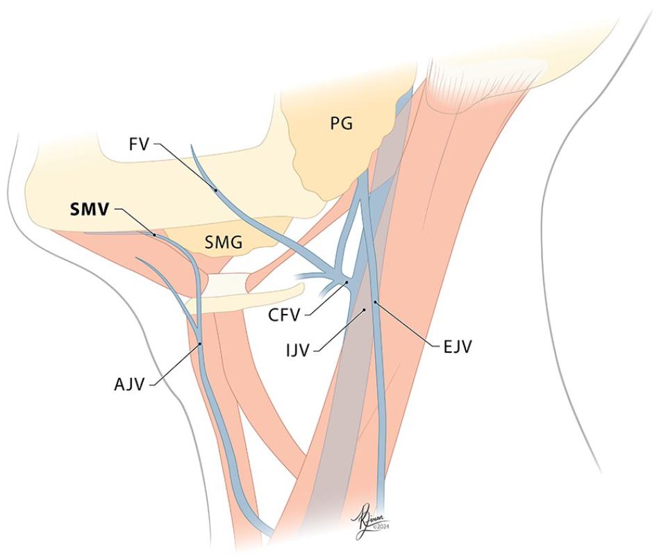
Figure of submental venous anatomy, Type 3—Submental vein branches off of the anterior jugular vein (Created by Roxanne Ziman, used with permission).
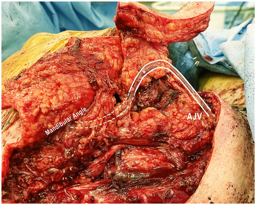
Intraoperative photo of submental venous anatomy, Type 3—Submental vein branches off of the anterior jugular vein.
Methods
An IRB-approved retrospective review was performed to identify patients who had undergone submental island flap reconstruction at the University of New Mexico Hospital over the period from 2015 to 2023. Seventy-eight patients were identified, and details were compiled pertaining to each case, including the vascular anatomy described in the operative note, venous anastomoses performed, and the presence or absence of preoperative imaging of the head and neck region. Patients whose operative report did not clearly specify the submental venous anatomy and those without contrasted preoperative imaging were excluded. The final study pool was 22 cases; patient demographics are shown in Table 1.
Patient Demographics.
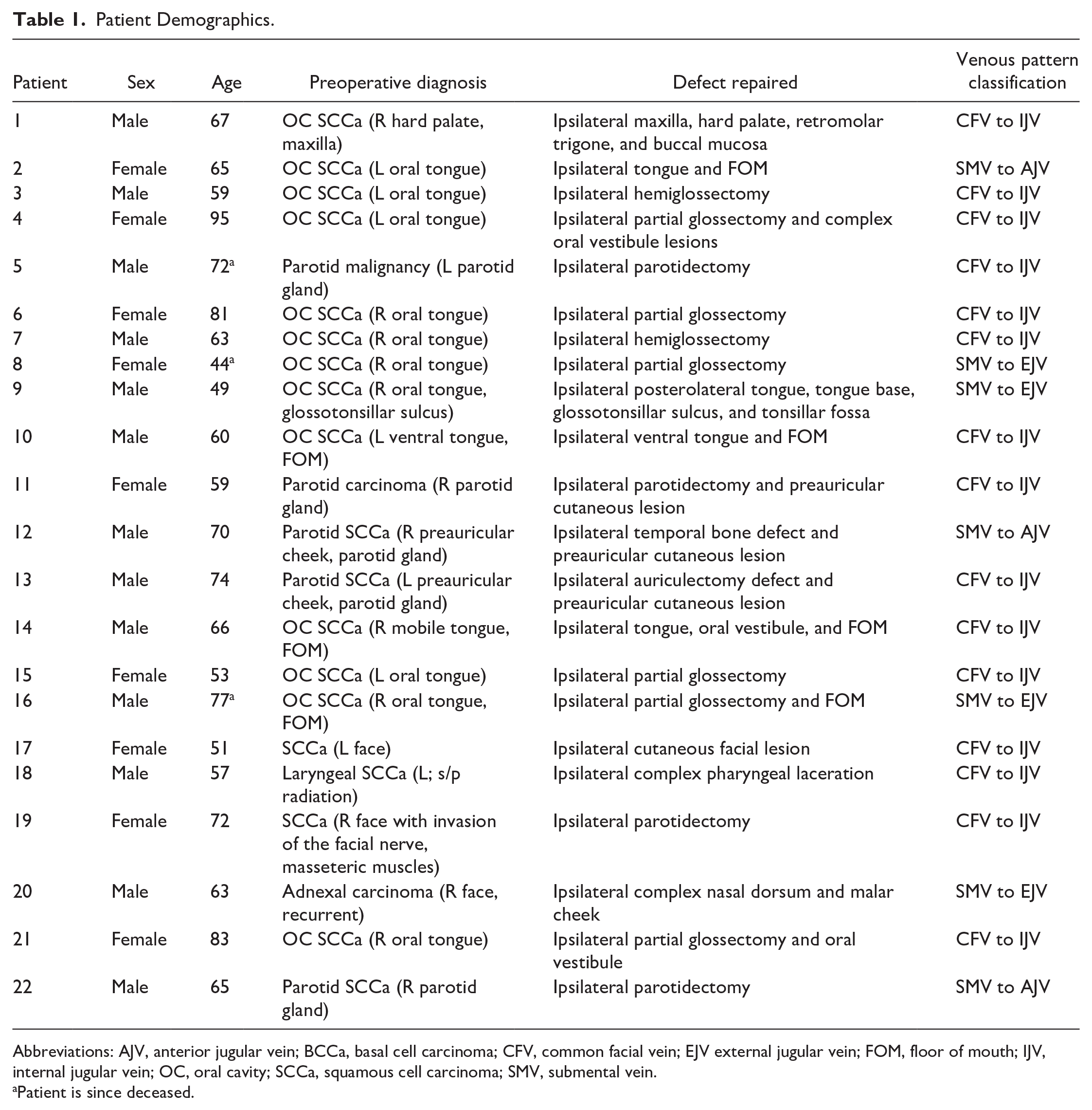
Abbreviations: AJV, anterior jugular vein; BCCa, basal cell carcinoma; CFV, common facial vein; EJV external jugular vein; FOM, floor of mouth; IJV, internal jugular vein; OC, oral cavity; SCCa, squamous cell carcinoma; SMV, submental vein.
Patient is since deceased.
We calculated the relative frequency of the most common submental venous variants. We then investigated the ability of the submental venous anatomy to be predicted by preoperative imaging analysis. Three head and neck reconstructive surgeons who regularly perform submental island flap procedures reviewed the preoperative imaging studies of study participants and were asked to categorize the venous anatomy to the internal, external, or anterior jugular system, without knowledge of the operative findings. Surgeon’s predictions based on imaging review were then compared to that identified at surgery. An additional comparison of vascular pattern identification between surgeons was analyzed using an inter-rater reliability analysis via Fleiss Kappa (k) calculation.
Results
During the study period, 22 patients underwent submental island flap reconstruction who had adequate preoperative imaging in the form of computed tomography (CT) of the head and neck with contrast and sufficient documentation of intraoperative findings to meet the study objectives. Patients ranged from 44 to 95 years old, with a mean of 66 years (SD: 12). Malignant neoplasm was the primary indication for surgery in 100% (22/22) of these patients, with squamous cell carcinoma accounting for 86.36% (19/22) of cases. Submental island flap reconstruction was applied to a variety of subsites within the head and neck: oral cavity in 59.09% (13/22) of patients, parotid bed in 5 (22.73%; 5/22) patients, laryngeal resection in 0.05% (1/22) of patients, and cutaneous defects in 13.64% (3/22) of patients. Concurrent selective neck dissection was performed in 54.55% (12/22) of the patients who underwent a submental island flap procedure. The most common venous drainage pattern identified for this flap was submental vein into the facial vein into the internal jugular vein and occurred in 68.18% (15/22) of patients. Four cases (18.18%; 4/22) showed the submental or common facial vein complex coursing into the external jugular vein. The remaining 3 (13.64%; 3/22) patients had the submental or common facial vein complex coursing into the anterior jugular vein.
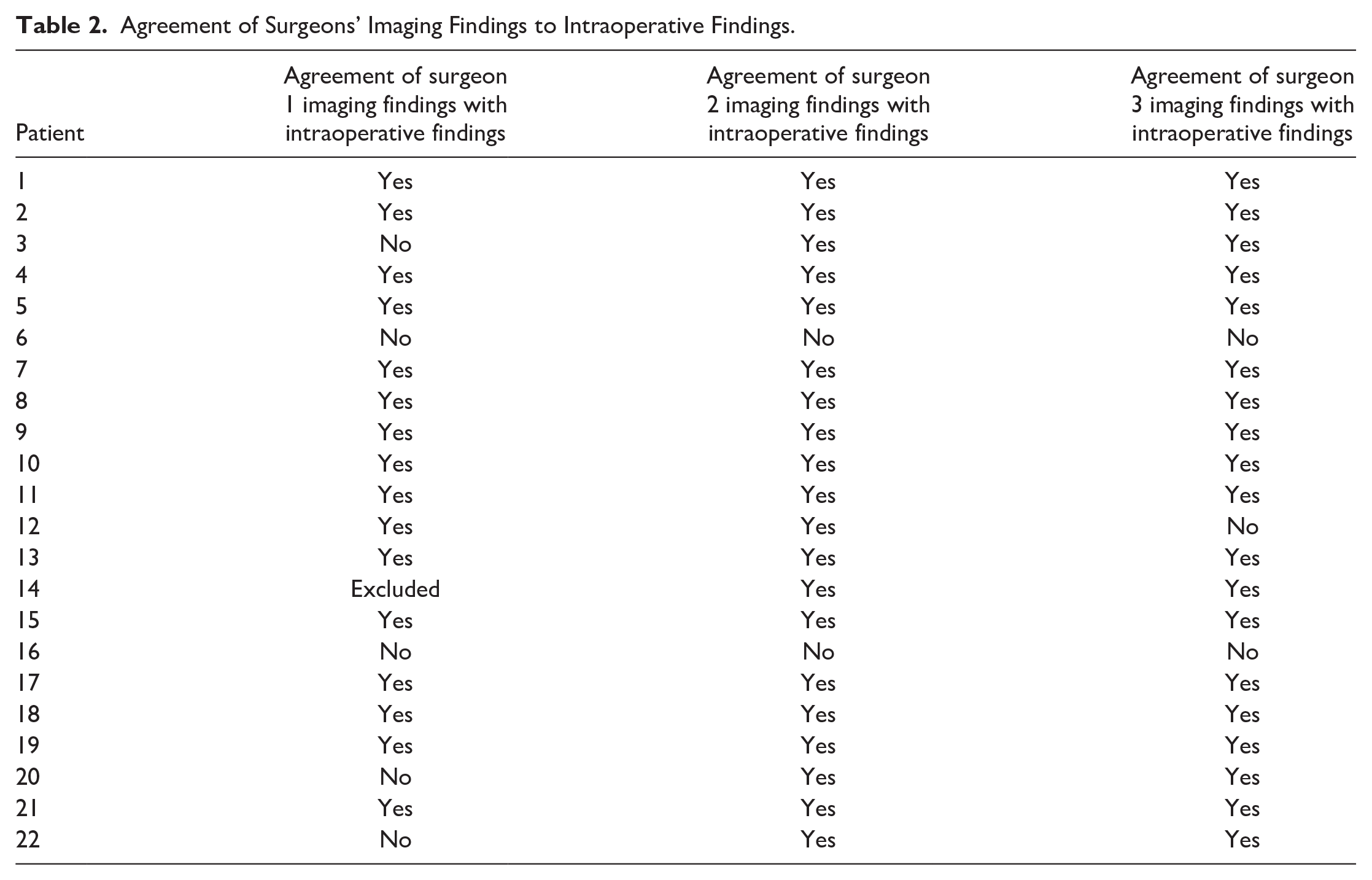
Three head and neck reconstructive surgeons who regularly perform submental island flap reconstruction were recruited to analyze preoperative imaging for all 22 study patients and to compare them to intraoperative findings. Surgeon 1 accurately predicted the intraoperative anatomy in 16 of the 22 cases (72.23%). Notably, this surgeon’s findings did not align with the intraoperative findings in 5 of the 22 cases (22.73%), with the surgeon choosing to withdraw themselves from imaging interpretation of the remaining 1 case due to uncertain conclusions drawn in the setting of degraded image quality, which they felt diminished their confidence. Surgeon 2 accurately predicted the intraoperative anatomy in 20 of the 22 cases (90.91%). The remaining 2 of 22 cases (9.09%) were negatively correlated. Surgeon 3 accurately predicted the intraoperative findings in 19 of 22 cases (86.36%). The surgeon provided a negative correlation in 3 of the 22 cases (13.64%). These data are tabulated in Table 2.
Agreement of Surgeons’ Imaging Findings to Intraoperative Findings.
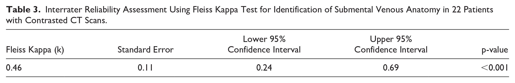
The Fleiss Kappa (k) test was performed to calculate inter-rater reliability between participating surgeons. Assessment of cases with preoperative CT scans with contrast yielded a Fleiss Kappa (k) value of 0.46 and a P-value of <.001.
Discussion
The submental island flap is a trusted method of reconstruction for a variety of head and neck defects due to its reliability, proximity to the head and neck, excellent cosmetic match,2-5 tissue bulk,9,10 and rapid harvest time, facilitating functional reconstruction without some drawbacks of free flaps.
The primary difficulty of harvesting a submental island flap is variability in venous anatomy. 23 The most common venous drainage pattern for this flap is generally understood to be the submental vein to the common facial vein to the internal jugular vein. 21 One of the goals of this study was to characterize the frequency of common variations in the venous drainage pathway and to determine if the drainage pathway can be accurately predicted from preoperative imaging. Our data confirm that variations in venous anatomy are indeed common, with variants comprising 31.82% (7/22) of our data and usually following 2 general patterns—the submental vein ultimately draining into the anterior jugular vein (13.64% or 3/22) or the external jugular vein (18.18% or 4/22), which may occur directly or via some anastomosis.24-30 In our review of 101 published works, only a small number make any mention of aberrant vascular anatomy. Miller et al 5 describe the submental vein passing posteriorly across the lateral submandibular gland to connect directly with the external jugular vein. Hayden et al 31 describe an instance of the submental vein never connecting with the facial vein and instead directly coursing into the internal jugular vein with a concurrent connection to the anterior jugular vein. Lin et al 27 address a few venous variations, one instance in which the submental vein drains into the common facial vein and then into the external jugular vein and another in which the submental vein drains into the common facial vein, which subsequently connected with both the external jugular and anterior jugular veins.
Anatomic variations make flap harvest more difficult and potentially more complicated. Several papers explore the use of Doppler ultrasound,23,25,29,31,32 near-infrared fluorescence (NIF) imaging, 33 and computed tomography angiography (CTA) 34 to identify variant vascular anatomy preoperatively. Other literature has suggested potential strategies for managing the variant anatomy during flap raising, including preserving major vessels during operation until the drainage pattern of the submental island flap is revealed.23,27,35
Anatomic venous variants may further complicate use of the submental island flap by diminishing the arc of rotation of the flap. 21 Preoperative imaging showing such drainage enables the surgeon to plan for a possible hybrid flap (venous re-anastomosis). This is especially important for flaps that drain into the anterior jugular vein and are planned for a lateral skull base or parotid reconstruction.
In some instances, the submental vein may drain into a confluence of more than 0 venous system. For example, a common facial vein may drain both into the external jugular vein and internal jugular vein. In cases of dual venous drainage, a single venous system is adequate for flap survival and the surgeon may choose whichever system allows for the ideal geometry for flap inset.
In our study, 3 subjects required creative adjustments to the standard flap technique to account for variant anatomy. In 1 case, a patient with squamous cell carcinoma of the right parotid gland required a right parotidectomy and associated right neck dissection with reconstructive efforts via submental island flap. In this case, the submental venous drainage flowed from the submental vein directly into the anterior jugular vein, bypassing the common facial and internal jugular veins, effectively limiting the arc of rotation. Venous microvascular anastomosis was performed, coupling the anterior jugular vein to the internal jugular vein system using a 4 mm coupler, thus extending the arc of rotation of the flap so the facial defect could be reconstructed with this method. Similar revisions were made in the other 2 patient cases.
Given the importance of surgical preparation, efficiency, and safety, our study aimed to determine if preoperative imaging would be viable in depicting submental venous vasculature and if surgeons who interpret these studies can do so consistently and accurately. We excluded patients with non-contrasted preoperative imaging modalities, as differentiating vascular anatomy from other surrounding structures without contrast present is unreliable. Table 3 demonstrates the inter-rater reliability of all participants analyzing all 22 preoperative CT scans with contrast, where a Fleiss Kappa (k) value of 0.46 represents a moderate agreement between surgeons 1, 2, and 3, with a low probability that the observed agreement is due to random chance, given by a P-value of <.001. From these data, it is evident that there is utility in assessing the submental vasculature on contrasted imaging prior to performing a submental island flap procedure. Establishing a preoperative imaging order and review process could enable adjustments to the surgical plan in advance, reducing the risk of extended operative times and preserving flap viability by accounting for unexpected anatomical variations. This also has the potential to eliminate the need for extensively experienced surgeons to conduct submental island flap procedures, broadening the scope of successful flap reconstruction to encompass those with less experience.
Interrater Reliability Assessment Using Fleiss Kappa Test for Identification of Submental Venous Anatomy in 22 Patients with Contrasted CT Scans.
Limitations encountered in this study include imaging not always being performed at the same institution as procedures were conducted, the quality of some scans, and the fact that this is a retrospective case series.
Conclusion
The submental island flap is a versatile, reliable option for reconstructive head and neck procedures. It is not uncommon to encounter variant submental venous vasculature that, if not anticipated, could alter the course of the procedure, potentially compromising flap survival or utility. Predicting venous anatomy via preoperative contrasted imaging analysis can be used to mitigate flap complications, ensure adequate time for surgical preparation in the case of anatomical variants, maintain shorter operative times, and increase the utility of submental island flap procedures.
Footnotes
Acknowledgements
Figures 1, 2, and 4 were created by Roxanne Ziman, biomedical illustrator.
Consent to Participate
Consent to participate in this retrospective chart review was not required under our IRB approval by the University of New Mexico Health Sciences IRB.
Consent for Publication
Not applicable. This manuscript does not contain any identifying information.
Data Availability
Original data will be made accessible upon reasonable request.
Declaration of Conflicting Interests
The author(s) declared no potential conflicts of interest with respect to the research, authorship, and/or publication of this article.
Funding
The author(s) received no financial support for the research, authorship, and/or publication of this article.
Ethical Considerations
This study received ethical approval from the University of New Mexico Health Sciences IRB (approval #23-442) on December 7, 2023. This is an IRB-approved retrospective study; all patient information was de-identified, and patient consent was not required. Patient data will not be shared with third parties.