
Abstract
Water-dispersed Cu@Ag core–shell nanoparticles (NPs) with 15 nm-diameter Cu core and 5 nm-thick Ag shell can be synthesized by a facile one-step chemical reduction at room temperature without any protective atmosphere. To obtain a homogeneous Ag coating on Cu, the influence of [Cu/Ag] molar ratio was investigated. Transmission electron microscopy (TEM) and X-ray diffraction (XRD) confirmed that Ag formed a dense coating on the surface of Cu, and that phase-pure spherical Cu@Ag core–shell bimetallic NPs were prepared when the [Cu/Ag] molar ratio was between 1/0.5 and 1/0.75. The time dependence of ultraviolet-visible (UV-Vis) spectra and XRD patterns of six-month stored Cu@Ag NPs showed that the as-prepared Cu@Ag NPs have a long-term antioxidant activity. Also, the surface-enhanced Raman scattering (SERS) signals had a high stability and reproducibility for the substrates. Hence, the as-prepared Cu@Ag nanostructures can be used as an efficient substrate for SERS signals.
Introduction
Bimetallic core–shell nanostructures have stimulated great interest in the past decade because of their low material costs along with enhanced optical, electronic, magnetic, biological, catalytic, and surface-enhanced Raman scattering (SERS) properties in comparison with their monometallic counterparts.1–4 On one hand, the shell layer can protect the core materials from oxidation and improve their compatibility and stability; on the other hand, the outer layer materials may also provide a platform for surface modification to obtain multifunctional materials.5,6 Among these, Cu@Ag bimetallic nanomaterials instead of single copper nanoparticles (CuNPs) or silver nanoparticles (AgNPs) are considered to be a desired option to overcome the high cost of Ag and to prevent the spontaneous oxidation of Cu. 7
Currently, various methods have been developed for preparing of Cu@Ag core–shell particles, such as physical vapor deposition, 8 the pulsed wire evaporation method, 9 electroless plating (also called spontaneous displacement reaction), 10 thermal evaporation under ultrahigh vacuum, 11 the redox-trans-metalation method in aqueous solution, 12 the solvothermal method, 13 the polyol process,14,15 etc. Unfortunately, some of these synthetic processes require harsh conditions (such as an inert atmosphere), expensive equipment, complicated procedures, or even low production yield. The most successful method to prepare Cu@Ag nanoparticles (NPs) is wet chemical reduction as it provides a facile and diverse way to control the particle size and morphology.
In our research, sodium hyposulfite (Na2S2O4), a cheap reducing agent much used in industry, was used to replace other reducing agents, such as sodium borohydride (NaBH4), hydrazine hydrate (N2H4.H2O), and sodium citrate (Na3C6H5O7). Gelatin has served as a dispersant to replace poly(acrylic acid) (PAA), hexadecyltrimethylammonium bromide (CTAB), polyvinylpyrrolidone (PVP), and ethylenediaminetetraacetic acid (EDTA) in many chemical reductions. 16 However, the synthesis of bimetallic Cu@Ag core–shell nanostructures of high purity (no oxide layer) and concentration by chemical reduction, in which Na2S2O4 works as the reducing agent and gelatin serves as the capping agent, is still a challenge.
Surface-enhanced Raman scattering (SERS) spectroscopy has become a valuable tool in the surface science, spectroscopy, and analytical chemistry fields. However, SERS, as a detection technique, should have a high stability and reproducibility for the substrates, and design and facile fabrication of reproducible SERS substrates for highly sensitive detection remain a critical challenge. 17 From the viewpoint of application, it is important for the preparation of reproducible SERS substrates with large and stable enhancement of the Raman signal. It is well known that the SERS enhancement of colloidal AgNPs is intrinsically much higher than that of CuNPs. 12 Thus, it is expected that the reproducibility and the SERS performance of colloidal CuNPs could be further improved by coating a dense silver shell onto the surface of CuNPs to form a core–shell structure.
Here, we report a facile, low-cost, and one-step synthetic method to synthesize monodisperse Cu@Ag bimetallic colloidal NPs in aqueous solution at room temperature without any protective atmosphere. The uniform and dense Ag shell can be deposited on the surfaces of the CuNPs via chemical reduction in which Na2S2O4 is utilized as a reduction agent and gelatin as a dispersant. Furthermore, the as-prepared Cu@Ag core–shell NPs have long-term stability, which is of great importance in practical application as SERS substrates.
Experimental section
Materials and Reagents
Copper(II) sulfate pentahydrate (CuSO4.5H2O), silver nitrate (AgNO3), Na2S2O4, sodium hydroxide (NaOH), and crystal violet (CV) were obtained from Sinopharm Chemical Reagent Co. Ltd., China. Gelatin was purchased from Guangdong Chem. Co. All chemicals were used directly without further purification.
Synthesis of Cu@Ag Core–Shell NPs
Firstly, CuNPs were synthesized according to the process in our previously published research.
16
Herein, 5 mL of CuSO4.5H2O (0.2 M) was added in 10 mL gelatin (0.01 g/mL) solution to form the oxidizing solution. Then a 5 mL aqueous solution containing 0.87 g Na2S2O4 and 1.3 mL NaOH (5 M), which functioned as reducing agent, was added rapidly to the solution, and the wine-reddish solution was stirred for 10 min to obtain the colloidal CuNPs solution. Subsequently, without any treatment, a 5 mL silver ammonia solution was directly added dropwise to the colloidal CuNPs solution, and the suspension was stirred mechanically for 15 min to ensure sufficient reaction. It should be pointed out that the reactor was kept in the aqueous solution at room temperature without any inert atmosphere. The chemical reduction reaction involved can be expressed as follows:


Via the above transmetalation reaction, a uniform and dense Ag shell can be coated onto the CuNPs surface by properly controlling the [Cu/Ag] molar ratio.18,19 Here, the [Cu/Ag] molar ratio was controlled by the concentration of the silver ammonia solution with a constant quantity of CuNPs.
Characterization
Ultraviolet-visible (UV-vis) absorbance spectra were recorded using a Shimadzu UV-2500 spectrophotometer in a 1 cm optical path quartz cuvette over the 200–800 nm range. Transmission electron microscopy (TEM) micrographs were obtained using a JEM-2000EX high-resolution transmission electron microscope operating at an acceleration voltage of 160 kV. The TEM samples were obtained by casting a drop of the as-prepared Cu@Ag NP suspension onto a carbon-coated copper grid and then drying them in air. The crystal structures of the as-prepared nanostructures were also obtained by Bruker D8 Advance X-ray diffraction (XRD) using Cu Kα radiation. Both ordinary Raman and SERS spectra were recorded on a Renishaw Invia Raman microscope excited by an argon ion laser beam (514.5 nm, 20 mW). To record the Raman spectrum of CV adsorbed on the Cu@Ag NPs, the Cu@Ag substrate aqueous solution (2 × 10⁻⁴ M) was mixed with an equal volume of CV aqueous solution (2 × 10⁻⁵ M). After 24 h of adsorption, the sample was measured. More details on the measurement can be found in our previous research. 20
Results and discussion
Characterization of Cu@Ag Core–Shell NPs
In our present study, we found that the homogeneous Ag shell on the CuNPs surface, which prevented the core material from being oxidized, was strongly affected by the [Cu/Ag] molar ratio. X-ray diffraction (XRD) analysis was performed to determine the composition and crystal structure of the synthetic Cu@Ag NPs. Figure 1 shows the XRD results of the as-prepared samples at different [Cu/Ag] molar ratios. The patterns obtained experimentally were identified through comparison with the standard Cu and Ag patterns. The diffraction peaks at 2θ values of 43.2°, 50.3°, and 74.1° are associated with the (111), (200), and (220) planes of face-centered cubic (FCC) Cu (JCPDS NO. 85-1326), and the peaks at 38.2°, 44.4°, and 64.6° correspond to the (111), (200), and (220) crystalline planes of Ag (JCPDS NO. 87-0719).10,21
XRD patterns of Cu@Ag NPs synthesized at different [Cu/Ag] molar ratios.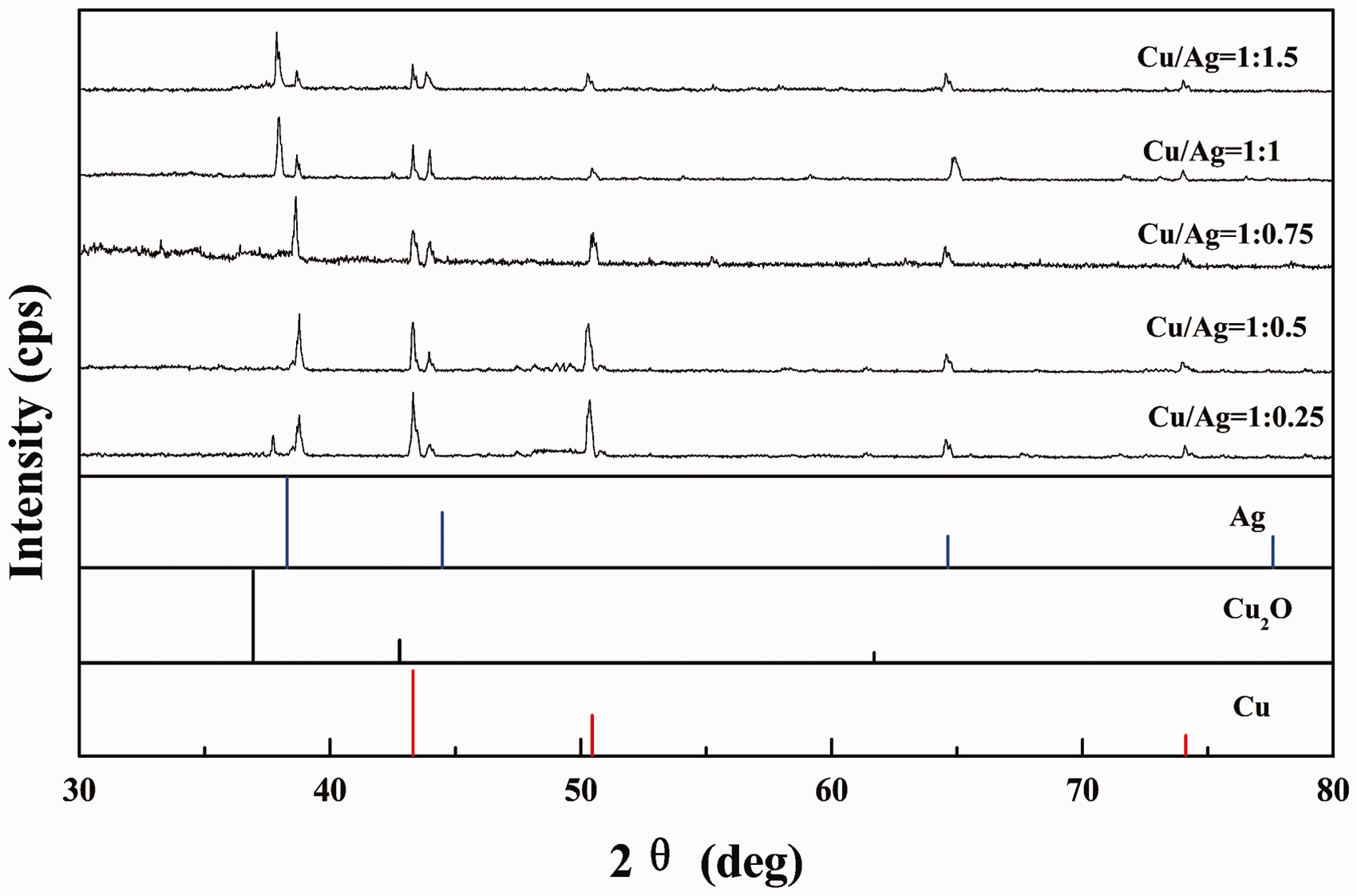
It can be seen that both Cu and Ag peaks can be found in all the samples, and the relatively intensities of the Cu(111) and Cu(200) peaks decreased compared to those of the Ag(111) and Ag(200) peaks with the decrease of [Cu/Ag] molar ratio from 1:0.25 to 1:0.75. This phenomenon can be explained by adding AgNO3 which helps to form the Ag shell coating on the CuNPs core surface. On the other hand, as shown in Figure 1, XRD patterns show no diffraction peaks of copper oxides except those of pure Cu and Ag when the [Cu/Ag] molar ratio is 1:0.5 and 1:0.75. Furthermore, no diffraction peaks of alloy are detected, which shows that the as-prepared nanostructures are core–shell bimetallic composites rather than Cu–Ag alloys.22,23 However, in the case of [Cu/Ag] molar ratios of 1:0.25, 1:1, and 1:1.5, the formation of Cu2O phase was observed in the XRD patterns, which showed that the as-prepared samples had been oxidized.
In order to gain more details about the microstructure of the Cu@Ag core–shell NPs, typical TEM and high-resolution TEM (HRTEM) images were obtained from the samples prepared at different [Cu/Ag] molar ratios, as illustrated in Figure 2. As can be seen clearly from Figure 2a, for samples prepared at a [Cu/Ag] molar ratio of 1:0.25, the core CuNP surfaces were incompletely coated by Ag shells. This is attributable to the fact that the generated AgNPs are not sufficient to fully cover the CuNP surfaces. With a decrease of the [Cu/Ag] molar ratio to 1:0.5 and 1:0.75, the formation of a dense and uniform Ag shell was revealed on the CuNP surface (Figure 2b and c), which is also in agreement with the XRD results. However, on further decrease of the [Cu/Ag] molar ratio to 1:1, the core CuNP surfaces were only partially coated by Ag shells, as shown in Figure 2d. This can be explained by the fact that when an excess amount of AgNO3 is added, the Ag+ ions are then reduced to AgNPs on the surfaces of the CuNPs core with the aid of CuNPs and Na2S2O4 which acts as reducing agent. The AgNPs thus generated quickly and agglomerated to form island structures as a result of surface, interface, and strain energies of the Ag film.
24
These observations indicate that the CuNP core is partially oxidized with decreasing [Cu/Ag] molar ratio, and the formation of Cu2O phase was experimentally confirmed using XRD analysis (Figure 1d and 1e). Moreover, the experiment of Chang et al. proceeded to show that,
25
when the [Cu/Ag] molar ratio was further decreased to 1:2, the NPs comprised a Ag core and a Cu shell.
25
So the optimum condition of the [Cu/Ag] molar ratio was determined to between 1/0.5 and 0.75.
TEM images of Cu@Ag NPs prepared with different [Cu/Ag] molar ratios: (a) 1:0.25; (b) 1:0.5; (c) 1:0.75; (d) 1:1.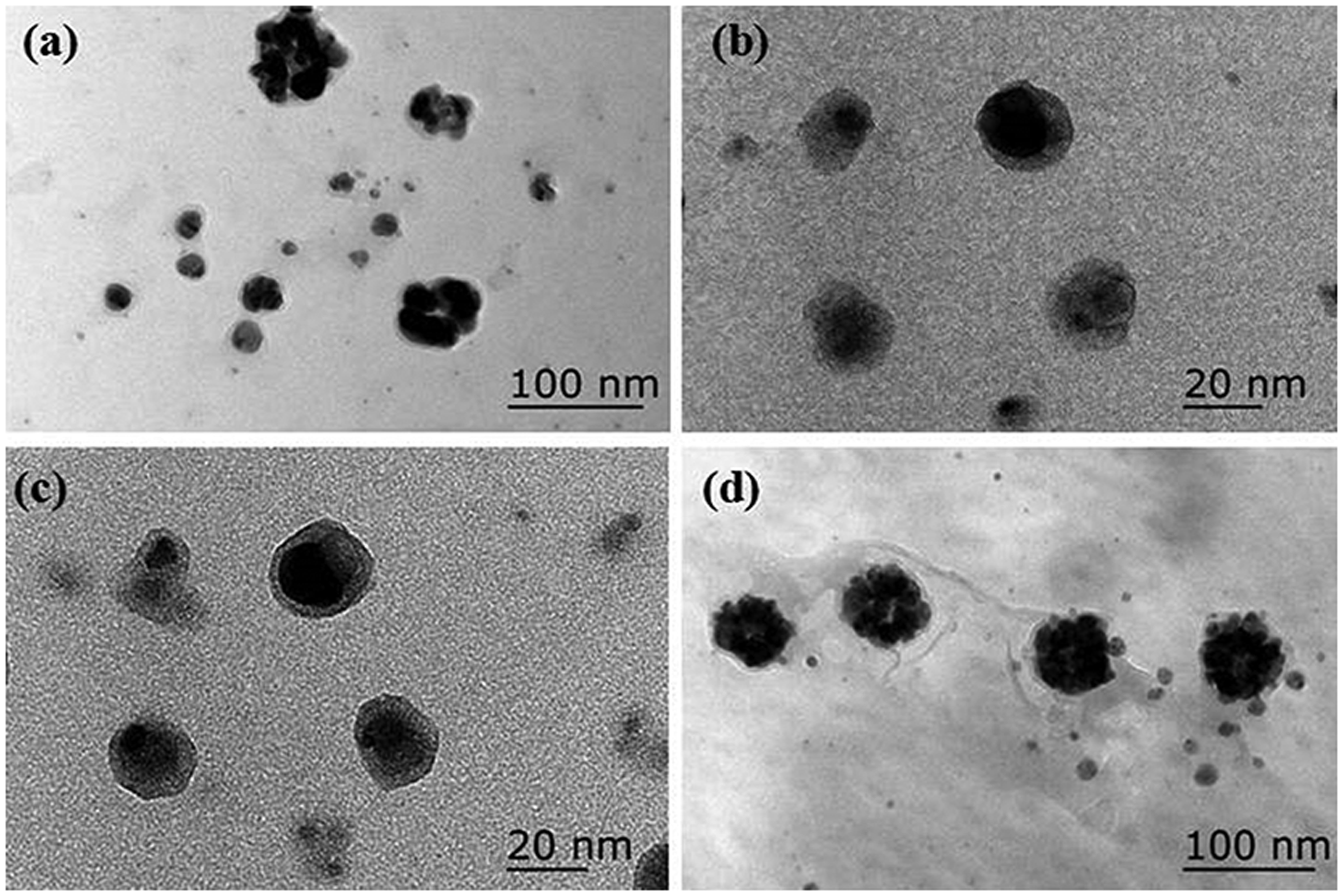
To further identify the structural nature of the Cu@Ag core–shell NPs prepared at a [Cu/Ag] molar ratio of 1:0.75, further insights into the morphology and microstructure of the samples were obtained using TEM and HRTEM, and the corresponding images are shown in Figure 3. As shown in Figure 3a, the as-prepared Cu@Ag core–shell NPs were found to be spherical in shape with an average particle size of 35 nm, and roughly monodispersed without any aggregation. Figure 3b and 3c shows the HRTEM images of a single Cu@Ag core–shell structure with about 15 nm-diameter Cu core and 5 nm-thick Ag shell. From the HRTEM lattice image of the Cu–Ag sample (Figure 3d), the as-prepared sample was crystalline rather than a single crystal, and the as-marked interplanar distance of the fringes were measured to be 0.236 nm and 0.209 nm, corresponding to the (111) lattice spacings of FCC Cu and FCC Ag, respectively.
22
These results are also consistent with the XRD data and those reported previously for Cu@Ag core–shell NPs prepared using different methods.
11
As-prepared Cu@Ag core–shell NPs using a [Cu/Ag] molar ratio of 1:0.75: (a) TEM image; (b, c) HRTEM images of a single Cu@Ag NP; (d) HRTEM lattice image.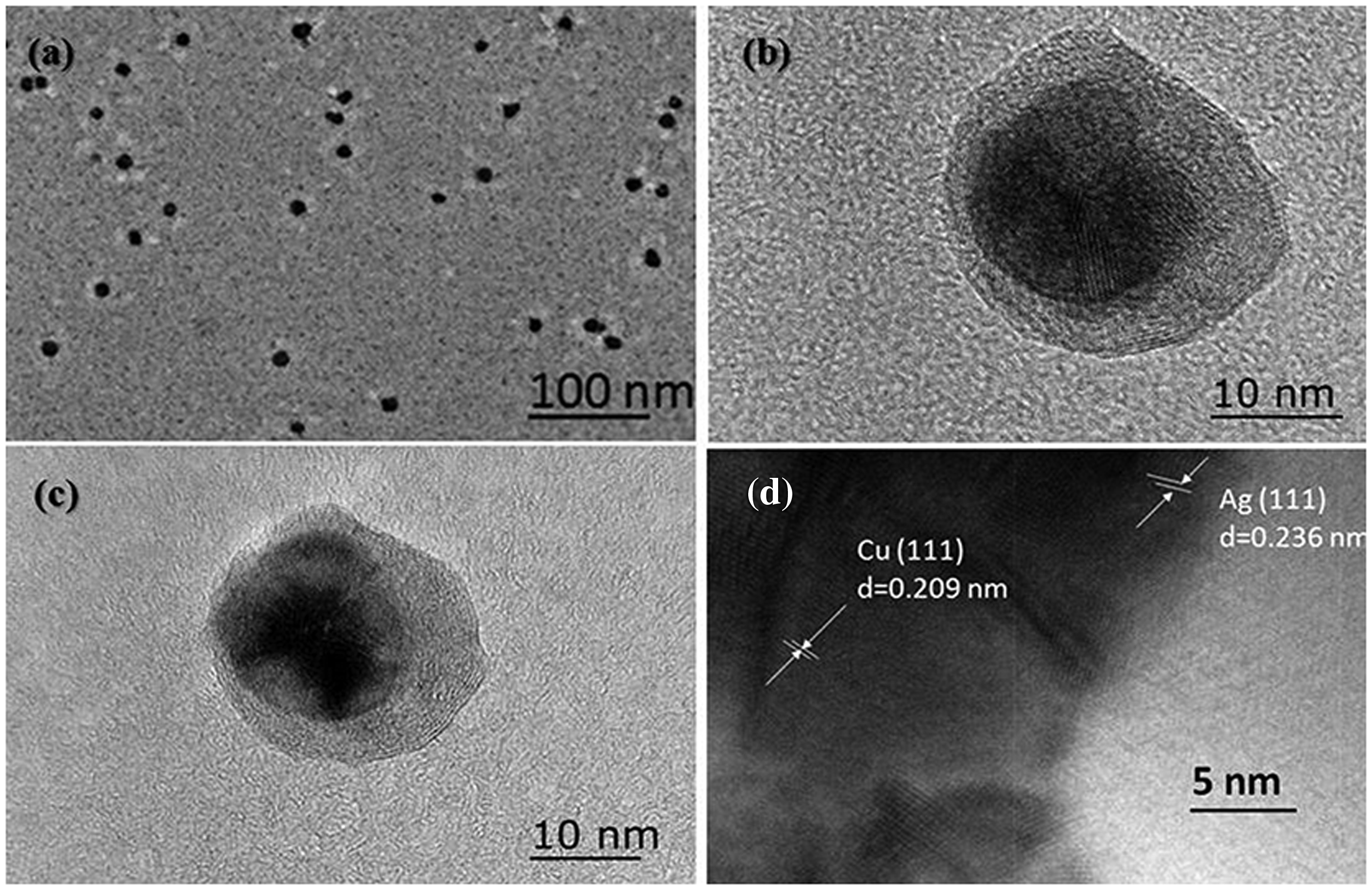
The above results confirm that the microstructure of the Cu@Ag core–shell NPs is strongly influenced by the [Cu/Ag] molar ratio, and the uniform Ag shell is fully coated on the CuNPs surfaces when the optimum condition of the [Cu/Ag] molar ratio is determined to between 1/0.5 and 0.75. When the optimum condition of the [Cu/Ag] molar ratio is between 1/0.5 and 0.75 the uniform Ag shell is fully coated on the CuNP surfaces, which prevents the CuNP core materials from being oxidized.
The XRD and UV-vis measurements were also performed after several months of storage to study the stability of the Cu@Ag core–shell NPs prepared at [Cu/Ag] molar ratio of 1:0.75. The results obtained for the stored samples are given in Figures 4 and 5. As can be seen in Figure 4, there are no diffraction peaks of copper oxides and alloy but only those of pure Cu and Ag after six-month storage.22,23
XRD patterns of freshly prepared and six-month stored Cu@Ag NPs: (a) freshly prepared Cu@Ag NPs; (b) six-month stored Cu@Ag NPs. UV-Vis absorption spectra of Cu@Ag NPs after several months storage.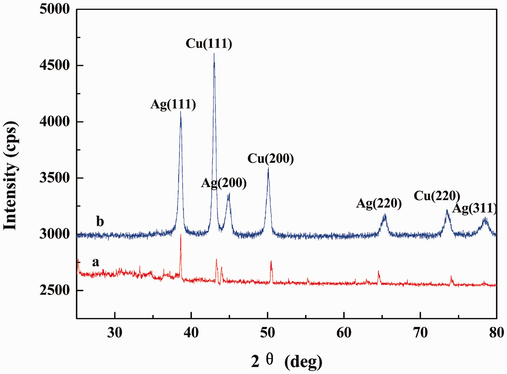
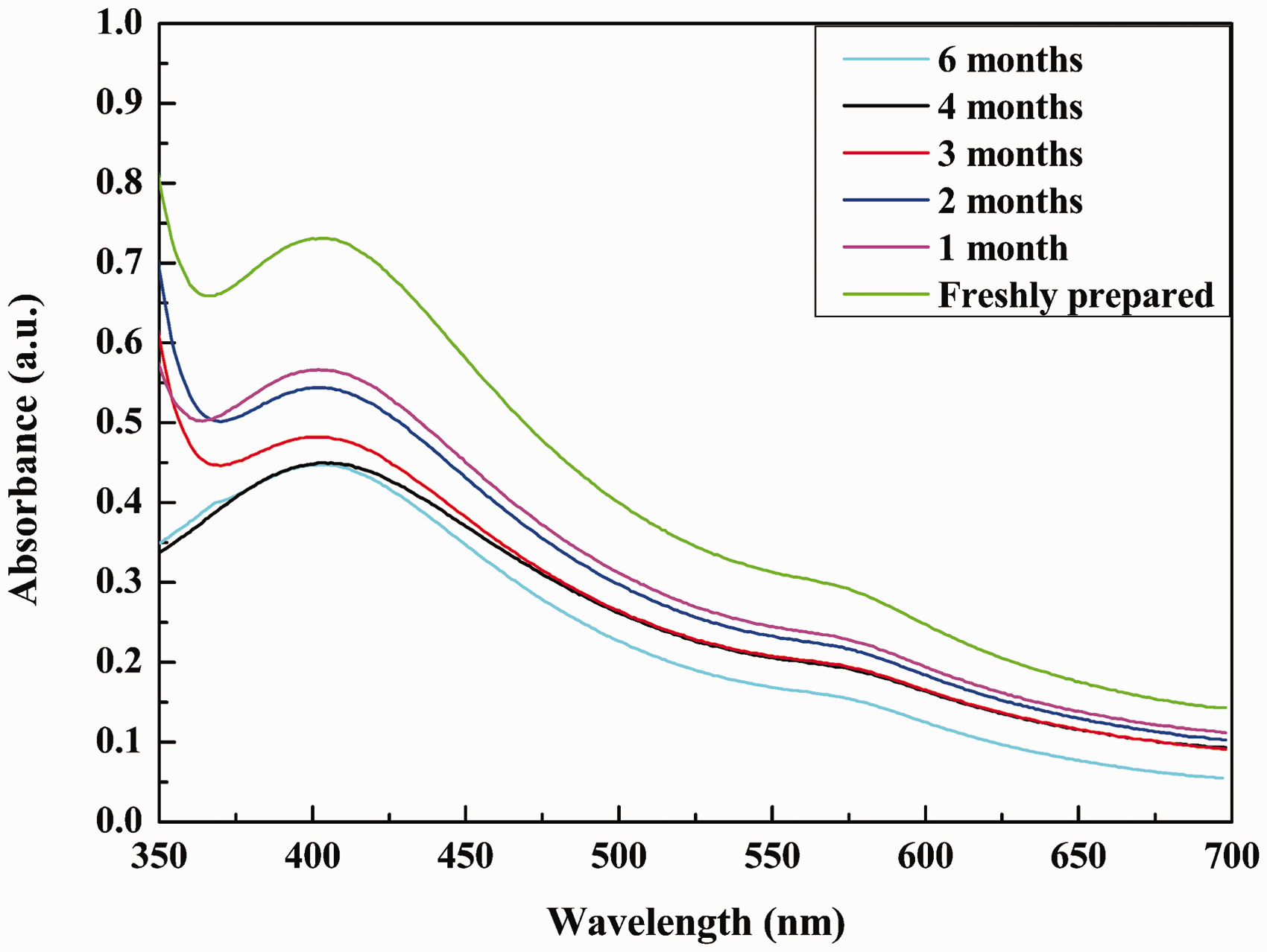
It is reported that spherical AgNPs typically exhibit localized surface plasmon resonance (LSPR) at around 410 nm. 26 In our previous report, the LSPR band of the CuNPs was centered at 575 nm. 16 Colloidal Cu@Ag particles display two bands corresponding to the LSPRs of Cu and Ag, respectively, and the Cu band is shifted to low wavelength.27,28 As can be seen in Figure 5, the Cu@Ag NPs have a maximum in their absorption at 550 nm (LSPR of Cu) and 400 nm (LSPR of Ag), respectively, and the Cu band is shifted from 575 nm to a lower wavelength (550 nm) as expected. This result further indicates that the as-prepared samples are Cu@Ag NPs.
As seen also in Figure 5, the absorption intensity of the samples decreases due to the slight aggregation of the Cu@Ag NPs during several months of storage. However, the two absorption peaks remained unchanged and no precipitation occurred after several months of storage under ambient conditions, clearly indicating that the Cu@Ag NPs have high stability. So the as-prepared Cu@Ag NPs have excellent antioxidant properties, which will not affect their potential application.
SERS Activity
Substrates with good stability and reproducibility are of crucial importance for the use of SERS as a routine detection technique. To test whether the as-prepared Cu@Ag colloidal NPs are able to give reproducible and deterministic SERS signals, a low concentration of the CV target molecule was used. The SERS spectra of CV molecules from three randomly selected positions on the Cu@Ag core–shell NP substrate, and also the SERS spectra of CV on Cu@Ag NPs after different storage times, are shown in Figure 6.
Comparison of SERS spectra of CV on freshly prepared and six-month stored Cu@Ag NPs (A – freshly prepared samples, B – six-month stored samples): (a) Raman spectra of CV with the intensities multiplied by 10; (b) a series of SERS spectra of CV collected on three randomly selected spots of Cu@Ag NPs.
Assignment and the SERS EF of several normal Raman (NR) peaks of CV on freshly prepared Cu@Ag NPs.
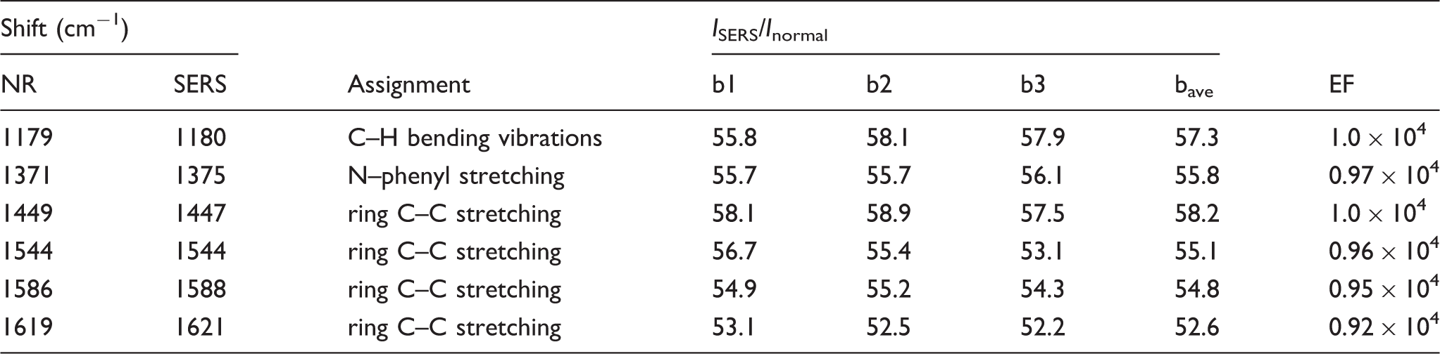
In order to estimate reproducibility of the SERS signals, the calculated ISERS/Inormal approximations of the freshly prepared samples are summarized in Table 1. The average ISERS/Inormal value is 55.6, and the relative standard deviation (RSD) of ISERS/Inormal of the bands at 1179, 1371, 1449, 1544, 1586, and 1619 cm−1 are all <20% at 3.1%, 0.3%, 4.7%, 0.9%, 1.4%, and 5.4%, respectively, again supporting the great enhancement effect and high reproducibility of this substrate. In addition, the ISERS/Inormal approximations of the six-month stored samples were calculated. The average ISERS/Inormal value is 51.9, and the RSD of ISERS/Inormal of all the bands increased, but all of them are all <20%. These results also indicate that the CV probe molecules are uniformly distributed on the surface of the Cu@Ag core–shell substrates, and the reproducibility of the substrate is relatively better than our previous research about the SERS of CuNPs. 16 So the colloidal Cu@Ag core–shell NPs can serve as an excellent SERS substrate to detect any analyte in trace amounts.
To evaluate the SERS activity of the as-prepared Cu@Ag core–shell colloids, the enhancement factor (EF) can be calculated using the following equation:
32
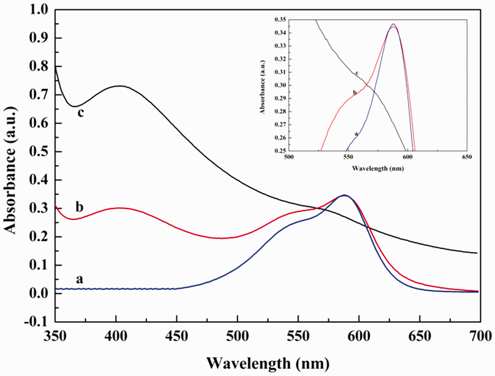
UV-vis absorption spectra of (a) CV, (b) CV solution in the presence of Cu@Ag NPs, and (c) Cu@Ag NPs.
Conclusions
In this work, Cu@Ag core–shell NPs were successfully prepared in a one-step process using successive sodium hyposulfite reduction of metal salts in aqueous solution at room temperature without any inert atmosphere. To obtain a homogeneous Ag coating on CuNPs, the influence of [Cu/Ag] molar ratio on coatings was investigated. In the synthesized process, [Cu/Ag] molar ratios play an important role in forming the uniform and dense Ag shell on the surface of the CuNPs. From the structure and the composition of the analyses of the samples prepared at different [Cu/Ag] molar ratios, the optimum condition of the [Cu/Ag] molar ratio varies between 1/0.5 and 1/0.75 for a 15 nm-diameter Cu core and 5 nm-thick uniform Ag shell structure. In these synthetic conditions, the as-prepared nanomaterials are shown to be stable over several months, and the long-term stability is of the great importance in practical application. The results of the SERS activity of the freshly prepared and six-month stored nanostructures indicate that the CV probe molecules are uniformly distributed on the substrate, and the reproducibility of SERS signals is very high. The average SERS enhancement factors were estimated to be up to 104, and the as-prepared Cu@Ag core–shell NPs can serve as an excellent SERS substrate. This study also has significant implications for the preparation of other bimetallic core–shell nanostructures, such as Au@Co, Au@Ni, and Au@Ni.
Footnotes
Conflict of Interest
None declared.
Funding
This work was supported by the Natural Science Foundation of Anhui Province (grant number 1508085SMB209), the Anhui Provincial Key Science Foundation for Outstanding Young Talent (grant number 2013SQRL022ZD), Zhejiang Province Key Laboratory of Soldering & Brazing Materials and Technology (grant number 1402), and the Graduate Innovation Foundation of Anhui University of Technology (grant number 2014075).