
Abstract
Background
A growing body of evidence suggests that male infertility is a precursor to future health problems. This study aimed to develop a unique circulating biomarker that could contribute to the diagnosis of male infertility.
Materials and methods
The study included 114 adult male participants aged 20–50 years who underwent sperm collection for in vitro fertilization and embryo transfer at Nagoya University Hospital, Gifu University Hospital, and Misao Ladies Hospital.
Results
Circulating VEGF-A and sFlt-1 levels did not differ significantly between men with normal and abnormal semen parameters. In contrast, proteomic analysis identified VEGFR1/2-related proteins associated with sperm motility, among which soluble MCAM/CD146 was significantly elevated in participants with abnormal semen parameters (84.0 [20.9–183.8] pg/mL vs 352.7 [115.8–1236.3] pg/mL, p < 0.0001), and in similar those with reduced sperm motility (106.5 [25.1–282.6] pg/mL vs 352.7 [125.5–1133.4] pg/mL, p < 0.0001). Receiver operating characteristic analysis demonstrated the diagnostic potential of soluble MCAM/CD146, with an area under the curve of 0.787, 66.7% sensitivity, and 78.6% specificity at the optimal threshold, supporting its role as a candidate biomarker for male infertility.
Conclusions
Circulating soluble MCAM/CD146 may represent a potential biomarker associated with abnormal semen parameters.
Introduction
Clinical infertility is the inability of a couple to conceive after 12 months of regular, unprotected intercourse. 1 Male-related factors are estimated to contribute to up to 50% of infertility cases, 2 and assisted reproductive technologies play a crucial role in managing male factor infertility. 3 The evaluation of male infertility includes detailed history-taking and physical examination and is primarily based on semen analysis, as blood-based screening tests remain insufficient and are not yet established. Treatments that lead to measurable improvements in fertility include lifestyle optimization, medical drug therapy, and surgical interventions. Although male infertility is recognized as a disease that impacts the quality of life of both members of the infertile couple, little data exist on its specific quantification and impact compared with other health-related conditions.
Although well-known physiologically and pathologically for regulating angiogenesis, vascular endothelial growth factor A (VEGF-A) and its receptors are present in the male and female reproductive fields. VEGF-A is also important for reproductive and bone angiogenesis. 4 It is present in spermatids, Sertoli cells, Leydig cells, and seminal plasma, and its varying concentrations are thought to play a role in implantation and fertility. 5 VEGF-A has recently been implicated in male fertility and in the maintenance of spermatogonial stem cells (SSCs). 6 Moreover, it enhances sperm motility in vitro in a concentration-dependent manner. 7
Melanoma cell adhesion molecules (MCAM), also known as CD146 or MUC18, were initially identified as biomarkers of melanoma progression. MCAM/CD146 is a transmembrane glycoprotein with a molecular weight of 113–130 kDa.8,9 Structurally, it comprises five immunoglobulin domains, 10 a transmembrane domain, and a cytoplasmic region. In normal adult tissues, MCAM/CD146 is primarily expressed in the vascular endothelium and smooth muscle. It functions as an important cell adhesion protein in vascular endothelial cell activity and angiogenesis and is present on circulating endothelial cells. In addition to its membrane-anchored form, MCAM/CD146 exists in a soluble form (soluble MCAM/CD146, 105 kDa), which is found in human plasma and in the supernatants of cultured human endothelial cell.10,11 Moreover, MCAM/CD146 has been identified as a coreceptor for VEGF receptor 2 (VEGFR2), mediating VEGF-A-induced angiogenesis both in vitro and in vivo. 12
Among the most critical factors inducing angiogenesis, the VEGF-A/VEGF receptor 2 (VEGFR2) pathway and MCAM/CD146 play key roles. However, the relationship between the VEGF-A-related proteins, MCAM/CD146, and semen parameters in male infertility remains unclear. This study examined the associations between semen parameters, VEGF-A-related protein levels, and soluble MCAM/CD146 levels to explore blood biomarkers as potential alternatives to semen analysis.
Materials and methods
Study participants
Adult males aged 20–50 years who underwent sperm collection for in vitro fertilization and embryo transfer at Nagoya University Hospital, Gifu University Hospital, and Misao Ladies Hospital were recruited. A total of 136 participants who provided informed consent were enrolled in the study. However, 22 participants did not undergo sperm collection for in vitro fertilization and embryo transfer; therefore, they were excluded from the study. Additionally, participants with known malignancies or clinically evident vascular diseases were excluded based on available medical records. Consequently, 114 participants who underwent both semen analysis and blood sampling were included in the study subjects. This study was approved by the Research Ethics Committee of Nagoya University Graduate School of Medicine (Approval No. 2020-0134) and the Gifu University Graduate School of Medicine (Approval No. 2023-312).
Analysis of three-dimensional vascular structure by clearing of biological tissue and immunostaining
Eight-week-old male BL6/ncr mice were purchased from Japan SLC (Shizuoka, Japan). The animals were housed in environmentally controlled rooms at the animal experimentation facility of Osaka University under standard conditions (22 ± 2°C, 12 h light/dark cycle, standard laboratory diet, and water ad libitum). Animals were maintained in individually ventilated cages (ISOrat900 N, Tecniplast, Buguggiate, Italy) with appropriate bedding, enrichment, and air filtration. All experiments were performed according to the guidelines of the Osaka University Committee for Animal and Recombinant DNA Experiments (Approval No. 4062). Mice were euthanized, and testicular tissues were excised using scissors. Testis samples were fixed with 4% paraformaldehyde in phosphate buffer (pH 7.4), washed with phosphate-buffered saline, and subjected to immunofluorescence staining with anti-mouse CD31 monoclonal antibody (BD Biosciences) and Alexa Fluor 546-conjugated goat anti-rat IgG polyclonal secondary antibody (Thermo Fisher Scientific). Following immunostaining, testis samples were immersed in RapiClear 1.52 reagent (SunJin Lab.) at 37°C with gentle shaking for 2 days to remove water and lipids. Finally, the refractive index was matched by placing the cleared sample in fresh RapiClear 1.52 reagent. These samples became sufficiently transparent to allow acquisition of inner fluorescence data using confocal microscopy (TCS/SP8 DM6000 CFS equipped with HC PL FLUOSTAR 5×/0.15 DRY and PL FLUOSTAR 10×/0.30 DRY objective lenses; Leica) and were processed using the Leica Application Suite (Leica).
Laboratory analysis
The patients’ clinical characteristics and data were retrospectively collected from their medical records. The central laboratory at Gifu University Hospital measured serum total protein, albumin, triglycerides, total cholesterol, low-density lipoprotein cholesterol, high-density lipoprotein cholesterol, C-reactive protein, and creatinine levels. Testosterone and free testosterone levels were measured by SRL, Inc. The estimated glomerular filtration rate (eGFR) and eGFR based on creatinine (eGFRcre) were assessed. eGFRcre was calculated using the following equation proposed by the Japanese Society of Nephrology: eGFRcre (mL/min/1.73 m2) = 194 × SCr −1.094 × age −0.287 × 0.739. 13 Serum samples were stored at −80°C until measurements were performed.
Semen collection and analysis
Semen collection and analysis were conducted at Nagoya University Hospital and Misao Ladies Hospital. Participants were instructed to observe a recommended sexual abstinence of 2 to 4 days. Semen samples were collected by masturbation into a sterile container. While collection was available in a designated room within the hospital, participants could also collect samples at home. For home collection, samples had to be delivered to the hospital within 3 h and maintained at room temperature. Sperm counts were performed using the Makler counting chamber method, in accordance with WHO Laboratory Manual recommendations. The abnormal semen parameter group was defined as having one or more semen parameters (semen volume, total sperm number, sperm concentration, or sperm motility) below the reference limits specified in the World Health Organization Laboratory Manual for the Examination and Processing of Human Semen (6th edition, 2021). Based on these criteria, participants were categorized into either the normal or abnormal group, as summarized in Supplemental Table 1.
Measurement of VEGF-A, sFlt-1 and soluble MCAM/CD146
Serum VEGF-A (sVEGF-A) levels were measured using the Human VEGF Quantikine ELISA Kit (DVE00; R&D Systems, Minneapolis, MN, USA) according to the manufacturer’s instructions. Serum sFlt-1 (sFlt-1) levels were measured using a Human VEGFR1/Flt-1 Quantikine ELISA Kit (DVR100 C; R&D Systems, Minneapolis, MN, USA) according to the manufacturer’s instructions. Serum soluble MCAM/CD146 levels were measured using the Human MCAM/CD146 DuoSet ELISA Kit (DY932-05; R&D Systems, Minneapolis, MN, USA) according to the manufacturer’s instructions.
Proteomic analysis
Proteomic analysis was performed on serum samples from seven control participants and nine participants with low sperm motility. These samples were randomly selected from eligible participants with available serum specimens. Twenty-five micrograms of protein from each serum sample was prepared using the phase-transfer surfactant (PTS) method. 14 Briefly, samples were solubilized, reduced, and alkylated in 5.4 M guanidine buffer (pH 8.5) containing 7.5 mM TCEP and 30 mM chloroacetamide at 95°C for 5 min, followed by sonication for 10 min. The total protein was then purified by chloroform-methanol precipitation and re-dissolved in PTS buffer (12 mM sodium deoxycholate, 12 mM sodium lauroyl sarcosinate, 100 mM Tris-HCl, pH 8.0), followed by heating at 95°C for 10 min. After overnight digestion using Lys-C and trypsin at 37°C, an equal volume of ethyl acetate was added, and the mixture was acidified with 20% TFA. After centrifugation at 15,600×g for 3 min, the samples were frozen at −80°C, and the upper layer was removed. The lower layer was vacuum-dried, re-dissolved in 0.1% TFA in 5% acetonitrile, and desalted using GL-Tip SDB. The resulting solution was vacuum-dried and resuspended in 0.1% TFA and 2% acetonitrile. Nano-LC-MS/MS was performed using a Dionex U3000 gradient pump (Thermo Fisher Scientific) coupled to a Q Exactive Hybrid Quadrupole-Orbitrap Mass Spectrometer (Thermo Fisher Scientific). The samples were concentrated on a C18 trap column (particle size, 5 µm; inner diameter, 300 μm; length, 5 mm; Chemical Evaluation and Research Institute, Tokyo, Japan) and separated on a C18 column (particle size, 3 µm; inner diameter, 100 µm; length, 125 mm; Nikkyo Technos, Tokyo, Japan) at a flow rate of 0.5 µL/min. The chromatographic mobile phases were as follows: solvent A (water containing 0.5% acetic acid) and solvent B (80% acetonitrile containing 0.5% acetic acid). The gradient program was as follows: 5–40% B over 100 min; 40–100% B over 1 min; held at 100% B for 3 min; returned to 5% B over 1 min; and re-equilibrated at 5% B for 10 min. Electrospray ionization was performed in the positive-ion mode, and the instrument was operated in DDA mode. Xcalibur 4.1.50 (Thermo Fisher Scientific) was used to record the peptide spectra. Full scans were acquired from 350 to 1800 m/z with a resolution of 17,500, an automatic gain control of 3 × 10⁶, and maximum injection time of 60 ms. MS/MS scans were performed with a resolution of 35,000, automatic gain control target of 1 × 10⁵, and a maximum injection time of 60 ms. The 10 highest-intensity precursor ions were isolated using a quadrupole analyzer within a window of 2.0 m/z and fragmented by higher-energy collisional dissociation with a normalized collision energy of 27%. Multiply charged peptides were selected for the MS/MS experiments. The dynamic exclusion time was set to 20 s. The Uniprot Homo sapiens (TaxID = 9606) protein sequence database was used, with cRAP included for contaminants (https://www.thegpm.org/crap/). MS/MS spectra were interpreted, and peak lists were generated using Proteome Discoverer 2.4.1.15 (Thermo Fisher Scientific). Searches were conducted using SEQUEST (Thermo Fisher Scientific) with the following parameters: enzyme specificity with up to two missed cleavage sites, peptide mass tolerance of 10 ppm, and MS/MS tolerance of 0.02 Da. A fixed modification of carbamidomethyl (Cys) and variable modification of oxidation (Met) were applied. Peptide identification was based on the significant Xcorr values using high-confidence filters. Peptide identification and modification data returned from SEQUEST were filtered at a false discovery rate of 1% using the Percolator node in Proteome Discoverer, resulting in confirmed peptide identifications and modification lists from HCD MS/MS.
Protein–protein interaction network functional enrichment analysis
To identify potential protein–protein interactions (PPIs) associated with the candidate proteins identified through proteomic analysis and with VEGFR1 and VEGFR2, we performed a functional enrichment analysis using the STRING database. 15 First, a list of candidate proteins identified from proteomic analysis was compiled. These proteins were then input into the STRING database to generate a network of predicted PPIs. STRING is a powerful tool that integrates known and predicted PPIs from various sources, including experimental data, computational predictions, and literature mining. We specifically focused on interactions between the candidate proteins and KDR (kinase insert domain receptor, also known as VEGFR2) and FLT1 (fms-related receptor tyrosine kinase 1, also known as VEGFR1). The STRING database was queried with VEGFR1 and VEGFR2 as seed proteins, and the interactions with the candidate proteins were analyzed.
Statistical analyses
Data are expressed as median (interquartile range) or n (%). Groups with normal and abnormal semen parameters were compared using the Wilcoxon rank-sum test and Fisher’s exact test. Receiver operating characteristic analysis was applied to evaluate the predictive performance of MCAM using the pROC package in R. 16 To determine the cutoff value, we used the Youden index, defined as sensitivity + specificity −1. The threshold corresponding to the maximum Youden index was selected as the optimal cutoff, as it provides the best balance between sensitivity and specificity. Multivariable analyses adjusting for potential confounders were not performed owing to the exploratory nature of this study. All P-values were two-tailed, and p-values <0.05 were considered statistically significant. All statistical analyses were performed using SAS software, version 9.4 (SAS Institute Inc.), and R, version 4.4 (https://www.r-project.org).
Results
Whole mount analysis of mouse testis and 3D immunofluorescent staining
VEGF-A has also been implicated in SSC maintenance.
6
Therefore, the vascular network in mouse testes was evaluated using the RapiClear technique to provide an anatomical context for angiogenesis-related signaling within the testicular environment. The testes comprised a dense vascular network (Figures 1(a) and (b)). While the functional significance of this vascular structure was not directly assessed in this study, these observations are presented as supportive background information for the potential involvement of angiogenic factors in sperm function. Vasculature and localization of vascular endothelial cells in the mouse testis.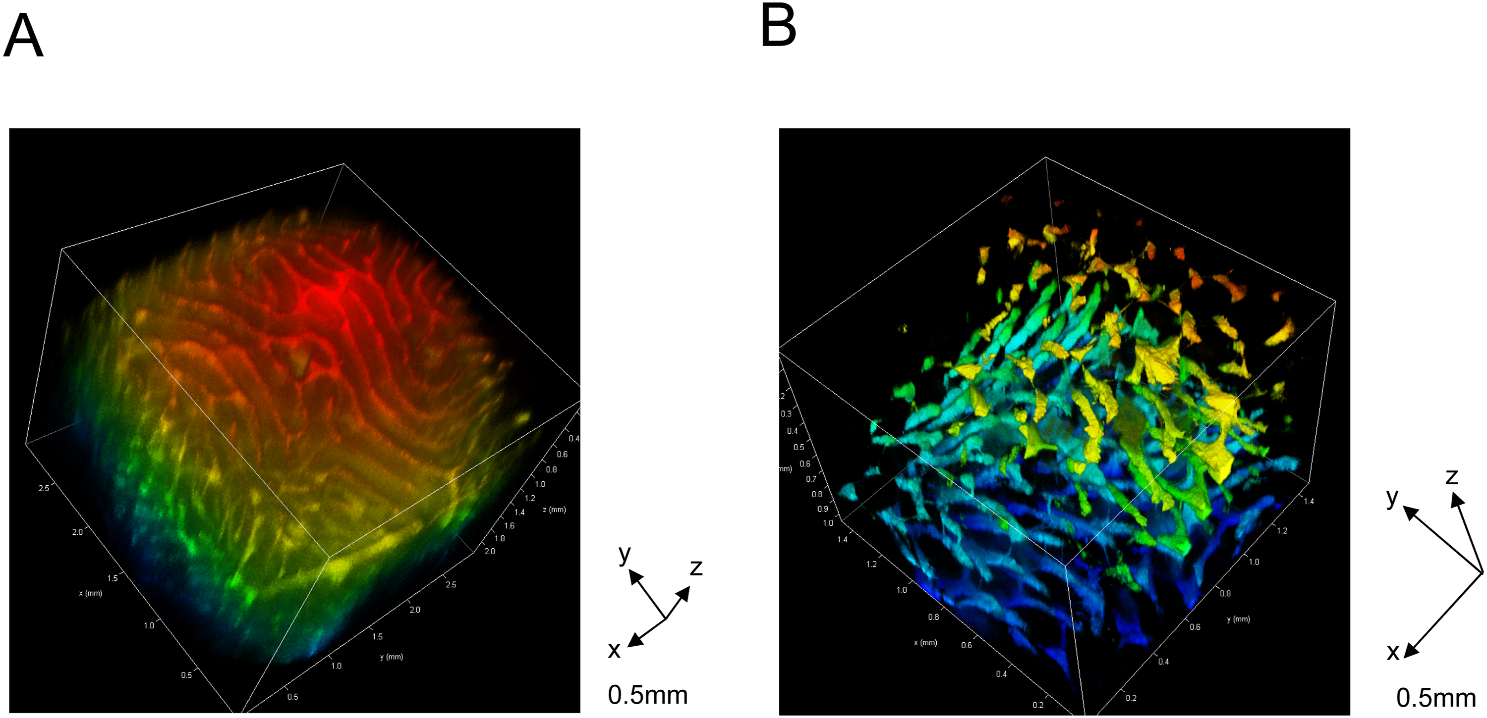
Circulating levels of VEGF-A and sFlt-1 in male infertility
Patient Characteristics.
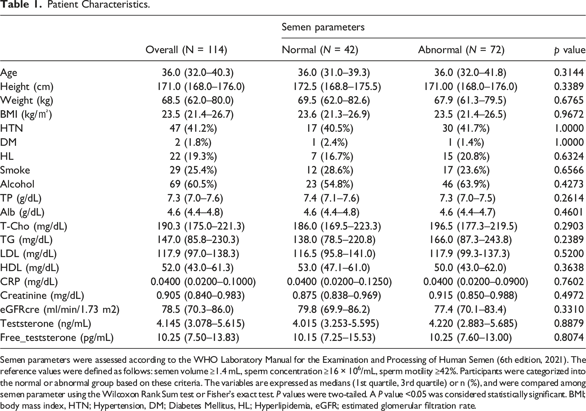
Semen parameters were assessed according to the WHO Laboratory Manual for the Examination and Processing of Human Semen (6th edition, 2021). The reference values were defined as follows: semen volume ≥1.4 mL, sperm concentration ≥16 × 106/mL, sperm motility ≥42%. Participants were categorized into the normal or abnormal group based on these criteria. The variables are expressed as medians (1st quartile, 3rd quartile) or n (%), and were compared among semen parameter using the Wilcoxon Rank Sum test or Fisher’s exact test. P values were two-tailed. A P value <0.05 was considered statistically significant. BMI; body mass index, HTN; Hypertension, DM; Diabetes Mellitus, HL; Hyperlipidemia, eGFR; estimated glomerular filtration rate.
Levels of Circulating of VEGF-A and sFlt-1 in Subjects With Abnormal semen Parameter.
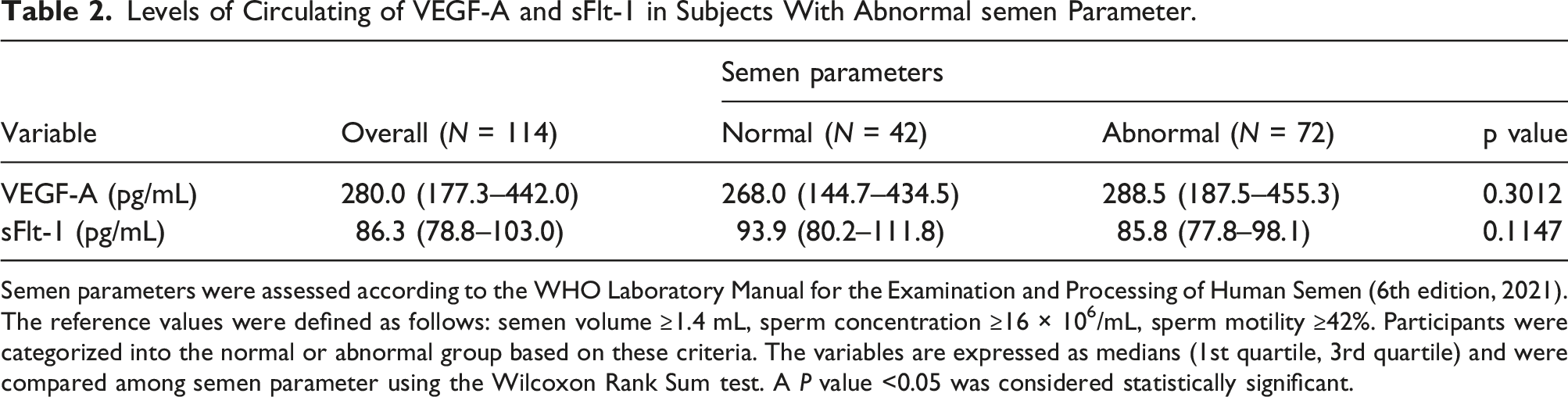
Semen parameters were assessed according to the WHO Laboratory Manual for the Examination and Processing of Human Semen (6th edition, 2021). The reference values were defined as follows: semen volume ≥1.4 mL, sperm concentration ≥16 × 106/mL, sperm motility ≥42%. Participants were categorized into the normal or abnormal group based on these criteria. The variables are expressed as medians (1st quartile, 3rd quartile) and were compared among semen parameter using the Wilcoxon Rank Sum test. A P value <0.05 was considered statistically significant.
Identification of VEGFR1- and VEGFR2-related proteins involved in sperm motility
Candidate Factors Related to VEGFR1 and VEGFR2.
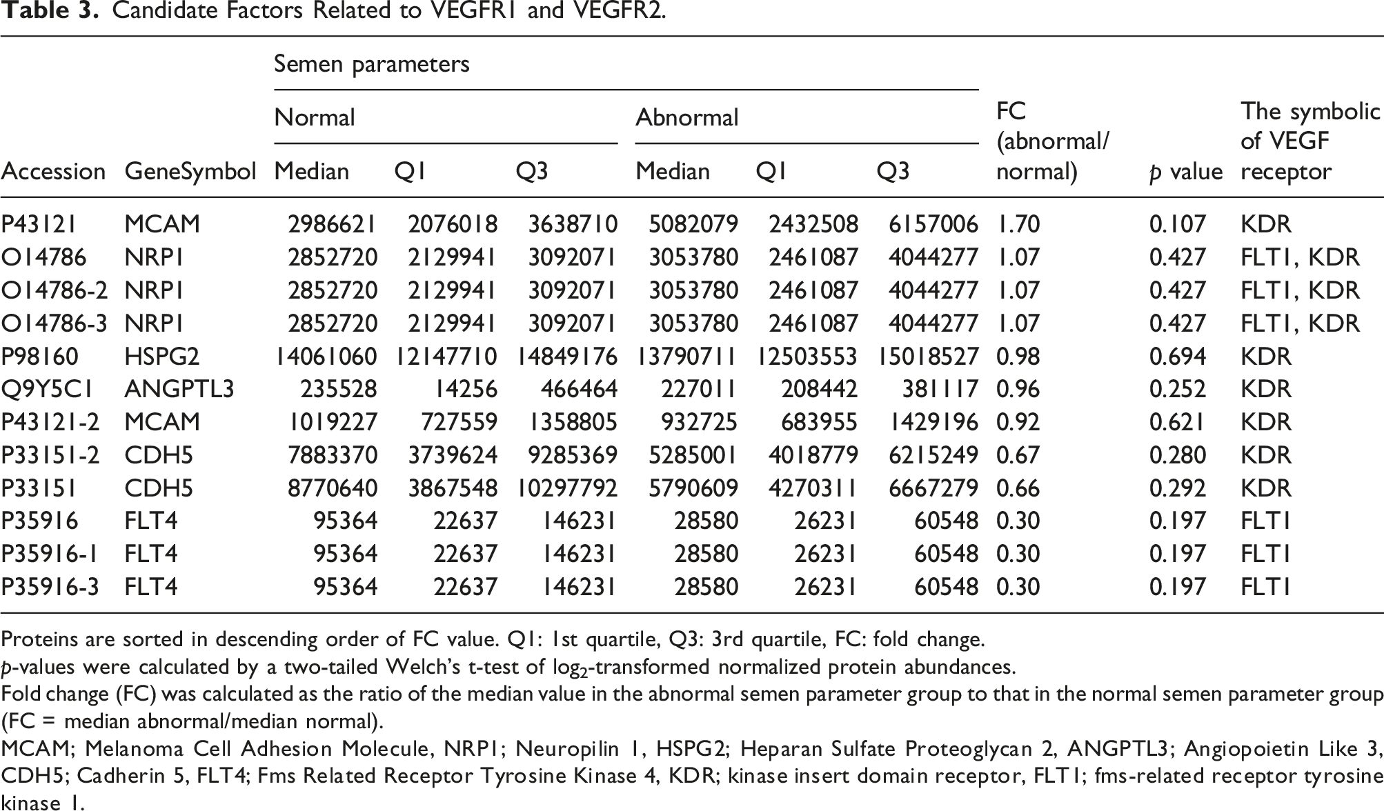
Proteins are sorted in descending order of FC value. Q1: 1st quartile, Q3: 3rd quartile, FC: fold change.
p-values were calculated by a two-tailed Welch’s t-test of log2-transformed normalized protein abundances.
Fold change (FC) was calculated as the ratio of the median value in the abnormal semen parameter group to that in the normal semen parameter group (FC = median abnormal/median normal).
MCAM; Melanoma Cell Adhesion Molecule, NRP1; Neuropilin 1, HSPG2; Heparan Sulfate Proteoglycan 2, ANGPTL3; Angiopoietin Like 3, CDH5; Cadherin 5, FLT4; Fms Related Receptor Tyrosine Kinase 4, KDR; kinase insert domain receptor, FLT1; fms-related receptor tyrosine kinase 1.
Relationship between circulating soluble MCAM/CD146 concentration and semen parameters
Evaluation of soluble MCAM/CD146 concentration in serum showed that it was significantly increased in participants with abnormal semen parameters compared with that in participants with normal semen parameters (84.0 [20.9–183.8] pg/mL vs 352.7 [115.8–1236.3] pg/mL, p < 0.0001, Figure 2). Furthermore, when focusing on sperm motility, the level of soluble MCAM/CD146 was significantly higher in participants with decreased sperm motility than in those with normal sperm motility (106.5 [25.1–282.6] pg/mL vs 352.7 [125.5–1133.4] pg/mL, p < 0.0001, Figure 3). The abnormal semen parameter group was defined as having one or more semen parameters (semen volume, total sperm number, sperm concentration, or sperm motility) below the reference limits specified in the World Health Organization Laboratory Manual for the Examination and Processing of Human Semen (6th edition, 2021). The variables are expressed as medians (1st quartile, 3rd quartile) and were compared between groups using the Mann–Whitney test. The p-values were two-tailed. Statistical significance was set at p < 0.05. Levels of circulating soluble MCAM/CD146 in participants with abnormal sperm motility. Normal participants were defined as having sperm motility ≥42%, and the abnormal group was defined as having sperm motility of <42%. The variables are expressed as medians (1st quartile, 3rd quartile) and were compared between groups using the Mann–Whitney test. The p-values were two-tailed. Statistical significance was set at p< 0.05.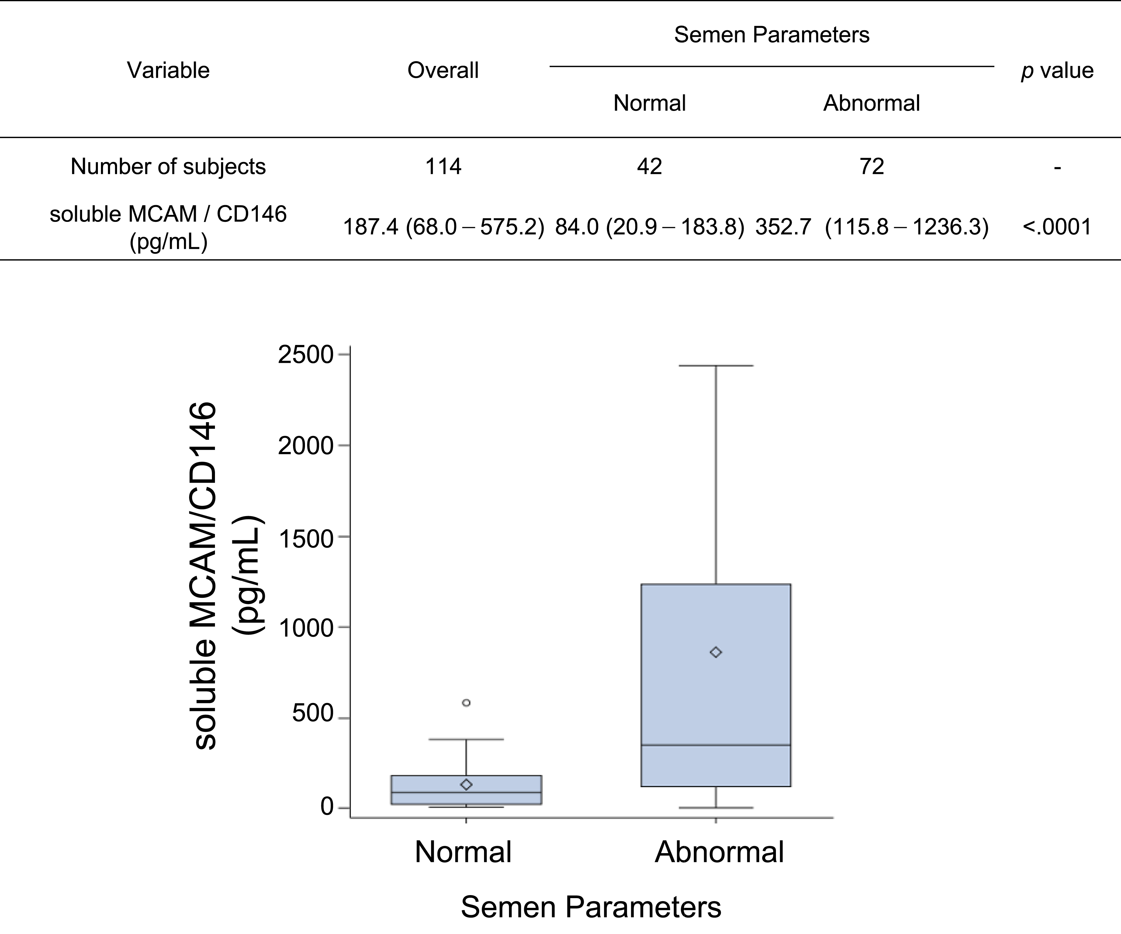

Receiver operating characteristic analysis of MCAM/CD146 for diagnostic accuracy
To evaluate the diagnostic utility of soluble MCAM/CD146 concentration as a biomarker, receiver operating characteristic analysis was performed in 114 participants (72 abnormal and 42 normal cases). The area under the curve (AUC) was 0.787, indicating moderate discriminatory ability (Figure 4(A)). The optimal cutoff value determined by the Youden index was >187.4 pg/mL (Figure 4(B), (C)). At this threshold, the sensitivity was 66.7% (95% CI: 54.6–77.3) and the specificity was 78.6% (95% CI: 63.2–89.7). The positive likelihood ratio at this threshold was 3.111. Receiver operating characteristic analysis of soluble MCAM/CD146 for abnormal semen parameters. Receiver operating characteristic curves were constructed to evaluate the diagnostic performance of serum soluble MCAM/CD146 in discriminating between men with normal and abnormal semen parameters. The area under the curve (AUC) was 0.787, indicating moderate discriminatory ability (a). Relationship between cutoff values and sensitivity/specificity (b). At a cutoff of >187.4 pg/mL, sensitivity was 66.7%, and specificity was 78.6% (c).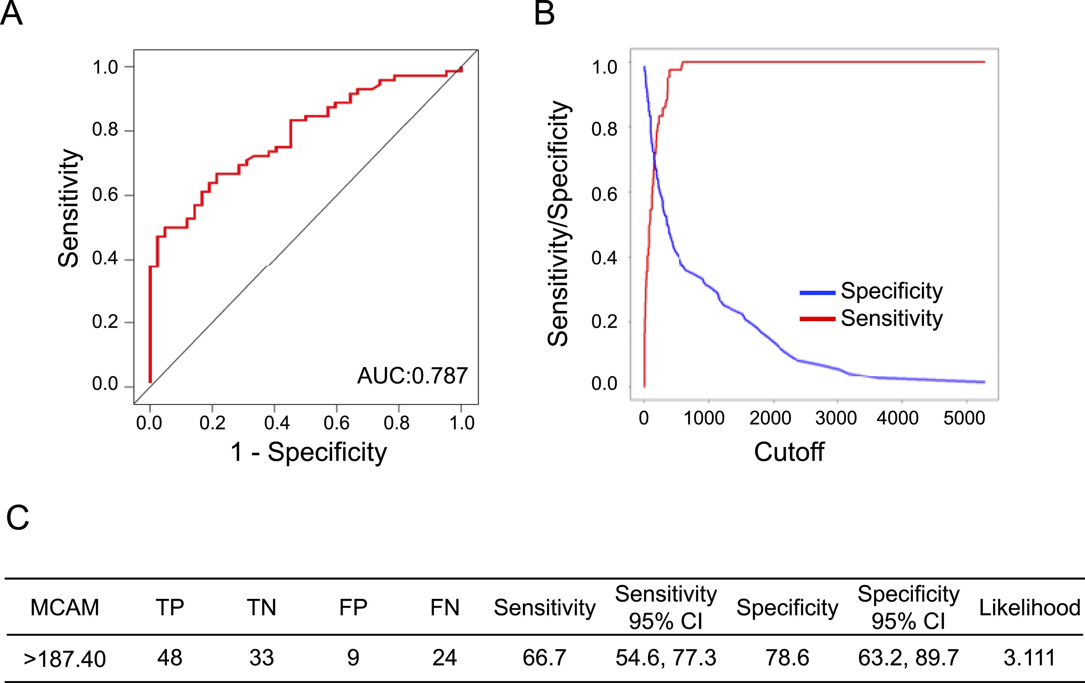
Discussion
Recent advances in assisted reproductive technologies have enabled many men with infertility to father children, including through intracytoplasmic sperm injection using ejaculated or surgically retrieved sperm. However, no fundamental treatment leading reliably to spontaneous pregnancy has been established in many cases of male infertility. To improve the evaluation of male infertility, blood-based biomarkers that may complement semen analysis and provide additional insight into the underlying pathophysiology need to be identified. This study aimed to clarify the relationship between male infertility and angiogenic factors and to develop a new testing method that contributes to the diagnosis of male infertility by identifying new target factors.
In some reports, angiogenic factors, such as VEGF-A, are involved in post-ovulatory luteal angiogenesis and pregnancy-associated luteal development in females. Additionally, in cases of ovarian hyperstimulation syndrome, VEGF-A levels in the blood are elevated in the mid-luteal phase. 17 However, only limited reproductive medical aspects of VEGF-A have been reported.6,7,18 For example, the in vitro effects of VEGF-A administration on human sperm motility have been described. VEGF-A has also been suggested to regulate germ cell proliferation in vitro and promote regeneration of mouse testes in vivo. These findings suggest that VEGF-A has direct or indirect effects in reproductive medicine; however, the precise mechanisms remain unknown. Therefore, we hypothesized that angiogenic factors influence sperm activity.
Assessment of the vascular network in mouse testes revealed multiple layers of vascular endothelial cells and various vascular migrations. However, serum levels of VEGF-A and sFlt-1 did not significantly differ between groups with normal and abnormal semen parameters. Therefore, we investigated the reduced responsiveness to VEGF-A receptors from the perspective of hormone refractoriness. The tyrosine kinases Flt-1 (VEGFR1) and Flk-1/KDR (VEGFR2) are high-affinity receptors for VEGF-A. We then attempted to extract proteins related to VEGFR1 and VEGFR2 using PPI network functional enrichment analysis and proteomic analysis of serum samples from participants with normal and abnormal semen parameters. Consequently, MCAM/CD146 was identified as a candidate factor.
MCAM/CD146 is a cell adhesion molecule that belongs to the immunoglobulin superfamily. It was originally described as a marker of tumor growth and metastasis in human melanoma. MCAM/CD146 has also been identified as a coreceptor for VEGFR2 in the endothelium, mediating VEGF-A-induced angiogenesis in vitro and in vivo.10,12 Accordingly, targeting MCAM/CD146 effectively inhibits angiogenesis in in vitro and in vivo experimental settings.19,20 In addition to its membrane-anchored form, a soluble form of MCAM/CD146 is generated by proteolytic cleavage of the membrane form through matrix metalloproteinase-dependent shedding. 21 The extracellular region of CD146 directly interacts with VEGFR2, leading to activation of the P38/MAPK and FAK pathways, which are involved in angiogenesis. 21
We hypothesized that the release of soluble MCAM/CD146 into the circulation may be associated with altered VEGF-A/VEGFR2 signaling. Because serum VEGF-A and sFlt-1 levels did not differ significantly between groups, we explored the possibility that altered VEGF-related signaling might be associated with abnormal semen parameters at the level of receptor-related pathways. Using proteomic analysis and STRING-based functional enrichment analysis, we identified MCAM/CD146 as an exploratory candidate factor related to VEGFR2 signaling. However, because this study did not directly examine seminal fluid, sperm cells, or testicular tissue, we cannot conclude that circulating soluble MCAM/CD146 reflects local receptor dysfunction in the testis. Therefore, this proposed mechanism should be regarded as speculative.
This study investigated the potential of soluble MCAM/CD146 as a diagnostic biomarker. While the ROC analysis demonstrated moderate discriminative ability (AUC = 0.787), the sensitivity and positive likelihood ratio observed in this study indicate that soluble MCAM/CD146 alone does not provide sufficient diagnostic accuracy for standalone clinical use. The optimal cutoff value was >187.4 pg/mL, yielding a sensitivity of 66.7% and a specificity of 78.6%. Although this level of performance may be acceptable in certain clinical contexts, the potential consequences of false negatives (33.3%) and false positives (21.4%) should be carefully considered. Additionally, the positive likelihood ratio of 3.111 indicates only a moderate increase in the post-test probability of identifying a true positive case. Therefore, soluble MCAM/CD146 should currently be regarded as a candidate adjunctive biomarker rather than a standalone diagnostic tool. Furthermore, the relatively wide confidence intervals for sensitivity and specificity at the selected cutoff suggest some uncertainty in the precision of these estimates.
This study has several limitations. First, it was a preliminary exploratory study, and no formal sample size estimation was conducted. Second, because of the cross-sectional design, clinical outcomes such as pregnancy and live birth were not available. Third, no external validation was performed for the ROC analysis, and the same study subjects were used for both cutoff determination and performance evaluation. Fourth, multivariable analyses were not conducted; therefore, the potential influence of confounding factors cannot be excluded. Fifth, detailed etiologic classification of infertility, including factors such as varicocele and female-only infertility factors within the couple, was not comprehensively available in this study subjects. Sixth, participants were not systematically excluded for all comorbid conditions that may affect circulating soluble MCAM/CD146 levels, such as malignancy or vascular disease. Finally, this study evaluated only serum samples, and the mechanisms underlying the release of soluble MCAM/CD146 into the blood remain unclear. Future studies should analyze soluble MCAM/CD146 in seminal fluid and reproductive tissues and validate these findings in larger independent another study subjects.
Conclusion
Serum soluble MCAM/CD146 may represent a candidate adjunctive biomarker associated with abnormal semen parameters. Given the exploratory nature of this study and the lack of independent validation, these findings should be interpreted cautiously. Further studies are needed to confirm the reproducibility, clinical utility, and biological significance of soluble MCAM/CD146 in male infertility.
Supplemental material
Supplemental material—Proteomic profiling identifies circulating soluble MCAM/CD146 as a candidate biomarker for abnormal semen parameters in male infertilitys
Supplemental material for Proteomic profiling identifies circulating soluble MCAM/CD146 as a candidate biomarker for abnormal semen parameters in male infertility by Ryosuke Kikuchi, Takahiro Shibata, Masahiro Nakatochi, Haruka Sei, Yumiko Kobayashi, Hiroyasu Kidoya, Fumitaka Muramatsu, Naomi Furusawa, Keiko Kano, Emi Mishiro-Sato, Hidekazu Ishida, Yasuo Katagiri, Atsuo Suzuki, Hirokazu Matsubara, Kyohei Takano, Tatsuro Furui, Hiroaki Kajiyama, Hiroyuki Okura in Annals of Clinical Biochemistry
Footnotes
Acknowledgement
The authors would like to thank and express their sincere appreciation to all patients, collaborating physicians, and other medical staff.
Funding
The authors disclosed receipt of the following financial support for the research, authorship, and/or publication of this article: This work was supported by JSPS KAKENHI, JAPAN (grant number; 22K09443 and 22H04923).
Declaration of conflicting interests
The authors declared no potential conflicts of interest with respect to the research, authorship, and/or publication of this article.
Ethical approval
■■■.
Guarantor
■■■.
Contributorship
All authors have accepted responsibility for the entire content of this submitted manuscript and have approved its submission.
Supplemental material
Supplemental material for this article is available online.
References
Supplementary Material
Please find the following supplemental material available below.
For Open Access articles published under a Creative Commons License, all supplemental material carries the same license as the article it is associated with.
For non-Open Access articles published, all supplemental material carries a non-exclusive license, and permission requests for re-use of supplemental material or any part of supplemental material shall be sent directly to the copyright owner as specified in the copyright notice associated with the article.