
Abstract
Background
The plant stem is composed of epidermal and inner tissues that are under tension and compression, respectively. Therefore, the adhesion of both tissues is considered to be involved in the structural integrity of the stem. However, the role of tissue adhesion in stem structure is unclear.
Objective
This study aimed to develop a method for quantitatively measuring the adhesive strength between the epidermal and inner tissues using a tensile tester to determine the possible role of tissue adhesion in stem integrity.
Methods
The epidermal tissue was partially peeled from the segment of pea epicotyls using forceps to create a peeling arm. The peeling arm and the segment region of the partially removed epidermal tissue were fixed to the upper and lower clamps, respectively. By raising the upper clamp at various speeds, the epidermal tissue was peeled from the segment, and the peeling force was recorded.
Results
Adhesive strength was defined as the peeling force normalized by the width of the peeled epidermal tissue. The peeling rate was determined as 100 mm/min. The adhesive strength in the elongation region of the stem was substantially smaller than in the non-elongation region.
Conclusions
A method for quantitatively measuring the adhesive strength between the epidermal and inner tissues was developed. Analysis using this method suggests that adhesive strength may be involved in regulating stem growth.
Introduction
Plant cells that constitute organs, such as stems, are surrounded by a mechanically rigid cell wall. The plant cell wall is composed mainly of cellulose, hemicellulose, pectin, and glycoproteins. 1 In dicotyledonous plants, xyloglucan, the major component of hemicellulose, is thought to cover the surface of cellulose microfibrils and form cross-links between them. 2 The outermost layer of the cell wall, the middle lamella, is primarily composed of pectin, which is believed to be involved in the adhesion of adjacent cells. 3 The cell wall plays a pivotal role in determining the size and shape of plant cells. Numerous methods, such as the bending method4,5 and the plasmometric method, 6 have been developed to measure the mechanical properties of cell walls. Several methods to measure the mechanical properties of plant cell walls using a tensile tester have also been developed.7–9 Numerous studies, especially using a tensile tester, have shown that the ability of the cell wall to extend (cell wall extensibility) is an important factor in determining the growth rate of plant cells.10–12
The plant stem is composed of two distinct tissue types: the epidermal tissue, which is under tension, and the inner tissue, which is under compression.13–15 Thus, the interaction of these tissues is considered to be important for the structural integrity or growth of the stem. Consequently, the structural integrity of the stem necessitates the adhesion of both tissues via cell walls. Nevertheless, a quantitative measure of the adhesive strength between the epidermal and inner tissues of the stem has not been conducted yet.
In materials engineering, the peeling test is one of the most frequently used methods for characterizing material toughness and adhesion between materials. 16 Various peeling test methods, such as 90°, 180°, and T peeling, have been developed and used depending on the materials and purposes. Goodman et al. (2002) reported that the 180° peeling test, in which the cortical tissue, including the fiber bundles, is peeled from the flax stem, can monitor the progress of retting. 17 This suggests that the peeling test can quantitatively measure the force required to peel tissue from the stem, as with industrial products such as glue or adhesive tape. As mentioned above, the adhesion between the epidermal and inner tissues is considered to be crucial for the structural integrity of the stems. In this study, a peeling test method using a tensile tester was developed for quantitative measuring the adhesive strength between the epidermal and inner tissues of plant stem. Furthermore, the relationship between stem growth and the adhesive strength of these tissues in the stem of etiolated pea seedlings was investigated.
Materials and methods
Plant materials
The cell wall of the epidermal tissue in stems is markedly thicker than that of the inner tissue. 18 Consequently, the epidermal tissue is more rigid than the inner tissue, allowing it to be easily peeled off with the use of forceps. Some plants, such as peas, possess a single-cell layer of epidermal tissue, whereas other plants possess multiple-cell layers. Also, the interactions between epidermal and inner tissues in growth regulation have been extensively studied in pea epicotyls using the split test. 19 Therefore, the epicotyls (stems) of peas (Pisum sativum L. cv. Alaska) were selected as the plant material in this study. Pea seeds were obtained from Watanabe Seed Co. (Miyagi, Japan). Seeds were soaked in running tap water for 1 day at 20°C and allowed to germinate on a plastic dish filled with water at 25°C in the dark. After 6 days, seedlings with a 35- to 40-mm-long second internode were selected. The 20 mm segment of the second internode (5–25 mm below the hook tip) and that of the first internode (5–25 mm below the first node) were excised from etiolated seedlings as the elongation and non-elongation regions, respectively.
Peeling test
A tensile tester (Tensilon STB-1225S; A&D, Tokyo, Japan) with the upper and lower clamps (fiber filament tensile grips 2711-006; Instron, Norwood, MA, USA) was used to measure the peeling force required to peel the epidermal tissue from the inner tissue. A peeling arm was created by partially peeling the epidermal tissue in a width of 0.5 to 1.0 mm from the basal side of the stem segment with forceps (Figure 1). The peeling arm and the segment region of the partially removed epidermal tissue were fixed to the upper and lower clamps of a tensile tester, respectively. To prevent contact between the epidermal tissue of the peeling arm and the epidermal tissue before peeling during the peeling test, the stem segment was fixed to the lower clamp with the peeling angle set to 170°. The epidermal tissue (∼10 mm long) was peeled from the stem segment by raising the upper clamp, and the peeling force was recorded. The peeling rate varied between 10 and 800 mm/min, depending on the test.
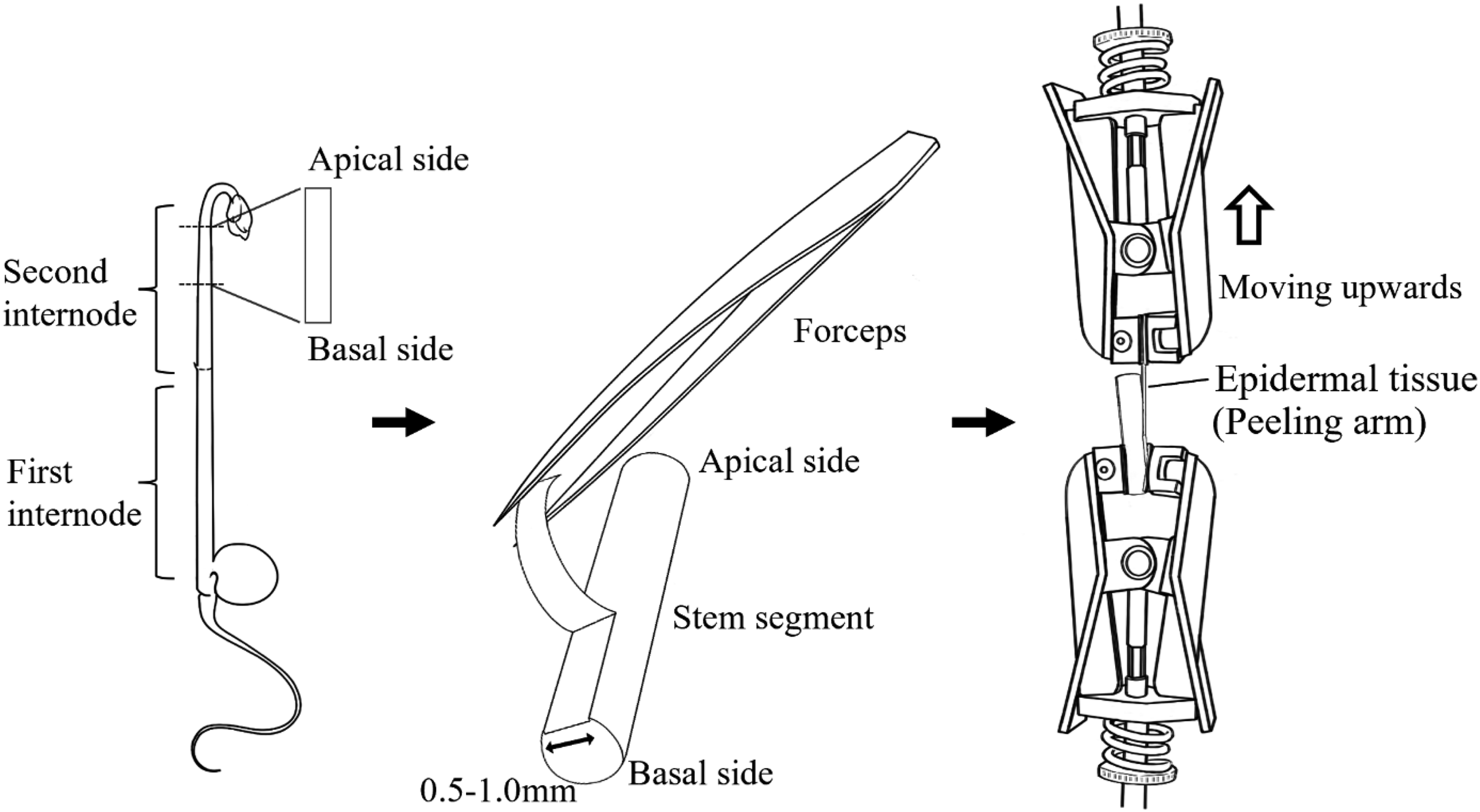
Schematic illustration of the measurement of the peeling force between the epidermal and inner tissues of pea stems. The epidermal tissue was partially peeled from the stem segment using forceps to create a peeling arm. The peeling arm was fixed to the upper clamp of a tensile tester, and the segment region with the removed epidermal tissue was fixed to the lower clamp. The epidermal tissue was peeled from the segment by raising the upper clamp, and the peeling force was recorded.
To ascertain the effect of epidermal tissue tearing on the force required to peel the epidermal tissue from the inner tissue, the peeling test was conducted on stem segments from which the epidermal tissue adjacent to the epidermal tissue to be tested was removed. The peeling test was conducted at a peeling speed of 100 mm/min, and the other conditions were as described above.
Microscopy
After the peeling test, the peeled epidermal tissue was mounted on glass slides, and the image was captured using a microscope (Axio Imager. A1; Carl Zeiss, Göttingen, Germany) equipped with a digital camera (DP74; Olympus, Tokyo, Japan). The width of the peeled epidermal tissue was measured using bundled image processing software (cellSens Imaging Software; Olympus).
To ascertain that only the epidermal tissue could be peeled from the inner tissue, the stem segment was observed with a microscope after the epidermal tissue was peeled. The stem segment was fixed in 4% (w/v) paraformaldehyde in 0.1 M phosphate buffer (pH 7.2). The fixed segment was embedded in 5% (w/v) agar and sectioned transversely at 150 µm with a vibratome (VT1000S; Leica, Nussloch, Germany). The section was mounted on glass slides, and the image was captured using a microscope (Axio Imager. A1) equipped with a digital camera (DP74).
Statistical analysis
The correlations between the peeling force and the width of the peeled epidermal tissue were analyzed using Student's t-test. The means and standard errors (SEs) were calculated for the adhesive strength. The significance of the data obtained from the experiments on the tissue tearing and stem region was analyzed using Student's t-test, while that of the peeling rate was analyzed using Dunnett's test.
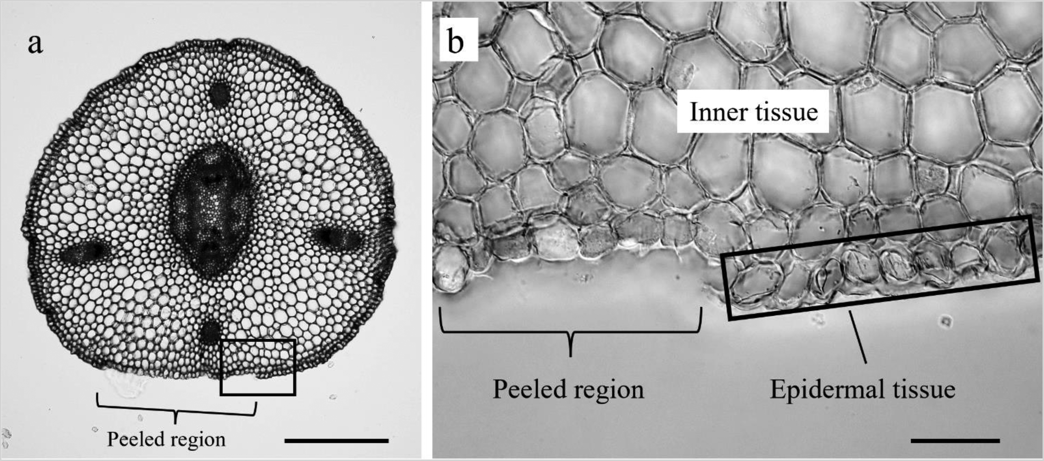
Cross-section of a stem with a portion of the epidermal tissue peeled off using a tensile tester. A cross-section was obtained from the middle of the epidermal tissue-peeled stem segment. Images of the stem as a whole (a) and a magnified view of the region where the epidermal tissue was peeled off (b) are presented. The scale bars of the entire image and the magnified image are 500 and 50 µm, respectively.
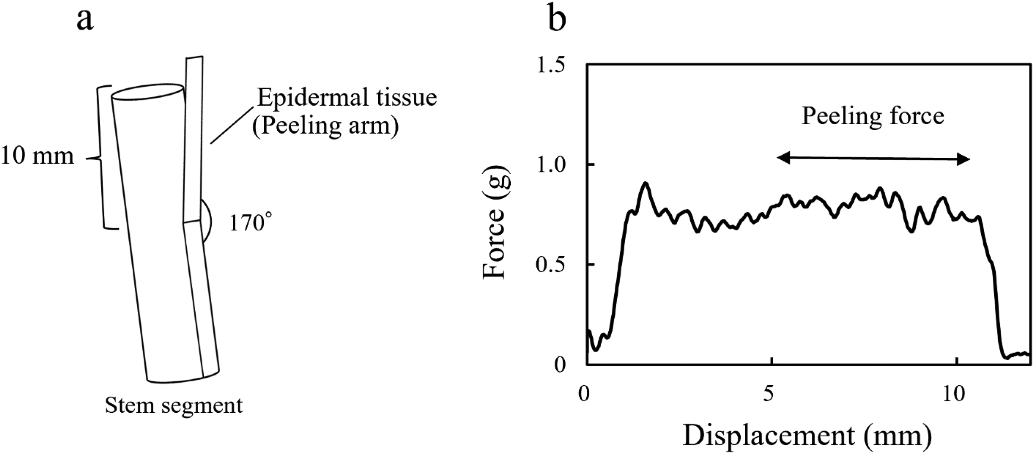
Schematic illustration of a stem segment and typical force-displacement curve in the peeling test. The peeling test was conducted at a peeling angle of 170° (a). A total of ∼10 mm of peeling was performed, and the mean peeling force for the latter 5 mm was calculated (b).
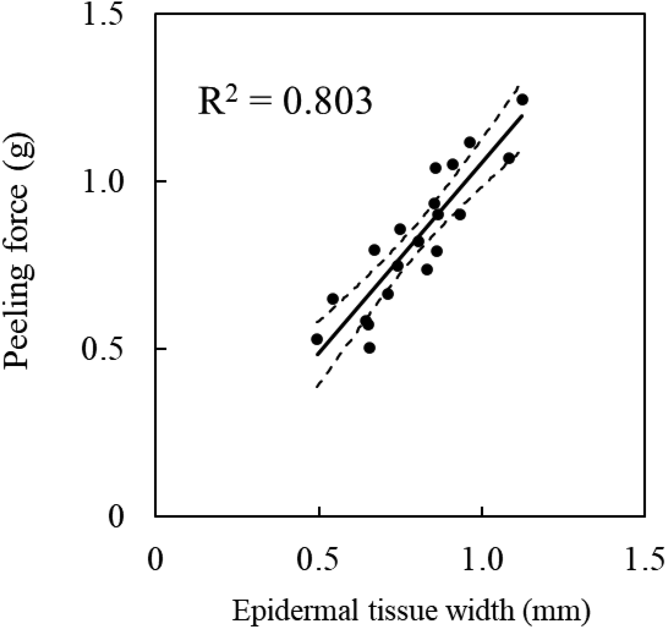
Relationship between the measured peeling force and the width of the peeled epidermal tissue. The regression line is shown as a solid line, and the confidence interval (95% confidence interval to slope) is shown as a dashed line. The drawing is based on the value of 20 samples.
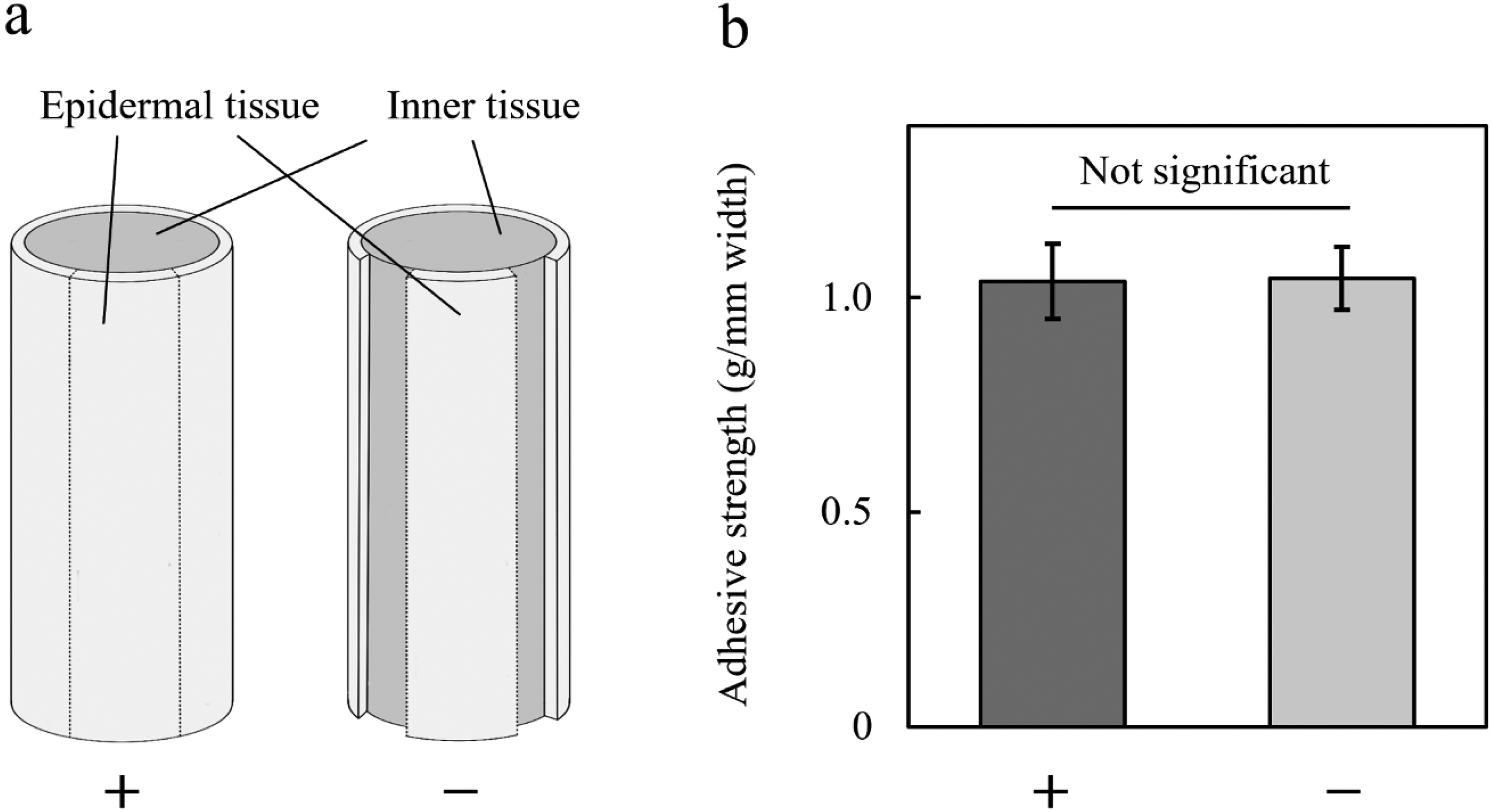
Effect of epidermal tissue tearing on the measurement of adhesive strength. Stem segments were prepared in the presence (+) and absence (–) of the adjacent epidermal tissue (a), and the adhesive strength between the epidermal and inner tissue was measured in each segment (b). Values are the mean with SEs (n = 10).
Results and discussion
Peeling of the epidermal tissue using a tensile tester
To develop a method for measuring the adhesive strength between the epidermal and inner tissues, whether the epidermal tissue could be peeled from the stem segment using a tensile tester was first ascertained (Figure 1). Figure 2 shows a photograph of a cross-section of a stem, with a portion of the epidermal tissue peeled off using a tensile tester at peeling rate of 100 mm/min. The epidermal tissue of pea epicotyls consisted of a single-cell layer. Upon peeling the epidermal tissue using a tensile tester, the epidermal tissue consisting of a single-cell layer was peeled without any cell breakage (Figure 2). The peeling rate varied from 10 to 800 mm/min, yet the epidermal tissue could be peeled off without cell breakage, as was the case at 100 mm/min. These results indicate that, in pea stems, the epidermal tissue can be peeled off using a tensile tester to measure the adhesive strength between the epidermal and inner tissues.
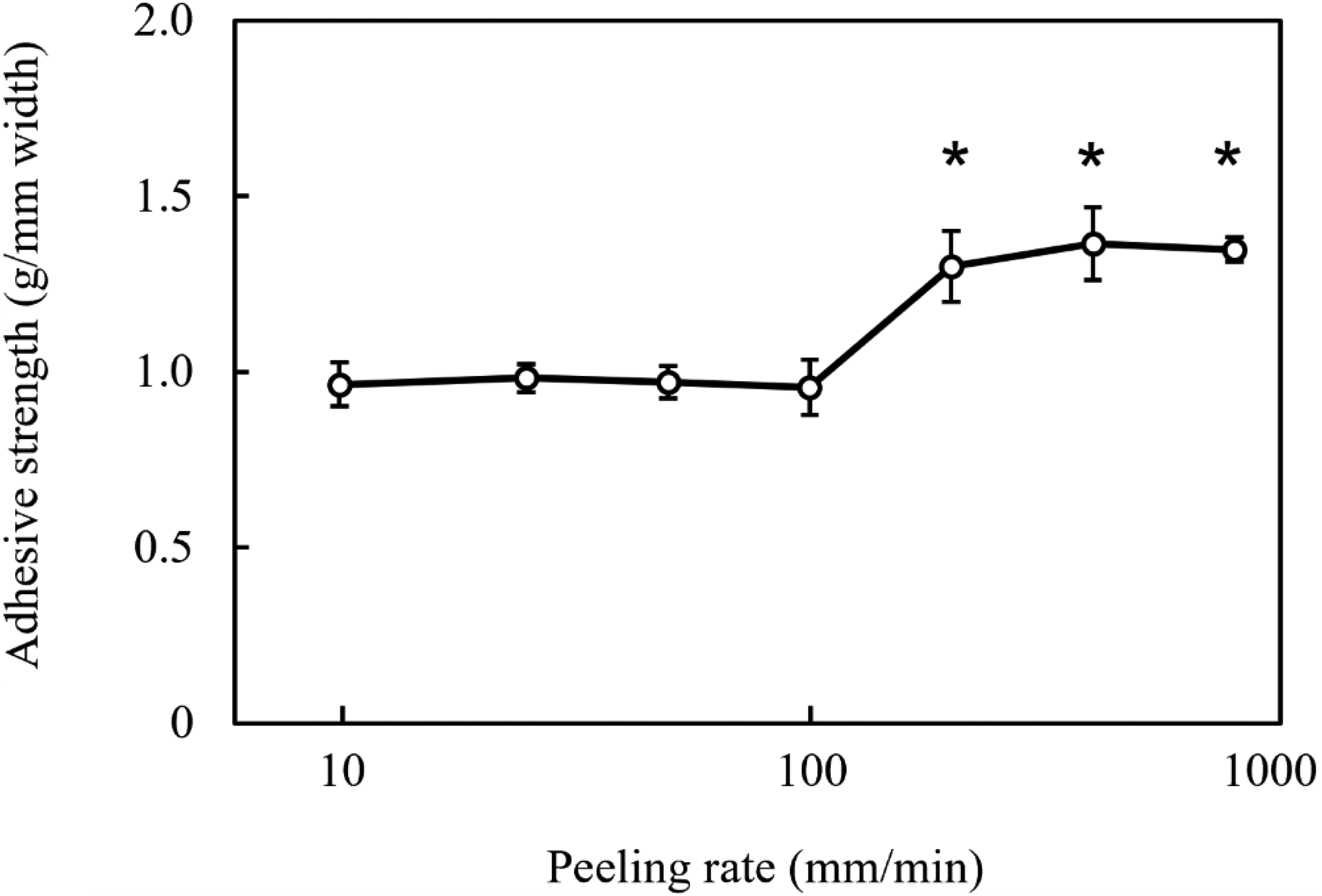
Effect of the peeling rate on adhesive strength between the epidermal and inner tissues. Values are the mean with SEs (n = 5). Asterisks denote significant differences from the adhesive strength at a peeling speed of 10 mm/min (p < 0.05, Dunnett's test).
Measurement of the peeling force
Figure 3 shows the force-displacement curve when the epidermis was peeled ∼10 mm from the stem at a peeling angle of 170°. In measuring the peeling force in plastic film bonding by an adhesive, the peeling angle is often tested at 180°. 16 However, in this study, the peeling angle was set at 170° because the peeling force between the epidermal and inner tissues was considerably smaller than that by the adhesive, and it was expected that the frictional force between epidermal tissues would influence the measurement of the peeling force. The force increased rapidly at the beginning of the peeling test, remained nearly constant as the epidermal tissue began to peel, although oscillations were observed, and decreased rapidly to zero as the peeling was completed (Figure 3). The force oscillations during peeling became smaller as the epidermal tissue was peeled. Therefore, the force to peel the last 5 mm of ∼10 mm of peeling was averaged and defined as the peeling force.
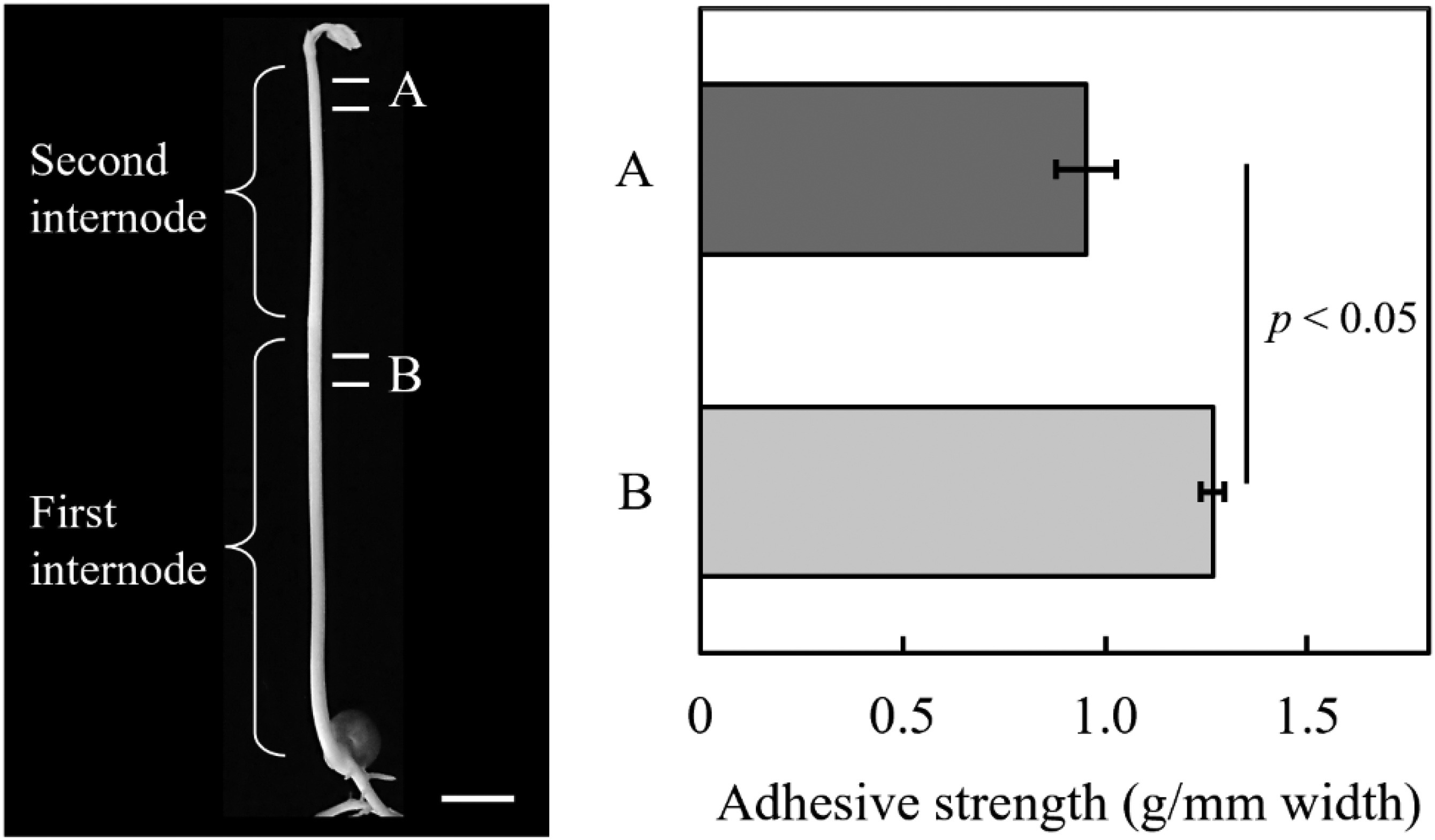
Adhesive strength in elongation and non-elongation regions of pea stems. The photograph shows pea seedlings (left), whereas the graph shows the adhesive strength between the epidermal and inner tissues (right). The adhesive strength was measured in the elongation region designated as A in the second internode and the non-elongation region designated as B in the first internode of 6-day-old etiolated pea seedlings. Bar in the photograph indicates 10 mm. Values are the mean with SEs (n = 10).
If the specimen is stretched significantly during the peeling test, the correct peeling force cannot be measured. The extent to which epidermal tissue is stretched when the force is applied to it was examined using a tensile tester. When the epidermal tissue with a width of 1 mm was subjected to a tensile strain rate of 20 mm/min, the epidermal tissue exhibited an elongation of 45.9 ± 3.5 μm at a load of 1 g per 1 mm length of the epidermal tissue. This value was sufficiently small compared to the 10 mm peeling length to have only a negligible effect on the measurement of the peeling force.
Adhesive strength
In general, adhesive tapes and other materials bonded using adhesives have a greater peeling force when the specimen width is increased.16,20 Therefore, in materials engineering, the peeling test is conducted by aligning the specimen width or normalizing for the peeling force by the specimen width. When peeling the epidermal tissue from the stem, the width of the peeled tissue is between 0.5 and 1.0 mm, with some variation. Figure 4 shows the relationship between the measured peeling force and the width of the peeled epidermal tissue. As the width of the peeled epidermal tissue increased, the peeling force exhibited a corresponding increase (p < 0.05, Student's t-test), indicating that the peeling force per unit width was almost constant. Accordingly, the results indicate that the measured peeling force needs to be normalized for the width of each epidermal tissue. The linearity between the sample width and the peeling force provides evidence of the reliability of the results obtained from the peeling test, 21 indicating that the method developed in this study can adequately measure the peeling force. Consequently, in this study, the peeling force normalized by the peeled epidermal width was defined as the adhesive strength.
Tearing of epidermal tissue
As shown Figure 1, tearing between the epidermal tissue occurs when epidermal tissue is peeled from the stem segment. The effect of epidermal tissue tearing on the measurement of adhesive strength was ascertained. Figure 5 shows the adhesive strength between the epidermal and inner tissues in the presence and absence of the adjacent epidermal tissue. The adhesive strength was not affected by the presence or absence of adjacent epidermal tissue. The results indicate that the lateral adhesive force between cells of the epidermal tissue is less than the adhesive force between cells of epidermal and inner tissues. Goodman et al. (2002) also reported that the peeling force of the cortical tissue, including the epidermal tissue, from flax stems in the process of retting was ∼10 times greater than the tearing force of the peeled cortical tissue. 17 These facts indicate that the adhesive force between the epidermal and inner tissues can be measured even when the adjacent epidermal tissue is present.
Peeling rate
The adhesive strength between the epidermal and inner tissues was measured by varying the peeling speed from 10 to 800 mm/min (Figure 6). Compare to the adhesive strength measured at a peeling speed of 10 mm/min, the adhesive strength measured at a peeling speed of ≥200 mm/min was significantly greater (p < 0.05, Dunnett's test). Réquilé et al. (2018) conducted the 90° peeling test on hemp fiber tissue to monitor the retting process and showed that peeling at a rate of 100 mm/min resulted in a considerably higher peeling force than peeling at a rate of 50 mm/min or less. 22 In other words, the aforementioned facts indicate that in the peeling test of plant tissue, the peeling force increases remarkably above a certain peeling rate. The adhesive strength of viscoelastic materials, such as adhesives, depends on the peeling speed. 23 The epidermal and inner tissues of plants adhere to each other via cell walls, that exhibit viscoelastic properties.10–12 These facts suggest that the peeling force depended on the peeling rate because the plant cell wall exhibited viscoelastic properties. The molecular bonding strength is measured at the peeling speed before the onset of the adhesive strength increases. 23 Therefore, the peeling speed was set to 100 mm/min in subsequent measurements.
Adhesive strength in elongation and non-elongation regions
The elongation growth rate of plant stems varies depending on the stem region. In pea seedlings of the age used in this study, the upper region of the second internode shows vigorous elongation growth, whereas the upper region of the first internode does not show elongation growth.24,25 To ascertain whether there is a difference in the adhesive strength between the epidermal and inner tissues in the elongation and non-elongation regions, the adhesive strength of region A (the elongation region) and region B (the non-elongation region) was measured (Figure 7). The adhesive strength in the non-elongation region was significantly greater than that in the elongation region (p < 0.05, Student's t-test). The findings suggest a potential correlation between the elongation growth rate and the adhesive strength between the epidermal and inner tissues.
Upon separation of the epidermal and inner tissues of stem segments and incubation in water, it is observed that the length of the epidermal tissue decreases while the length of the inner tissue increases. In other words, in the stem, the epidermal tissue is subjected to tension, while the inner tissue is subjected to compression.13–15 This observation leads to the conclusion that epidermal tissue plays a pivotal role in determining the elongation growth rate of the entire stem. This is called the “tissue tension concept”. 15 This study showed that the adhesive strength in the non-elongation region is greater than in the elongation region (Figure 7). The findings suggest that, in the non-elongation region, the epidermal and inner tissues are more strongly adherent than in the elongation region, suppressing the elongation growth of the inner tissue, thereby preventing the elongation and growth of the entire stem.
Conclusion
In this study, a method for quantitatively measuring the adhesive strength between the epidermal and inner tissues in plant stems using a tensile tester was developed. The analysis conducted using this method indicated that adhesive strength may be involved in regulating stem growth. The growth of plant stems is influenced by environmental factors, including light and gravity. Therefore, it is necessary to investigate whether these factors affect the adhesive strength. Furthermore, it is important to elucidate the mechanism regulating the adhesive strength. These studies are expected to provide new insights into plant growth regulation and to be useful for agriculture.