
Abstract
Aims. The aims of this study were to detect the infection rates of DNA viruses in liver tissue of biliary atresia and to investigate the effect of perinatal infection of cytomegalovirus in biliary atresia. Methods. A total of 85 liver biopsies (taken during Kasai portoenterostomy) were tested by fluorescence quantitative polymerase chain reaction for DNA viruses (herpes simplex virus [HSV], Epstein–Barr virus [EBV], varicella zoster virus [VZV], cytomegalovirus [HCMV], and adenovirus). Immunocytochemical detection of CMV-pp65 antigenemia assay was used to detect the presence of viral protein in liver samples. Human intrahepatic biliary epithelial cells was infected by the laboratory strain AD169 of HCMV in vitro. Results. Virus DNA was found in the biopsies (51/85 HCMV, 5/85 ADV, 3/85 EBV). The biopsies of 2 patients were tested positive for 2 viruses simultaneously. They include one case of HCMV in combination with ADV and one case of ASV in combination with EBV. CMV-pp65 antigenemia were distributed in hepatocyte, vascular endothelial cell, and biliary duct endothelial cell. The cytopathic effect and apoptosis were observed after HCMVAD169 infected human intrahepatic biliary epithelial cells at 6 days. Conclusion. Human intrahepatic biliary epithelial cell is the target cell of HCMV. The etiology of biliary atresia is probably multifactorial. The perinatal infection of HCMV is one of the important etiologies for biliary atresia in China.
Biliary atresia (BA) is a severe neonatal disease of the hepatobiliary system, characterized by a progressive inflammatory obliteration of the extra- and intrahepatic bile ducts.1,2 BA is a rare condition that occurs in about 1:15000 live births (1.5 times more common in females). 3 Since the introduction of hepatoportoenterostomy (HPE), Kasai portoenterostomy (KPE) remains the first surgical option in these patients and should be offered early to all patients. A surgical technique to treat uncorrectable type of BA, there have been encouraging results in treating this disease by KPE.4,5 In the long term, most patients develop end-stage liver cirrhosis and, thus, BA is the most frequent indication for liver transplantation in childhood. 6 The etiology of BA remains unclear, so that there is as yet no means of addressing the underlying pathology. Hypotheses about the pathogenesis have included viral infection, autoimmunity, inflammation, abnormal development, and genetics.1,7 As a potential initiator of this immune process, a viral infection has been considered. This hypothesis has been supported by findings of individual viral strains in BA patients. 8
However, there are data which suggest that the known hepatotropic viruses do not play a major role in the etiology and progression of BA. The viral infection is only a secondary phenomenon. 9 In China, the perinatal infection of virus is seen frequently. But the relationship between the hepatotropic viruses and BA is unknown. The purposes of this study were to describe the incidence of hepatotropic DNA viruses in BA undergoing the Kasai procedure and to analyze the effect of perinatal infection of cytomegalovirus in BA.
Materials and Methods
Patient Samples
A total of 85 patients with BA (50 males and 35 females) treated at the Guangzhou Women and Children’s Medical Centre, China, between January 2004 and December 2009 were retrospectively included in the study. The diagnosis of BA was established by examining the bile ducts at the time of surgery and by histologic study of the extrahepatic bile duct remnants. Liver biopsies were taken during the Kasai procedure.
The mean age at operation was 56 ± 28.5 days (range 12-180 days). Specimens were fixed in 4% buffered formalin for immunostaining, and other specimens immediately snap-frozen in liquid N2 and stored at −80°C for polymerase chain reaction (PCR) analysis. Because of ethical considerations, it was not possible to obtain liver biopsies from patients in this age group without hepatic disease. In all, 10 liver biopsies were performed in patients’ embryo of 6 months gestation age suffering from induction delivery.
Cell Lines and Culture Conditions
The cells studied were cultured. Human intrahepatic biliary epithelial cells (HIBECs) purchased from ScienCell (San Diego, CA) and human embryonic lung fibroblast (HELs) were obtained from American Type Culture Collection. HIBECs were cultured in sterile medium supplemented with 2% fetal bovine serum (FBS), epithelial cell growth supplement (ScienCell, San Diego, CA), and 1% penicillin in cell culture flasks coated with poly-
Viruses, Infection Conditions, and Virus Titration
The human cytomegalovirus (HCMV) strain AD169 was purchased from Wuhan Research Institute (University of Wuhan, Hubei, China). Virus stocks were prepared from supernatants of infected HELs when a marked cytopathic effect (CPE) was seen. Infectious supernatants were harvested when 100% of cells showed CPE. Supernatants were cleared of cellular debris by centrifugation for 10 minutes at 3500g and were stored directly at −80°C. Virus titers were determined by standard plaque assay; the titer of each virus stock was between approximately 1 × 10⁷ and 5 × 10⁷ plaque forming units (PFU)/mL. HCMV infection was enhanced by centrifugation at 800g for 30 minutes. HIBECs were infected with HCMV strain AD169 at a multiplicity of infection of 5 to observe CPE with inverted phase contrast microscope and electron microscope.
DNA Viruses Polymerase Chain Reaction
Liver samples were screened for the following DNA viruses: herpes simplex virus (HSV), cytomegalovirus (CMV), Epstein–Barr virus (EBV), adenovirus (ADV), and hepatitis B virus (HBV). The DNA of 50 mg of frozen liver tissue was extracted using a commercial kit (QIAamp DNA Mini Kit, QIAGEN, Valencia, CA), and the resuspended DNA was stored at −20°C. The real-time PCR assays were performed using an iCycler iQ real-time PCR detection system (Bio-Rad Lab, Hercules, CA). The PCR conditions are given in Table 1.
Polymerase Chain Reaction Conditions
Abbreviations: HCMV, human cytomegalovirus; ADV, adenovirus; HSV, herpes simplex virus; EBV, Epstein–Barr virus; HBV, hepatitis B virus.
Immunohistochemistry
Immunohistochemical staining was performed on formalin-fixed, paraffin-embedded sections using the following monoclonal antibodies: anti-CMV (clones CCH2 and DDG9, dilution 1:200, USCNLIFE, USA), which is a cocktail of 2 antibodies that react with a 76-kDa HCMV early protein and the delayed early DNA binding protein p52, and anti-pp65 (clones 2 and 6, dilution 1:50, USCNLIFE, USA), which is specific for CMV pp65 protein. Slides were pretreated by proteolytic digestion with proteinase K (DAKO, Glostrup, Denmark) for 5 minutes (anti-CMV) or by steam heating slides in citrate buffer (pH 6.0) in a steamer (Black and Decker, Shelton, CT) for 20 minutes (anti-pp65). Staining was performed using an automated immunostainer (DAKO), followed by antibody detection using the DAKO EnVision+ System and 3,3′-diaminobenzidine as a chromogen. The slides were counterstained with hematoxylin and coverslipped.
Results
DNA Viruses
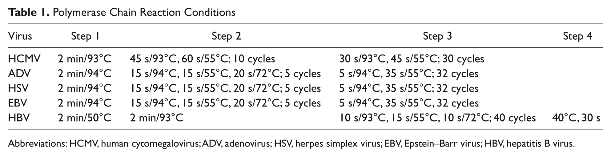
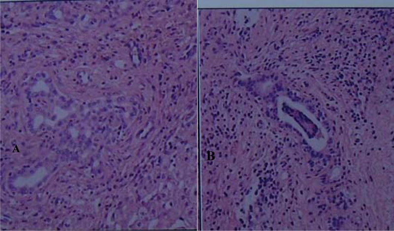
DNA was extracted from 85 liver biopsies. Virus DNA was found in the biopsies (51/85 HCMV, 5/85 ADV, 3/85 EBV). The biopsies of 2 patients were tested positive for 2 viruses simultaneously. They include one case of HCMV in combination with ADV and one case of HCMV in combination with EBV, and the histological changes were more marked in liver. None of the other tested DNA viruses could be detected (Figures 1 and 2).
Liver organization showing pathological changes of human cytomegalovirus infection in combination with adenovirus
Liver organization showing pathological changes of human cytomegalovirus infection
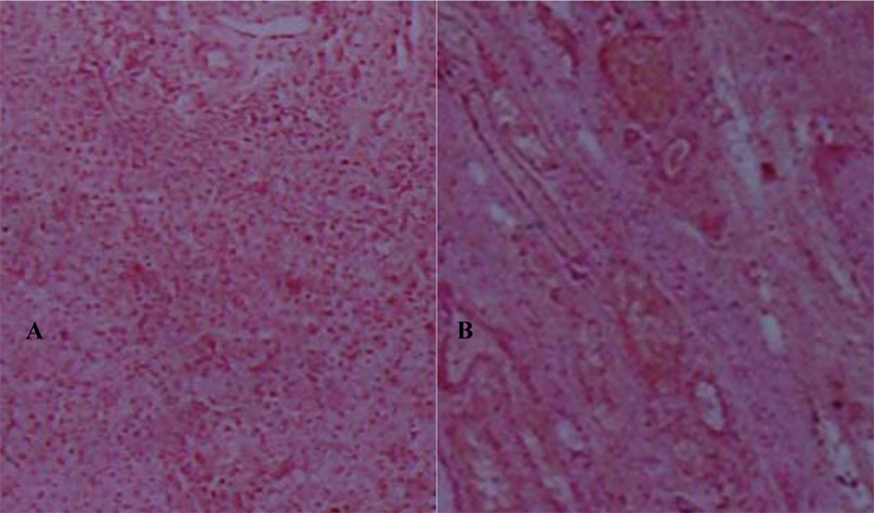
The Cytopathic Effect
The cytopathic effect and apoptosis were observed after HCMVAD169 infected human intrahepatic biliary epithelial Cells at 6 days (Figures 3 and 4).
The cytopathic effect
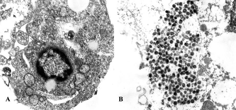
Apoptosis of human intrahepatic biliary epithelial cells.
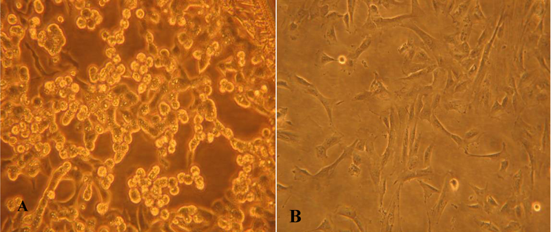
Immunocytochemical Detection of CMV-pp65 Antigenemia
Immunocytochemical detection of CMV-pp65 antigenemia assay was used to detect the presence of viral protein in liver samples. CMV-pp65 antigenemia detection was positive in HCMV(+) fluorescence quantitative (FQ)-PCR patients with BA. CMV-pp65 antigenemia were distributed in hepatocyte, vascular endothelial cell and biliary ducts endothelial cell. CMV-pp65 antigenemia detection was negative in HCMV(−) FQ-PCR patients with BA (Figure 5).
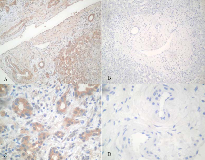
Immunocytochemical detection of CMV-pp65 antigenemia
Discussion
Biliary atresia is a progressive inflammatory disease that affects the intrahepatic and extrahepatic biliary tract. Over the past 30 years, this disease has changed from being fatal to being a disorder for which effective palliative surgery or curative liver transplantation (or both) is available. Good outcomes for infants depend on early referral and timely Kasai portoenterostomy, and its pathogenesis remains unclear. BA affects 1/10 000 to 1/20 000 live births worldwide. Asian infants have a higher incidence (Japan, 0.8 to 1.1 per 10 000; Taiwan, 2 per 10 000), whereas Europe and North America have lower incidences of 0.6 to 0.8 per 10 000.10-12 BA is the single most common cause of chronic cholestasis in children and is the leading indication for liver transplantation worldwide. It results from an inflammatory and fibrosing obstruction of the extrahepatic bile ducts in the first few months of life. Early diagnosis and timely surgical portoenterostomy are necessary for improved biliary drainage, but the liver disease progresses to end-stage biliary cirrhosis in most patients. Although the pathogenesis of BA is largely unknown, in 1974, Landing 8 hypothesized that BA could represent the result of a virally induced process of the liver and the hepatobiliary tree. Recent patient- and animal-based experiments indicate that interactions between infectious agents and inflammatory circuits may be important pathogenic mechanisms of the disease.13,14
Therefore, we hypothesized that, to support Landing’s theory, we should detect one virus in almost every liver biopsy if testing simultaneously for the most common hepatotropic DNA viruses, and we had a sufficient number of patients in our study. We, indeed, observed a higher incidence of viral infection in our study. Virus DNA was found in the biopsies (51/85 HCMV, 5/85 ADV, 3/85 EBV), and the biopsies of 2 patients were tested positive for 2 viruses simultaneously. They include one case of HCMV in combination with ADV and one case of HCMV in combination with EBV. None of the other tested DNA viruses could be detected. The results of our study show that a majority of BA patients (60%, 51/85 patients) tested positive for HCMV. Immunocytochemical detection of CMV-pp65 antigenemia assay showed that CMV-pp65 antigenemia detection was positive in HCMV(+) FQ-PCR patients with BA. CMV-pp65 antigenemia were distributed in hepatocyte, vascular endothelial cell, and biliary duct endothelial cell. CMV-pp65 antigenemia detection was negative in HCMV(−) FQ-PCR patients with BA. The question now arises whether the perinatal infection of cytomegalovirus can survive the IBECs and whether they evoke cell immunologic injury?
Human intrahepatic biliary epithelial cells were infected with HCMV strain AD169 in vitro. CPE and apoptosis were observed after HCMVAD169 infected HIBECs at 6 days. Apoptotic cells were identified with the following electron microscope criteria: high nuclear density, chromatin condensation, and nuclear fragmentation. HIBECs are the target cells of HCMV.
Human cytomegalovirus is a member of the herpes virus family, and HCMV infection is ranked as one of the most common infections in adults, with the seropositive rates ranging from 60% to 99% globally.15,16 Primary HCMV infection in immunocompetent individuals presents most commonly as an asymptomatic illness or as a benign febrile infectious mononucleosis-like syndrome. When HCMV infection occurs in individuals with compromised immunity, such as neonates and liver transplant recipients, clinical disease with high morbidity may develop and, in some cases, this may lead to death.17-19 Once acquired, the infection persists lifelong and may undergo periodic reactivation. Infection with HCMV is associated with BA. 20 Our data show that CMV infection markedly downmodulated the expression of cytokine IFN-γ and transcriptional factor T-bet, and significantly upregulated the expression of cytokine IL-4 and transcriptional factor GATA-3, indicating that CMV infection led to the disequilibrium of Th1/Th2 cells differentiation and their cytokines expression, which is likely to suppress the functions of cellular immunity in the infected host. This is also likely to be one of the reasons of CMV escaping the attack of body’s specific cellular immunity, causing persistent or latent infections and even leading to diseases under certain conditions. 21
The observation that a progressive fibrosing inflammation of the liver causes the obliteration of the bile ducts in our patients led to the theory that BA is caused by an immune response to an unknown triggering event.23,24,25 Our study in China supports the theory that an HCMV infection is a potential initiator of this immune process.
In summary, our study demonstrates a high incidence of HCMV in BA. BA is caused by an immune response by reactivation of a latently triggered HCMV infection. BA is a rare disease of infancy, and many of its aspects, including its pathogenesis, are poorly understood. There are several other hypotheses implicating genetic predisposition, dysregulation of immunity, and so on, but the cause is probably multifactorial, with obliterative extrahepatic cholangiopathy as the common endpoint. Further studies are needed to assess these factors, which might become therapeutic targets to halt the inevitable development of cirrhosis and need for liver transplantation. The perinatal infection of cytomegalovirus is one of the important etiologies for BA in China.
Footnotes
The authors declared no conflicts of interest with respect to the research, authorship, and/or publication of this article.
The research was supported by Guangzhou Municipal Health Bureau grant (2008-YB-64).