






Abstract
In children, most colonic polyps are juvenile polyps with negligible risk for malignant transformation. The exception is juvenile polyposis syndrome (JPS) where there is a risk for developing colon cancer. The authors studied differences in clinical features and colonoscopic findings in children with solitary juvenile polyps (SJP), multiple juvenile polyps (MJP), and JPS. Methods. Children were identified as SJP (1 polyp), MJP (2-4 polyps), or JPS (>5 polyps). Demographic data, laboratory values, family history, and colonoscopic findings were recorded. Results. Children having polypectomy had juvenile polyps (93%), adenomatous polyps (5%), and Peutz–Jegher syndrome (3%). Juvenile polyps were classified as SJP (67%), MJP (16%), and JPS (17%). Children with SJP were younger, were more likely to have polyps limited to the rectosigmoid colon, and had larger polyps than children with MJP and JPS. Anemia was more common in JPS than MJP and SJP. Conclusion. Clinical and endoscopic findings differ between SJP, MJP, and JPS.
Introduction
Colonic polyps are a common finding in children, representing one of the most common causes of rectal bleeding in this age group. 1 Historically, the vast majority of childhood polyps are solitary, located in the distal colon, and are histologically classified as juvenile polyps with negligible risk for malignant transformation.1-4 However, advances in colonoscopic techniques have changed our understanding of childhood colonic polyps. Employment of pancolonoscopy has revealed that, while more commonly located in the restosigmoid, polyps are distributed throughout the colon, are often multiple, recur in some children, and may be associated with premalignant changes.5-16 Although the malignant potential for solitary juvenile polyps (SJP) in children is remains exceedingly low, there is a significant malignant potential in juvenile polyposis syndrome (JPS).12,14 There are data to indicate differences in genetic abnormalities for individuals with JPS, specifically, abnormalities in tumor suppressor genes. To date, 3 tumor suppressor genes have been identified in individuals with JPS: SMAD4, BMPR1A, and PTEN.17-23 SMAD4 and BMPR1A are both found in nonsyndromic juvenile polyposis and account for 15% and 25% of said cases, respectively. 20 Individuals with SMAD4 gene defects are more apt to have gastric polyps than individuals with defects in BMPR1A. 21 Abnormalities in PTEN have been found in Cowden’s disease and Bannayan–Ruvalcaba–Riley syndrome22,23; both syndromes have extraintestinal findings that define the respective syndrome. It has been postulated that these 2 syndromes represent a spectrum of the same disease.
We reviewed a series of children undergoing polypectomy and compared clinical features, polyp distribution, and polyp size (volume) to determine if difference existed between children with SJP and those with JPS.
Methods
Medical records of children who underwent colonoscopy with polypectomy from September 1998 through January 2007 were reviewed and the following data were retrieved: age, gender, race, hemoglobin count, mean corpuscle volume, number of colonoscopies performed, number of polyps seen/removed, volume of the polyps removed, the type of polyp(s) removed, location of the polyps within the colon, and family history of polyps or colon cancer. The type and cumulative number of polyps were used to categorize patients into groups. Familial adenomatous polyposis (FAP) was defined by the presence of adenomatous polyps and a family history of FAP. Peutz–Jeghers syndrome (PJS) was defined by the presence of colonic, small bowel, and/or gastric polyps with arborization of smooth muscle in the polyp, a positive family history, and cutaneous manifestations. SJP was defined as the presence of a single juvenile polyp. Multiple juvenile polyp (MJP) was defined as the presence of 2 to 4 juvenile polyps limited to the colon. JPS was defined by the presence of more than 5 juvenile polyps located anywhere in the gastrointestinal tract, the presence of any number of gastrointestinal polyps with a positive family history of JPS, or the presence of gastrointestinal polyps in combination with extracolonic hamartomatous polyps. 24 Comparison was made between SJP, MJP, and JPS to determine if there was a statistical difference in the colonic distribution and size of polyps.
Descriptive analyses were performed using frequency distributions, proportions, means, standard deviations, medians, and percentiles. After testing normality of the data using the Shapiro–Wilk test, group comparisons were made using analysis of variance or the Kruskal–Wallis test as appropriate. Post hoc comparisons were made using Tukey’s test for the multiple comparisons. Association between categorical variables was tested using χ2 or Fisher’s exact test as appropriate. SPSS 17.0 (SPSS Inc, Chicago, IL) was used for the statistical analyses, and significance level was set at .05.
Results
During the 100-month study period, we identified a total of 948 lower endoscopic procedures performed in 758 children distributed as follows: colonoscopy (54.5%), flexible sigmoidoscopy (32.0%), colonoscopy with polypectomy (12.7%), and flexible sigmoidoscopy with polypectomy (0.8%). Looking just at lower endoscopy with polypectomy, 100 children underwent 127 procedures (range = 1-6). The diagnoses for the children were familial adenomatous polyposis (FAP; n = 5), Peutz–Jegher syndrome (PJS; n = 2), and juvenile polyps (n = 93). Children with FAP and PJS were excluded from the study assessing location of polyps, as polyps are known to be distributed throughout the colon in these conditions. Children with juvenile polyps were further characterized as having SJP (n = 62), MJP (n = 15), or JPS (n = 16). Five children with juvenile polyps had endoscopic evaluation limited to flexible sigmoidoscopy. None of these children had further rectal bleeding during a follow-up period ranging from 2.9 to 8.0 years and were thus classified as having SJP.
Among the 93 children with juvenile polyps, the average age of presentation was 6.4 ± 3.6 years with a range of 1.3 to 16.4 years. Comparison of the various juvenile polyp groups showed that children with SJP presented younger (5.5 ± 2. 9 years) than both MJP (7.2 ± 3.5 years) and JPS (9.2 ± 4.5 years; Table 1). Gender and race were also compared for children with juvenile polyps. Overall, there were more males (59%) than females. No significant difference was seen in the percentage of males in the various juvenile polyp groups; SJP (57%), MJP (60%), and JPS (69%; Table 1). Overall, Caucasians (51%) and African Americans (47%) were equally represented among the children; American Indians accounted for the remainder (2%). Children with JPS were more likely to be Caucasians, most likely related to overrepresentation by a single family; 3 of the children in this study are related to a previously reported family with a JPS. 25
Demographics of Children With Juvenile Polyps

Abbreviations: SJP, solitary juvenile polyps; MJP, multiple juvenile polyps; JPS, juvenile popyposis syndrome; MCV, mean corpuscular volume.
Significant difference was seen in age for JPS compared with SJP (P < .01).
No statistical difference.
Significant difference was seen in hemoglobin for JPS compared with SJP and MJP (P = .002).
Significant difference was seen in MCV for JPS compared with SJP and MJP (P = .003).
Significant difference was seen in anemia for JPS compared with SJP and MJP (P < .001).
The number (Table 2) and distribution of juvenile polyps (Figure 1) according to the previously described classification is shown. The majority of children with juvenile polyps had SJP (66.7%), with the remaining children being nearly evenly distributed between MJP and JPS. There was an obvious difference in the distribution of polyps dependent on the classification. In SJP, 98% of the polyps were located in the rectosigmoid region, whereas significantly fewer rectosigmoid polyps were seen in MJP (64.0%) and JPS (44.6%; Figure 1). Overall, median polyp volume was significantly larger in SJP (median = 540.8 mm3, interquartile range [IQR] = 174-1872), than MJP (74.9 mm3, IQR = 15-452) and JPS (87.4 mm3, IQR = 8.6-411; P < .001). This difference was more pronounced for rectosigmoid polyps: SJP (540.8 mm3, IQR = 175-1872) compared with MJP (132.0 mm3, IQR = 55-872) and JPS (12.5 mm3, IQR = 6.2-139; P < .001; Figure 2).
Classification of Children With Juvenile Polyps

Abbreviations: SJP, solitary juvenile polyps; MJP, multiple juvenile polyps; JPS, juvenile popyposis syndrome; NA, not applicable.
One child with a inflammatory gastric and duodenal polyps and strong family history of juvenile polyposis had only 3 colonic polyps.
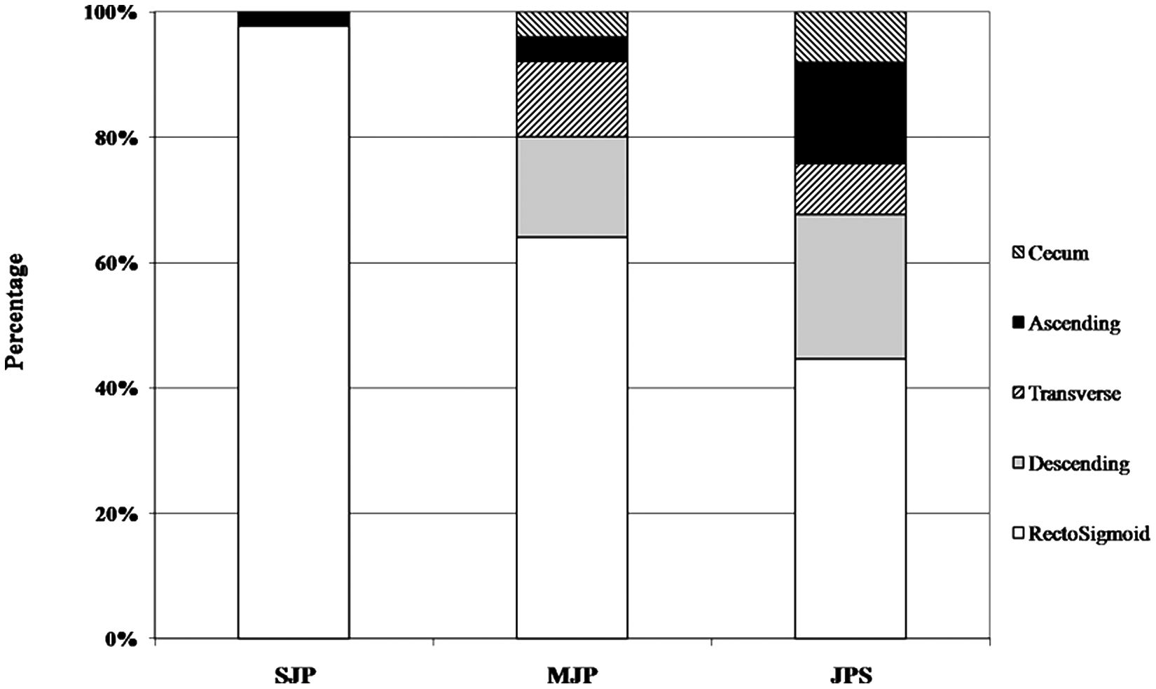
Distribution of colonic polyps according to the classification of juvenile polyp type
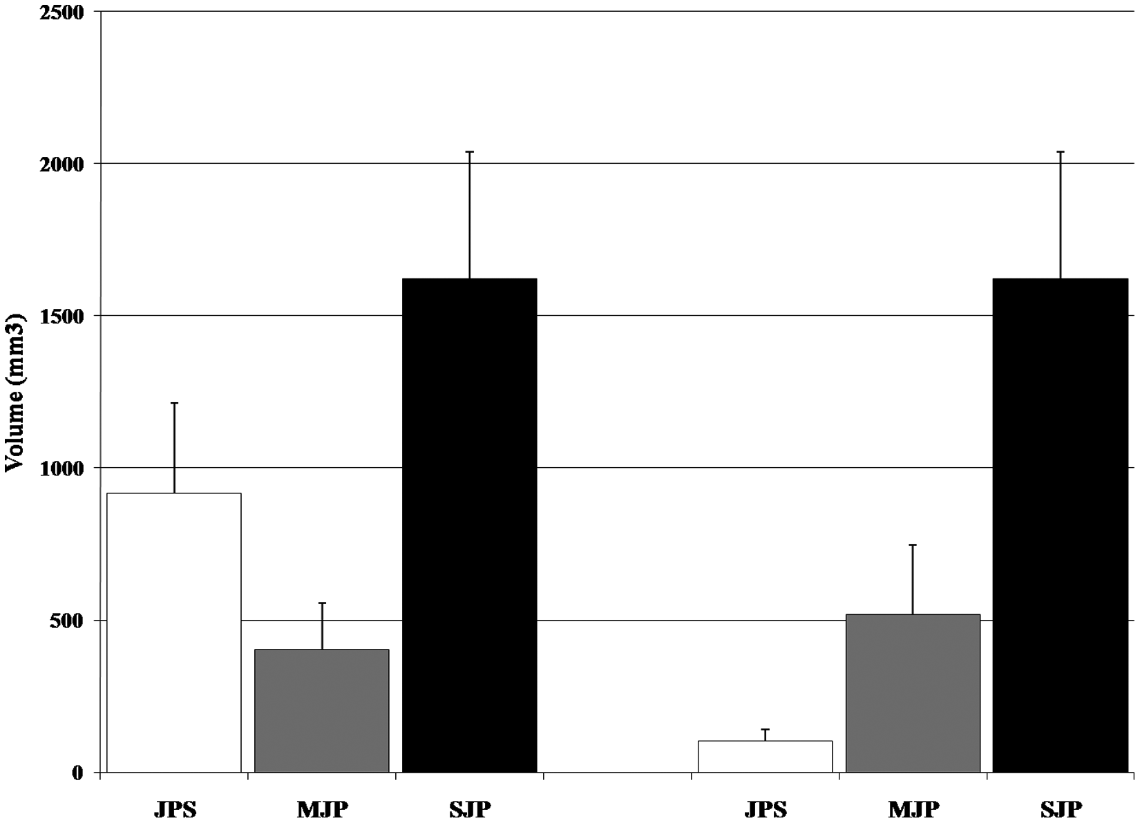
Polyp volume according to the classification of juvenile polyp type
Hemoglobin and mean corpuscle volume (MCV) data were available within 1 month of initial colonoscopy for 76% of SJP, 87% of MJP, and 94% of JPS children. We defined anemia as a hemoglobin ≤10.5 g/dL. Mean hemoglobin was significantly lower in JPS (10.6 ± 2.3 g/dL) than MJP (12.4 ± 0.9 g/dL) and SJP (12.0 ± 1.2 g/dL; P = 0.002). Similarly, MCV was significantly lower in JPS (74.2 ± 10.0 fL) than MJP (81.5 ± 6.3 fL) and SJP (80.9 ± 4.6 fL; P = .003). Anemia was more common in JPS (53.8%) than MJP (0%) and SJP (8.5%; P < .001).
Discussion
Colonic polyps are a common cause of painless rectal bleeding in children, the overwhelming majority of which are represented by juvenile polyps. Less commonly, children may have polyps as part of a syndrome, such as FAP or PJS, both conditions with a known malignant risk. The risk for malignant transformation in juvenile polyps is negligible in the absence of an underlying JPS. We compared clinical features and endoscopic findings of children with SJP, MJP, and JPS. Furthermore, we evaluated polyps with a battery of immunohistochemical stains to determine if differences existed among the various juvenile polyp groups.
In our series of children having colonoscopy with polypectomy, 93% had juvenile polyps whereas the remaining had either FAP (5%) or PJS (2%). These findings are in keeping with previous reports where juvenile polyps accounted for 88% to 100% of polyps removed at colonoscopy in children.5-8 Children with juvenile polyps were further divided according to the number of polyps into SJP (67%), MJP (16%), and JPS (17%). The percentage of children with JPS in our series was higher than previously reported (2.4% to 8.9%),5-8 which may be accounted for by founder effect as we have a large pedigree family with JPS that is cared for at our institution. 25
Review of demographic data showed that the majority of children in this study were male (59%), which is in keeping with previous reports where males accounted for 58% to 78% of children requiring polypectomy.5,11,13,15 We noted no difference in the percentage of males with SJP, MJP, or JPS. Data also showed that children with JPS were older at initial presentation than children with SJP and MJP, a finding that has not been previously reported. Reasons for this difference are speculative. Nonsyndromic juvenile polyps tend to occur in children in response to an inflammatory process, typically in early childhood. The mechanism for polyp formation in JPS may be related, in part, to genetic factors that do not require inflammation. To date, defects in two gene have been associated with JPS, SMAD4 (also referred to as MADH4 [mothers against decapentaplegic homolog 4]) and bone morphogenetic protein receptor 1A (BMPR1A)17,18; together defects in these genes account for less than 50% of JPS.19,21 Both SMAD4 and BMPR1A belong to the TGF-β superfamily and are considered tumor suppressor genes. 26 Despite being tumor suppressor genes, the mechanism by which polyps are formed have not been established. 20
Alternatively, delayed presentation in JPS may be related to differences in polyp size and location compared with nonsyndromic juvenile polyps. Our data show that JPS polyps are more evenly distributed throughout the colon and are smaller in size than polyps of SJP and MJP. Juvenile polyps most often present with rectal bleeding, which is more apt to happen with large, distally located polyps that experience traumatic ulceration from passage of formed stool. Smaller, more proximally located polyps are less likely to be damaged by liquid and semiformed stool. However, if these polyps are injured and bleed, the blood can be incorporated into the stool and pass unnoticed by the child and parent, delaying presentation.
The utilization of pancolonoscopy for the evaluation of rectal bleeding has expanded our knowledge of the true distribution of juvenile polyps. Combined data retrieved from previous studies5-16 show that the majority of juvenile polyps (76.4%) are located in the rectosigmoid colon, with the remaining polyps being equally distributed between the left colon (8.6%), transverse colon (8.4%), and right colon (6.6%; Figure 1). Our data combining all juvenile polyps are similar, with the majority of juvenile polyps (64.1%) located in the rectosigmoid colon, with the remaining polyps being equally distributed between the left colon (14.8%), transverse colon (11.4%), and right colon (11.8%). However, when separated by groups, significant differences are seen in the distribution of polyps. SJPs are almost exclusively (98%) found in the rectosigmoid colon. The percentage of rectosigmoid polyps is decreased in MJP (64%) and further decreased in JPS (45%). The numbers are too small for analysis, but there appears to be no difference in the distribution of polyps outside the rectosigmoid colon between MJP and JPS.
We also assessed polyp volume to determine if differences existed among the various juvenile polyp groups. Including all polyps within each group, the mean (±SE) polyp volume was smallest for MJP (404 ± 152 mm3), followed by JPS (918 ± 294 mm3) and SJP (1622 ± 541 mm3). Statistical significance was only established for the difference between MJP and SJP (P < .05). Because of the wide range in polyp volumes within the groups we compared the median volume for each group. JPS had the smallest median polyp volume (87 mm3), followed by MJP (117 mm3) and SJP (541 mm3). Addressing only rectosigmoid polyps, we found that the average for JPS polyps was significantly less (102 ± 39 mm3) than for MJP (229 ± 132 mm3; P < .05) and SJP (1622 ± 541 mm3; P < .05). Similarly, the median polyp volume for JPS (13 mm3) was significantly less than for MJP (132 mm3) and SJP (541 mm3). We can explain these findings as follows. Polyps in JPS are in various stages of development throughout the lower intestine. Polyps in the right colon can grow to a larger size before presenting with rectal bleeding because they are not exposed to the irritating force of formed stool. When more distal polyps grow to a size that allows for clinical presentation, colonoscopy will reveal polyps at various stages of development and size. Also, repeat surveillance colonoscopy is more likely to detect smaller developing polyps. Children with MJP had either multiple polyps simultaneously or recurrent polyps. Similar to JPS, in simultaneously detected polyps, proximally located polyps may grow until more distally located polyps become symptomatic. In cases of recurrent polyps, parental knowledge of previous polyps as a cause for rectal bleeding may lead them to seek care earlier. In children with SJP, the polyps have grown to a size that makes them symptomatic, prompting colonoscopy.
In our series, children with JPS were more likely to have iron-deficient anemia than children with MJP or SJP, in agreement with a previous report. 14 The average hemoglobin and mean corpuscular volume were significantly lower in JPS. Also, the prevalence of anemia in JPS (54%) was higher than SJP (9%). As stated above, occult bleeding from multiple small proximally located polyps may persist for some time before development of a polyp large enough to result in bright red rectal bleeding, allowing development of anemia prior to clinical presentation.
In conclusion, the distribution of juvenile polyps in children with SJP differs from that of children with MJP and JPS. Children with SJP have polyps found almost exclusively in the rectosigmoid colon of children with SJP; whereas polyps are more diffusely distributed in children with MJP and JPS. Rectosigmoid polyps tend to be larger in SJP than in MJP and PJS. In this study, children with JPS were older and were more likely to have anemia at the time of diagnosis than were children with MJP or SJP. Despite the risk of colon cancer in JPS, we found no difference in markers for replication or malignancy among the various groups of children with juvenile polyps.
Footnotes
The author(s) declared no potential conflicts of interest with respect to the research, authorship, and/or publication of this article.
The author(s) received no financial support for the research, authorship, and/or publication of this article.