






Abstract
Congenital muscular torticollis (CMT) is a common newborn pediatric muscular deformity of the neck. The purpose of this article is to suggest a clinical algorithm for pediatric clinicians to promote prompt identification and intervention for infants with CMT. Early intervention for a child with CMT at less than 1 month of age yields a 98% success rate by 2.5 months of age, with the infant achieving near normal range of motion. Intervention initiated at 6 months of age or later can require 9 to 10 months of therapy with less success in achieving full range of motion of the cervical musculature. The clinical algorithm proposed here incorporates the American Physical Therapy Association guideline for CMT to optimize outcomes for the child and reduce health care expenditures. Current evidence and guidelines demonstrate that primary care providers are the primary diagnostic clinicians, while physical therapists are the preferred provider for the treatment of CMT.
Introduction
Congenital muscular torticollis (CMT) is the third most common congenital pediatric musculoskeletal deformity next to hip dysplasia and talipes equinovarus.1,2 CMT is characterized by fibrosis, or shortening, of the sternocleidomastoid muscle, which results in lateral sidebending of the head to the involved side and rotation to the opposite side. The estimated incidence of CMT is reported to be between 0.3% and 2% of newborns, with a possible occurrence as high as 16% of live births.3-5 The actual etiology of CMT is unknown, but evidence suggests it is closely related to positioning in utero, or trauma during delivery.1,3,6 CMT is associated with multiple congenital conditions including plagiocephaly,3,5,7,8 hip dysplasia,3,7-10 C1-C2 subluxation and c-spine annomalies,1,11,12 brachial plexus injury,1,3,13 ocular disorders,1,11,12,14,15 metatarsal adductus, 1 upper extremity deformities, 3 temporomandibular joint dysfunction,3,16 talipes equinovarus,1,3 early persistent developmental delay,3,6,12 and cosmesis relative to facial asymmetry.3,6,12,17 Additionally, secondary sequelae related to the presence of CMT include plagiocephaly, facial asymmetry, and developmental delay.3,6,17 While 82% of cases of CMT are muscular in origin, 18% are nonmuscular1,13,15,16; therefore, early identification and differential diagnosis is critical to assess for underlying issues such as neurogenic pathology and skeletal anomalies.1,3,11,18 Identification and intervention of CMT by 2.5 months of age has a successful outcome of 98% with the infant achieving near normal range of motion if the intervention is initiated at 1 month of age or sooner.3,17 Due to the high risk of developing secondary sequelae, and the effectiveness of early intervention, it is imperative for clinicians to implement, as part of any physical exam of a newborn through the age of 4 months, screening for CMT. This article proposes a clinical algorithm for early identification and intervention of CMT in the primary care office or other pediatric early-care centers to ensure optimal outcomes and the prevention of secondary sequelae for infants birth to age 4 months. The cost-effectiveness of this approach can save time and resources, while improving effectiveness of treatment.
Background
CMT occurs at birth, or soon after, but is not typically identified until 4 months of age or later at a normal well-baby examination.14,18 A delay in identification can directly affect the child’s long-term outcome with increased health care services and costs. Evidence demonstrates that early intervention of physical therapy services for a child less than 1 month of age yields a 98% success rate by 2.5 months of age, with the infant achieving near normal range of motion. Comparatively, intervention initiated at 6 months of age or later can require 9 to 10 months of therapy with less success in achieving full range of motion of the cervical musculature.3,17 Early intervention is hypothesized as more successful since once the infant is mobile it becomes very difficult to perform the necessary stretching exercises and positioning techniques. Early treatment normally consists of noninvasive positioning and handling techniques, cervical stretching exercises, and parental education and instruction. Delayed intervention is met with child resistance and avoidance of treatment, which affects successful outcomes. Preliminary evidence with a small sample suggests that treatment supervised by a physical therapist may be more effective than unsupervised parent intervention. 19
CMT has a significant association with plagiocephaly, which is present in 80% to 90% of cases. 7 Plagiocephaly is a condition of cranial flattening and is known to place the child at high risk for developmental delays, requiring direct corrective action. 14 Additional correlations are found between CMT and hip dysplasia, present in 8% to 17% of cases depending on severity.1,3,7-9 C1-C2 subluxation, spinal anomalies, brachial plexus injury, ocular disorders (resulting in lateral head tilt as a compensatory strategy for visual accommodation), and central nervous system (CNS) pathology, such as syringomyelia, are associated at a rate of 2% to 9.4% of infants identified with CMT. 1 Metatarsal adductus, upper extremity deformities, temporomandibular joint dysfunction, and talipes equinovarus are also associated with CMT relative to uterine positioning and skeletal anomalies. Early persistent developmental delay and cosmesis relative to facial asymmetry due to the musculoskeletal imbalances are associated characteristics of CMT requiring direct intervention. Therefore, if CMT is suspected during early screening, a complete assessment of possible associated, or underlying, issues should be completed.
Assessment for CMT
Based on the evidence, all infants from birth to 4 months should be screened for CMT during each postnatal interaction by pediatric clinicians (eg, lactation specialists, nurses, physicians, therapists). The signs of CMT have been identified previously by many authorities to include head and neck asymmetry, possible plagiocephaly, and decreased cervical spine range of motion affecting sidebending to one side and rotation to the opposite side.3-8,12,14,17 A thorough examination of the newborn’s head and neck symmetry, cervical spine range of motion, skeletal anomalies, palpable masses, and CNS dysfunction as discussed below will provide for the differential diagnosis of muscular CMT. 3 Once CMT is identified, and spinal anomalies have been ruled out, the caregiver should be given initial instructions in positioning and stretching techniques, followed by a complete and expedited referral to a pediatric physical therapist.
Algorithm for Identification and Treatment of CMT
The American Physical Therapy Association’s CMT clinical guideline strongly recommends all infants be screened for CMT throughout the first 3 to 4 months of life by pediatricians, 3 but a specific process by which this can occur has not been established. Utilizing available evidence and published guidelines, a screening and referral algorithm for CMT is described here (see Figure 1).3,20 This tool can be utilized for all newborns through 4 months of age by any pediatric clinician (eg, lactation specialists, nurses, physicians, therapists) who interacts with infants and caregivers. Early identification and intervention are critical for successful outcomes and to improve efficiency and effectiveness of care.
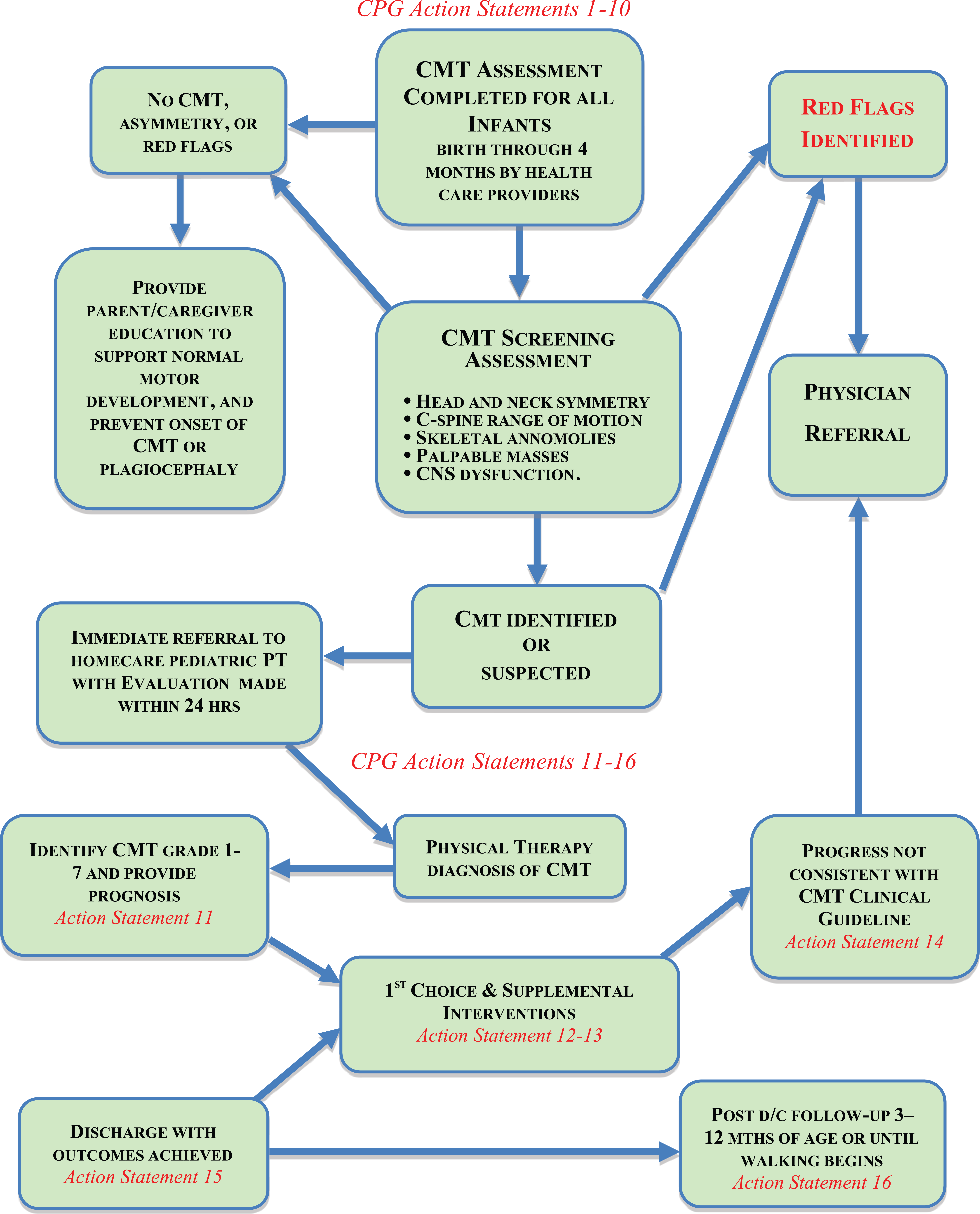
Clinical algorithm for identification and treatment of CMT.
Process for Early Identification and Intervention
Up to 4 months of age all infants need to be screened for CMT during each contact with a pediatric clinician (eg, lactation specialists, nurses, physicians, therapists). The screening examination will include an assessment of the newborn’s head and neck symmetry, cervical spine range of motion, possible skeletal anomalies, palpable masses, and CNS dysfunction.
Head and neck symmetry are assessed through observation and palpation. The normal observed shape of the head during infancy is round with a prominent occipital area that becomes less prominent as the child gets older. A flattening of one part of the head (and hair loss in one area), indicative of plagiocephaly, is a risk factor for asymmetry. Swelling and ecchymosis of the presenting part of the head in the first week postpartum is called caput succedaneum, and it should resolve in the first week of life. The position of the head and neck with the child in supine position on a flat surface should be centered on the trunk with the chin, nose, and vertical center of the throat in alignment. Any rotation of the head, as indicated by the chin being off-centered, or sidebending, as indicated by the nose being off-centered and/or curvature of the neck, should be considered asymmetrical and an indication for additional screening.3,21
Normally, cervical range of motion occurs in isolation with symmetry present between the right and left sides. Cervical range of motion is assessed by gently holding the head in midline from the base of the occiput, careful to not place pressure on the mandible. To assess rotation, the clinician slowly rotates the head toward one shoulder, and then the other. Sidebending, right or left, occurs without rotation with the clinician moving the head so the ear moves toward the shoulder. Flexion occurs with the chin moving toward the chest, while extension brings the occiput back toward C7. Assessment of cervical range of motion should monitor for resistance or compensatory limitations of sidebending with rotation, or shoulder elevation to either side, as well as for inequalities in mobility between the right and the left. Infants who exhibit decreased cervical range of motion in rotation or sidebending, or asymmetries of the head, neck, or shoulders, should be referred for additional screening and possible treatment by a physical therapist. 3
Skeletal alignment and symmetry should be assessed, with follow-up radiographic exam indicated if skeletal anomalies or asymmetries such as congenital scoliosis, cervical spine anomalies, or subluxation are suspected.
Identification of palpable masses of the head, neck, and trunk will require further diagnostic testing. Pseudotumor of infancy, which is an inflammatory response to birth trauma of the sternocleidomastoid muscle, is present in 28.2% to 47.2% of infants with CMT. This benign soft fluid filled tumor initially enlarges at about 2 weeks after birth, and then resolves between 5 and 21 months. 14
CNS dysfunction is identified by abnormal muscle tone or movement imbalances between extremities, abnormal ocular motor responses and asymmetries, evidence of abnormal reflexes (eg, clonus, tremors), and delays in the integration of primitive reflexes necessary for maturation.3,20,22
If neither muscular nor nonmuscular CMT are identified, or suspected, the clinician should educate the parent or caregiver on CMT, typical motor development and positioning related to symmetrical head control, and the prevention of plagiocephaly and muscular CMT. If CNS dysfunction or skeletal anomalies, including, but not limited to, palpable masses are identified the clinician should complete an immediate referral to the infant’s pediatrician. Alternatively, if muscular CMT is identified, or suspected, based on asymmetrical positioning of the head, or restricted cervical range of motion, a referral to a pediatric physical therapist should occur. Ideally, a pediatric physical therapy evaluation will occur within 24 hours following the referral. Physical therapy evaluation can determine the severity grade, treatment, expected outcomes, and prognosis for the individual child. The physical therapist will repeat a full infant motor evaluation and will initiate referral back to the pediatric primary care provider if any additional red flags, such as CNS dysfunction, skeletal anomalies, or palpable masses, are identified. 3
Common red flags identified by physical therapists during the evaluation or over a course of treatment can include any of the following:
Ocular motor asymmetry (eg, nystagmus, strabismus)
Palpable masses
Asymmetrical head positioning due to abnormal muscle tone, muscle tone imbalance, or absence of sternocleidomastoid muscle contracture
Palpable skeletal anomalies or asymmetries (eg, congenital scoliosis, cervical spine subluxation), which will require radiographic exam
Delayed development and integration of primitive reflexes
Presence of pathological reflexes (eg, clonus, tremors)
Absence of movement of any extremity, or the presence of any hand or leg dominance
If muscular CMT is confirmed in the evaluation, the therapist will implement at the very least the following 5 first-choice interventions 3 :
Neck passive range of motion
Neck and trunk range of motion
Development of symmetrical movement
Environmental adaptations (eg, positioning of infant in crib and infant carrier, placement of toys)
Parent and caregiver education (eg, home program)
The infant will be referred back to the primary care provider when indications of a more severe condition are identified with any of the following 3 :
No progress occurs after 4 to 6 weeks of intensive intervention
After 6 months of treatment there is only moderate recovery
If the infant is older than 7 months and there is a tight band or SCM (sternocleidomastoid) mass identified, or the torticollis changes sides
If the infant is older than 12 months on the initial evaluation and/or there is a facial asymmetry difference of 10° to 15° between the right and left sides
Successful resolution of CMT with resultant discontinuation of direct physical therapy services is measured by full passive range of motion within 5° of the nonaffected side; symmetrical active movement occurs throughout the available range of motion; age-appropriate motor development is present; no observable head tilt; and parent or caregiver verbalize understanding of key signs to monitor as the infant develops. The infant should be reassessed by the physical therapist in 3 to 12 months after discharge in order to monitor for reoccurrence of CMT and associated secondary sequelae, including developmental delay. Timing of the reassessment should occur prior to walking developing, with an earlier reassessment being most appropriate for those infants who were discharged between 4 and 6 months of age. 3
Summary
CMT is a common newborn pediatric muscular deformity of the neck, occurring in between 0.3% to 2%3,4 of live births, with reports indicating a possible incidence as high as 16%.3,5 Early intervention of physical therapy services for a child with CMT less than 1 month of age yields a 98% success rate by 2.5 months of age, with the infant achieving near normal range of motion. Comparatively, intervention initiated at 6 months of age or later can require 9 to 10 months of therapy with less success in achieving full range of motion of the cervical musculature.3,17 Therefore, early intervention is of the utmost importance to maximize benefit, prevent secondary sequelae, and minimize health care expenditures. The implementation of the clinical algorithm for early identification and intervention of CMT ensures optimal outcomes and prevention of secondary sequelae for infants birth to age 4 months, while subsequently reducing health care expenditures relative to the improved efficiency and effectiveness of treatment. Current evidence and guidelines have demonstrated that physical therapists are the preferred provider for the treatment of CMT.3,7,21,23 To ensure physical therapy intervention occurs expeditiously it is specifically recommended that referral be to a certified home care agency, which employs pediatric physical therapists, as they are in the unique position of responding in the home, within 24 hours.
Author Contribution
SN is the author and researcher of the article. Suzanne Brown professor in University of New England, served as direct advisor towards the article’s development, providing invaluable guidance and expertise.
Footnotes
Acknowledgements
Suzanne R. Brown, PhD, MPH, PT; Advisor.
Declaration of Conflicting Interests
The author declared no potential conflicts of interest with respect to the research, authorship, and/or publication of this article.
Funding
The author received no financial support for the research, authorship, and/or publication of this article.