
Abstract
Echinococcus granulosus, first reported by Goeze in 1782, is the causative parasite of cystic echinococcosis (hydatidosis) especially for countries that are endemic areas. Since the 1970s, the incidence of echinococcosis in Greece has been very high. Nevertheless, with the implementation of special prevention measures in the 1980s, a large reduction in the incidence of hydatidosis meant that it reached European levels. Therefore, we analyzed the demographics, multiple organ localizations of the parasite, diagnosis, and conservative and optimal surgical treatment over a total period of 39 years, especially for pulmonary and hepatic echinococcosis in children. The higher incidence of pulmonary echinococcosis compared with other localizations, male predominance, wide range of age, and various cystic sizes were some of the main demographics. Because cystic echinococcosis remains a main public health problem, we advocate a meticulous clinical investigation and treatment methodology to bridge the gap between knowledge and awareness of this important disease.
Introduction
Cystic echinococcosis is a zoonotic disease that is transmitted from animals to humans. The disease is caused by the tapeworm Echinococcus granulosus. It represents the most common type of human echinococcosis because it accounts for >95% of the estimated 2 to 3 million global cases. For these reasons, cystic echinococcosis (hydatidosis) is still a major medical, social, and very serious economic problem, especially for countries where there are endemic areas. The World Health Organization (WHO) has estimated that the financial burden, in order to manage the worldwide echinococcosis, is about 3 billion dollars per year. Also, according to a WHO survey for 2010, 1200 deaths caused by echinococcosis have been recorded worldwide. Also, a future WHO objective is the complete eradication of the disease, which is not easily achievable.1-3
Phylogenetically, the Echinococcus parasite is divided into 4 main types: E granulosus, E multicularis, E vogeli, and E oligarthrus. The mitochondrial DNA of E granulosus has been identified and classified into 10 distinct genotypes (G1-G10). In Greece, E granulosus is the dominant type, and 3 genotypes have been recorded. There is a main genotype G1 (sheep) and, rarely, G3 (cattle and buffalos) and G7 (pigs and goats).4,5
The first historical reference for the Echinococcus cyst is recorded in the fifth century
Goeze in 1782 was the first who reported the heads of E granulosus. The causal relationship between echinococcosis and parasite E granulosus was mainly described by August Batsch in 1786. There were also other types of Echinococcus, such as E multilocularis, which were first analyzed by R. Leuckart (1863). Later Vogel recorded E vogeli. Especially for E oligarthrus, the first observation was made in 1863 by K. M. Diesing. In 1972, Rausch and Bernstein recorded the differences between E vogeli and E oligarthrus.9-16
Cystic echinococcosis is endemic in areas related to livestock and pasture. Such areas are Africa, the Mediterranean Basin, the Middle East, Central Asia, South America, Australia, and New Zealand. The incidence of echinococcosis in Western countries is low: <1 case per 100 000 population. However, the incidence of the disease has increased over the past decades because of massive migration and high tourist traffic, especially in Europe and North America.5,17
In the past, various prevention programs have been implemented to reduce echinococcosis in Iceland in the 1950s, in New Zealand in 1959, in Tasmania in 1965, and in Cyprus in the 1990s. These programs have achieved a great reduction in echinococcosis cases. At the same time, systemic administration of anti-echinococcal agents (Praziquantel) reduced the rate of echinococcosis in some endemic areas, such as in Argentina in 1970 and in Chile and Uruguay in 1978. Moreover, the systematic use of the recombinant vaccine (EG95) offered protection against the parasite in canines, at a rate of 96% to 100%. This vaccine was widely used worldwide and especially in Australia and Argentina. However, despite the precautionary measures, since 1992, there has been an increase in echinococcal cystic cases, particularly in the former Soviet republics, Eastern Europe, Central Asia, and South America. This increased incidence of echinococcosis is probably a result of the relaxation of preventive measures.5,18-20
The present study analyzes the demographic data, diagnosis, and surgical management of a large number of children suffering from cystic echinococcosis (E granulosus) and originated from Northern and Central Greece. It should also be noted that in the 1970s, the incidence of echinococcosis in Greece was very high. But in the 1980s, because of the implementation of special prevention measures, a large reduction in the incidence of hydatidosis, reaching the usual levels of that in European Union countries, has been achieved.
Patients and Methods
Over a total period of 39 years, divided into 2 consecutive chronological periods, all our cases were managed in 2 separate departments of pediatric surgery of the Aristotle University of Thessaloniki. These 2 consecutive chronological periods were, first, in the department of pediatric surgery of G Gennimatas, 1970 to 2003, and second, in the department of pediatric surgery of the Papageorgiou Hospitals, 2004 to 2009. During this period, a total of 187 children with a variety of localized cystic echinococcosis received successful treatment.
Chronological Period
The 39-year-period is analyzed chronologically over a decade and is as follows: In the 1970s, 123 cases (65.77%) were dealt with; in the 1980s, the number of cases decreased to 54 (28.87%); in the period between 1990 and 2003, cases decreased to 8 (4.28%); and in the period 2004-2009, there were 2 recorded cases (1.08%; Figure 1A).
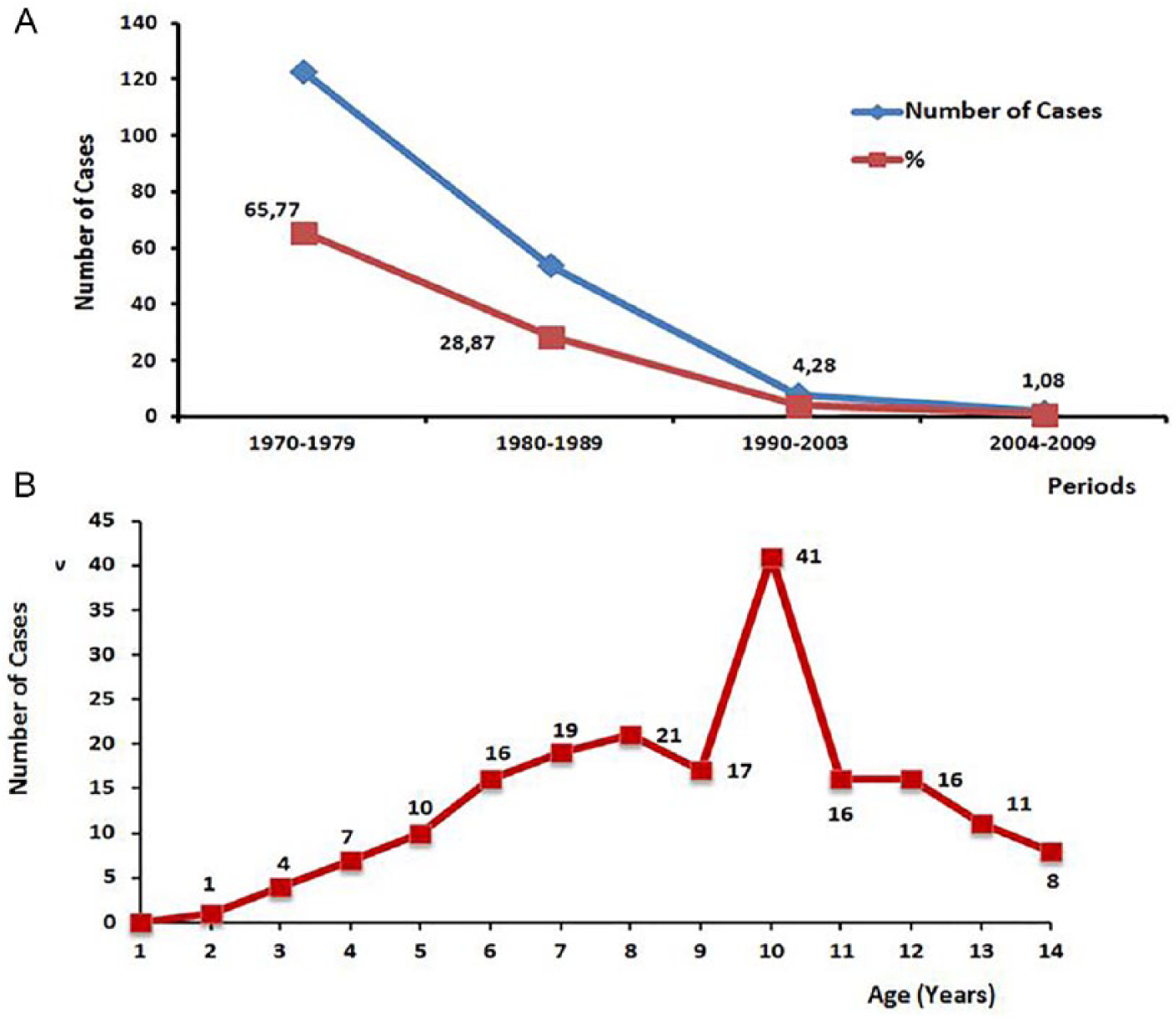
(a) Echinococcosis in childhood in the period 1970-2009 and (b) age distribution of echinococcosis in childhood.
Geographical Distribution
Of our 187 cases, 170 (91.01%) were from Northern Greece, including the following regions: eastern Macedonia and Thrace 71 cases (37.96%), central Macedonia 58 cases (31.02%), and Western Macedonia 41 (21.93%). Regarding central Greece, we recorded 17 cases (9.09%) that originated from Thessaly region. The majority of our young patients were from rural areas where livestock farming was well developed.
Age
This ranged from 2 to 14 years, with an average age of 9.2 years. Most cases were encountered between the ages of 7 and 13, with the most common age of 10 years. Our youngest patient was 23 months of age (Figure 1B).21-23
Gender
Of our 187 cases of echinococcosis, 101 (54.01%) cases were boys and 86 cases (45.99%) were girls.
Localization
Of the 187 cases, 96 cases (51.33%) were detected in the lung, 78 cases (41.71%) in the liver, and 13 cases (6.96%) in various organs (Table 1).
Localization.
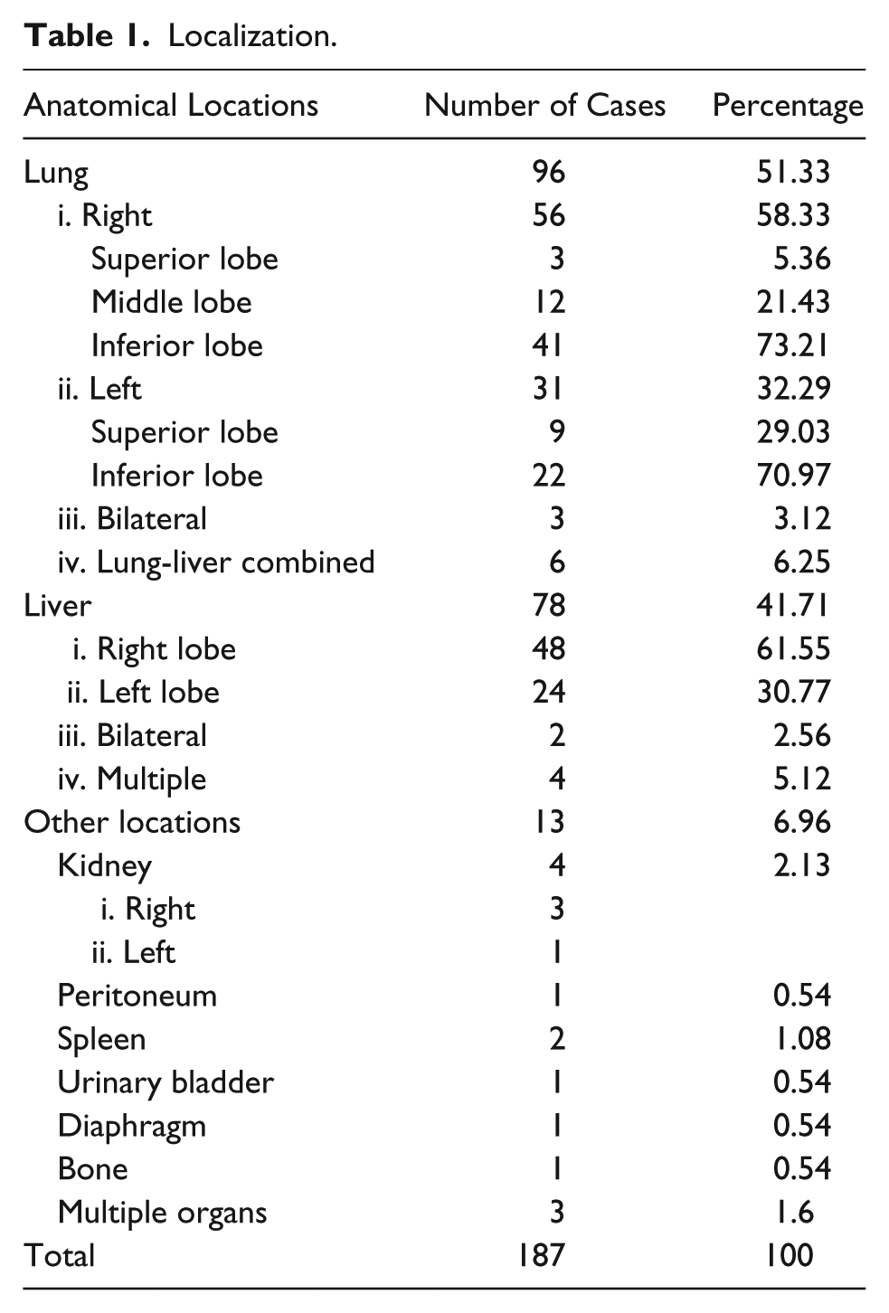
Lung
The echinococcal cyst, located in the right lung, involved a total of 56 (58.33%) cases, in which 43 cases (73.21%) were located in the lower lobe. In 31 cases (32.29%), they were found in the left lung, with anatomical localization mainly in the lower lobe in 22 cases (70.97%). Also, 3 cases (3.12%) that had bilateral localization in the lungs were treated, whereas the combined location in the lung and liver was treated in 6 cases (6.25%; Table 1).
Liver
Regarding the echinococcal cyst in the liver, the right lobe was the most frequently affected and included 48 cases (61.55%), whereas the left lobe was affected in 24 cases (30.77%). Two cases (2.56%) with bilateral localization and 4 cases (5.12%) with multiple cysts were also treated (Table 1).
Other Locations
Echinococcosis in other organs was very rare, and the frequency range was as follows: kidney, 4 cases (2.13%); multiple localization in at least 3 organs, 3 cases (1.6%); spleen, 2 cases (1.8%); other cases were related to other rare sites (Table 1).
Multiple Localization
Of the total 187 cases of children with echinococcosis, in 18 cases (9.62%), there were more than 2 locations in the same or in other organs. Among these 18 cases, 9 (50%) were located in the lung, 3 with bilateral localization (1 of them had 3 cysts), and 6 (33.33%) included combined localization in lungs and liver. In this group, 2 cases had multiple locations in the same lung (1 case with 2 and the other with 4 sites). Multiple localization in the liver involved a total of 6 cases, of which 2 cases were located in bilateral lobes and 6 (33.33%) had multiple locations (3-6).
Finally, multiple locations of the Echinococcus cyst in the various organs concerned 3 (16.66%) cases, where at the same time 3 were localized in the lung, 3 in the liver, 2 in the peritoneum, and 1 in the spleen. The total distribution of multiple localization in the various organs in 18 cases was as follows: lungs 18 cases (100%), liver 9 (50%), peritoneum 2 (11.11%), and spleen 1 (5.55%).
Symptoms
These were varied, depending on the location of the parasite in the various organs. Analytically, the main symptoms per organ were as discussed below.
Lung
Of the 96 cases of echinococcal cyst in the lungs, 51 cases (53,12%) were asymptomatic. The remaining cases showed the following symptoms in order of frequency: cough in 23 cases (23.95%), pain in 14 cases (14.58%), and fever in 11 cases (11.45%), whereas other less common symptoms were dyspnea and hemoptysis. Our cases also rarely developed thoracic empyema, hydrothorax, and hydatidemesia, which involved a total of 3 cases of complicated echinococcal cysts. In our cases, no serious allergic reaction was recorded (Figure 2A).
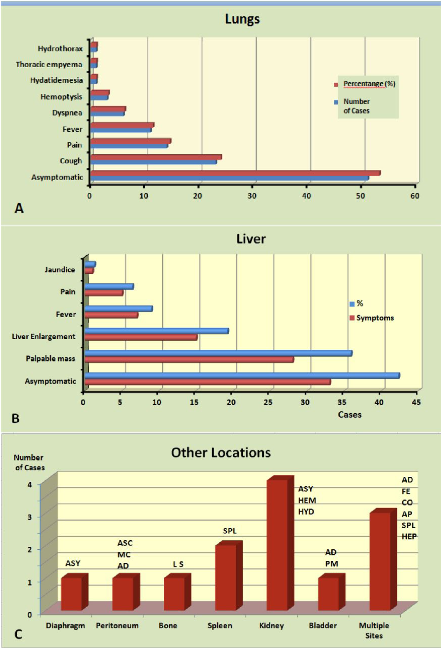
Main symptoms of echinococcal disease. (a) lungs, (b) liver and © other locations.
Liver
Of the 78 cases with hydatid disease, 33 (42.3%) were asymptomatic. The remaining cases showed the following symptoms: residual mass in 28 cases (35.89%) and hepatomegaly in 15 cases (19.23%); fever, pain, or jaundice rarely occurred. Additionally, we did not observe any symptoms of intense pruritus, severe allergic reaction, or acute surgical abdomen caused by spontaneous rupture of the hydatid cyst (Figure 2B).
Other Sites
In the 13 cases (6.96%) of echinococcosis in other anatomical sites, symptoms were dependent on site location. Analytically, in one case with left diaphragmatic localization, it was asymptomatic, whereas another case with location in the peritoneal cavity showed extensive abdominal distension and ascites. Also, in 1 case with bone location, local swelling of the affected upper limb occurred. In 2 cases with splenic localization, there was splenomegaly. In 4 cases with kidney hydatid disease, one experienced hematuria and pain and the other, swelling in the lumbar region. The remaining 2 cases, with location in the kidneys, were asymptomatic. In 1 case, a palpable mass was found in the abdomen by localization in the urinary bladder. In 3 cases with combined multiple sites, 2 cases concerned the lung, liver, and peritoneum and the other, lung, liver, and spleen, and symptoms were consistent with organ location (Figure 2C).
Echinococcal Cyst Size
Of the total 187 cases of Echinococcus cysts, the largest incidence, involving 169 cases (90.38%), was solitary, whereas 18 cases (9.62%) were multiple. During surgery, the echinococcal cysts were large, and their diameter varied from 7 to 38 cm; 19 (10.16%) had a diameter of <10 cm, whereas the usual diameter was >15 cm. Generally, the echinococcal cysts of the chest and liver were large at the time of diagnosis. As an exception to this rule, the cysts in the peritoneal cavity were smaller in size. Also, for the rare localizations of the Echinococcus cyst in other organs, such as the spleen, the bladder, the kidneys, and the diaphragm, they were usually medium or large, while in the bones, the cyst was small in size.
Diagnosis
Prior to the 1980s, older laboratory tests for the diagnosis of echinococcosis were used, such as Casoni’s dermoreaction and the Weinberg seroreaction. Other laboratory tests were the indirect hemagglutination test and the latex agglutination test (LAT). In contrast, more accurate diagnostic tests, such as the enzyme-linked immunosorbent assay (ELISA) and polymerase chain reaction (PCR) were applied. Alternatively, eosinophilia was an indicative finding of the laboratory diagnosis of echinococcosis.
Radiographic imaging of the Echinococcus cyst, in the first half of the period of our study, was based mainly on chest and abdominal X-rays or other positions. After this period, all our cases were studied in more detail by conducting various examinations such as ultrasound (US), computed tomography (CT), and magnetic resonance imaging (MRI).
Treatment
All our cases were treated with 2 main therapeutic options, discussed below.
Conservative
In the decade of 1970, specific antiechinococcal agents (mebendazole and albendazole) were not widely used. So we used them only exceptionally and in certain specific complicated cases, multiple locations, or large echinococcal cysts. At the beginning of 1980, mebendazole had been systematically administered, whereas in the mid-1980s, administration of albendazole followed, based on 2 sequential clinical protocols (1985 and 2004). In overall periods, this specific antiechinococcal chemotherapy was administered in 57 children, for a time range from 1 to 4 weeks preoperatively and 1 to 3 months postoperatively.22,24
Surgery
All our cases (187) of echinococcosis were surgically treated using the methods discussed below.
General principles
The basic surgical steps, which included removal of echinococcal cysts by “Lege Artis” cystectomy were as follows:
(a) Cystic puncture and aspiration of a small amount of echinococcal fluid was done.
(b) Injection of scolicidal agents as 15% NaCl solution was used in our cases for many years, which did not cause problems and generally had excellent results in children. The same solution was used to protect adjacent organs. These were preserved by gauzes that were impregnated with this hypertonic solution. Also, the same solution was used for the final cleaning of the various cavities such as the residual cavity of the Echinococcus cysts and the thoracic and abdominal areas. All the above precautions must be made to prevent local inoculation of the parasite.
(c) Full aspiration of echinococcal cystic fluid was done.
(d) The reactive pericyst was opened with a small incision.
(e) The empty echinococcal cyst was removed.
(f) The residual cavity of the cyst was washed out with a hypertonic solution (15%) NaCl.
(g) The size of the residual cavity was reduced by various techniques, according to the affected organ.
(h) The reactive pericystic opening was sutured with continuous absorbable suture (Vicryl, PDS, etc).
Exceptions to these rules are given below.
The Ugon method in the lung was characterized by a complete “enucleation” of the echinococcal cyst, through a small incision of the reactive pericystic capsule without puncture.
For complete or partial resections of various abdominal organs, such as the liver, spleen, kidney, diaphragm, bladder, bones, and so on, the echinococcal cyst is removed with the affected part of the organ.
For synchronous localizations of the echinococcal cyst, such as the lung and liver, removal of these cysts can be performed at the same surgical time or at 2 different surgical times.
Regarding the bilateral locations in the lungs, the removal of the cysts usually take place at 2 different surgical times.
In multiple locations in the same organ as the liver and lungs, removal of the cysts is intended to take place at the same surgical time. In situations where there are surgical difficulties, removal can be done at 2 surgical times.
Specific surgical techniques
Lungs
(a) Barret method: This method was applied in 35 cases (36.45%). This technique refers to a complete cystic removal (cystectomy) after a puncture of its contents through a closed circuit by injection of a scolicidal agent, such as the 15% hypertonic NaCl solution, which is recommended for children. The remaining residual cavity is sutured by successive sutures, from the bottom to the top (capitonnage), by avoiding the development of hematoma or exudate or the prevention of pneumothorax. Also, when there were open communications of bronchi with the residual cavity, we performed special sutures closing the communication (Posada’s Method). Büllow was placed in all of our cases.
(b) Ugon method: This was the surgical method applied in 26 (27.08%) of our cases. In the majority of our cases (22 cases, 84.62%), we left the residual cavity without sutures. In selective cases (4%-15.38%), we performed Capitonnage of the residual cavity according to the Barrett technique, with internal sutures and placement of Büllow catheter in all cases.
(c) Perez Fontana method: This method was applied to 13 cases (13.54%) and considered to be more radical than the Barret method. According to this technique, cystectomy and complete pericystectomy were applied. In our cases, it followed by reduction of the residual cavity with special internal sutures (Capitonnage, as in the Barret technique) and closure of the open bronchi communications with the residual cavity (as in Posada’s technique) and placement of Büllow catheter.
(d) Segmentectomy/Lobectomy: We performed this radical technique in 22 cases (22.93%). With this technique, younger patients recovered within a very short time, with minimal complications or recurrences (Figure 3).
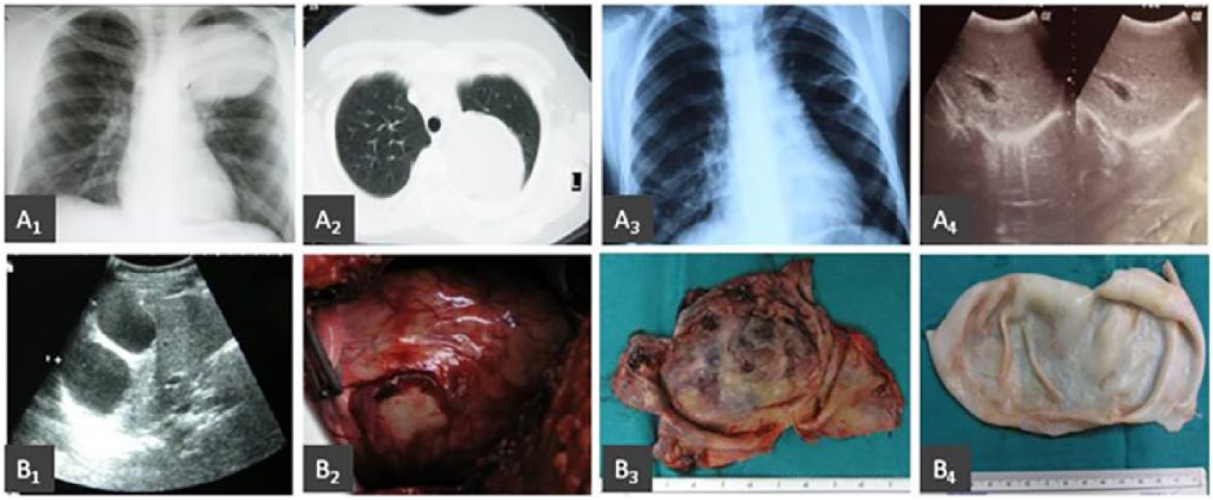
Lung hydatid disease. A1 to A4: echinococcal cyst of the left superior pulmonary lobe; cystectomy with Barret technique. A1: preoperative thoracic plain X-ray, reveals a spherical shadow in the left superior pulmonary lobe. A2: preoperative CT imaging of the cyst presented as a circular hyperdense lesion. A3, A4: postoperative plain X-ray and ultrasound (US) imaging, after cystic excision with pulmonary reconstruction leaving insignificant residual air cyst. B1 to B4: right lung segmentectomy of echinococcal cyst located in the right inferior pulmonary lobe. B1: preoperative US imaging of doubled spaced right pulmonary echinococcal cyst. B2: intraoperative image of segmentectomy “en bloc” with the doubled spaced cyst. B3: pathology specimen of pulmonary segmentectomy “en bloc” with the cyst. B4: pathology specimen of the echinococcal cyst.
These techniques are particularly popular and safe when they are combined with classic monopolar and bipolar diathermies and, later, other methods, such as radiofrequency intraoperative USs, argon, and so on, which are now the methods of choice.
Liver
(a) Simple cystectomy + Peritoneal fixation: This technique was used in 17 cases (21.79%), followed by all previous surgical times of the cystectomy. This technique was applied in cases where the cyst was intraparenchymal but closed to Glisson’s capsule. However, no external drainage was placed in the cystic cavity. Then, the roof opening is fixated on the posterior abdominal wall along the surgical incision in selective cases (segments II, III, V, VI). We performed this technique to control a potential postoperative bile leak or abscess formation.
(b) Cystectomy + Partial pericystectomy + Peritoneal fixation: This technique was applied to the majority of liver Echinococcus, in 52 cases (66.67%). This is an accepted surgical method, which is used today. This technique is characterized by cystectomy and partial pericystectomy. During partial pericystectomy, a large portion of the anterior surface of the fibrous reactive capsule is removed. This results in a relatively large reduction of the residual cavity. The cavity is then closed only externally on the front surface, with a continuous absorbable suture, without external drainage, as we previously mentioned. Then, the roof opening is fixated on the posterior abdominal wall along the surgical incision (Figure 4).
(c) Cystectomy + Partial pericystectomy + Peritoneal fixation + Cavity drainage: This method was applied in 5 cases (6.41%) associated with cystectomy and partial pericystectomy. Because of the obscure bile leak, the remaining residual cavity was externally drained by the insertion of a specific catheter, and closure of the cystic roof next to the catheter was carried out. Subsequently, we sutured the cystic roof as previously mentioned.
(d) Cystectomy + Partial pericystectomy + Epiplooplasty: This method is not used today but has been applied in our 4 earlier cases (5.13%) during the first decade of this study. Technically, the method consisted of cystectomy and suturing a portion of the greater omentum in the pericystic opening, within the residual cavity.
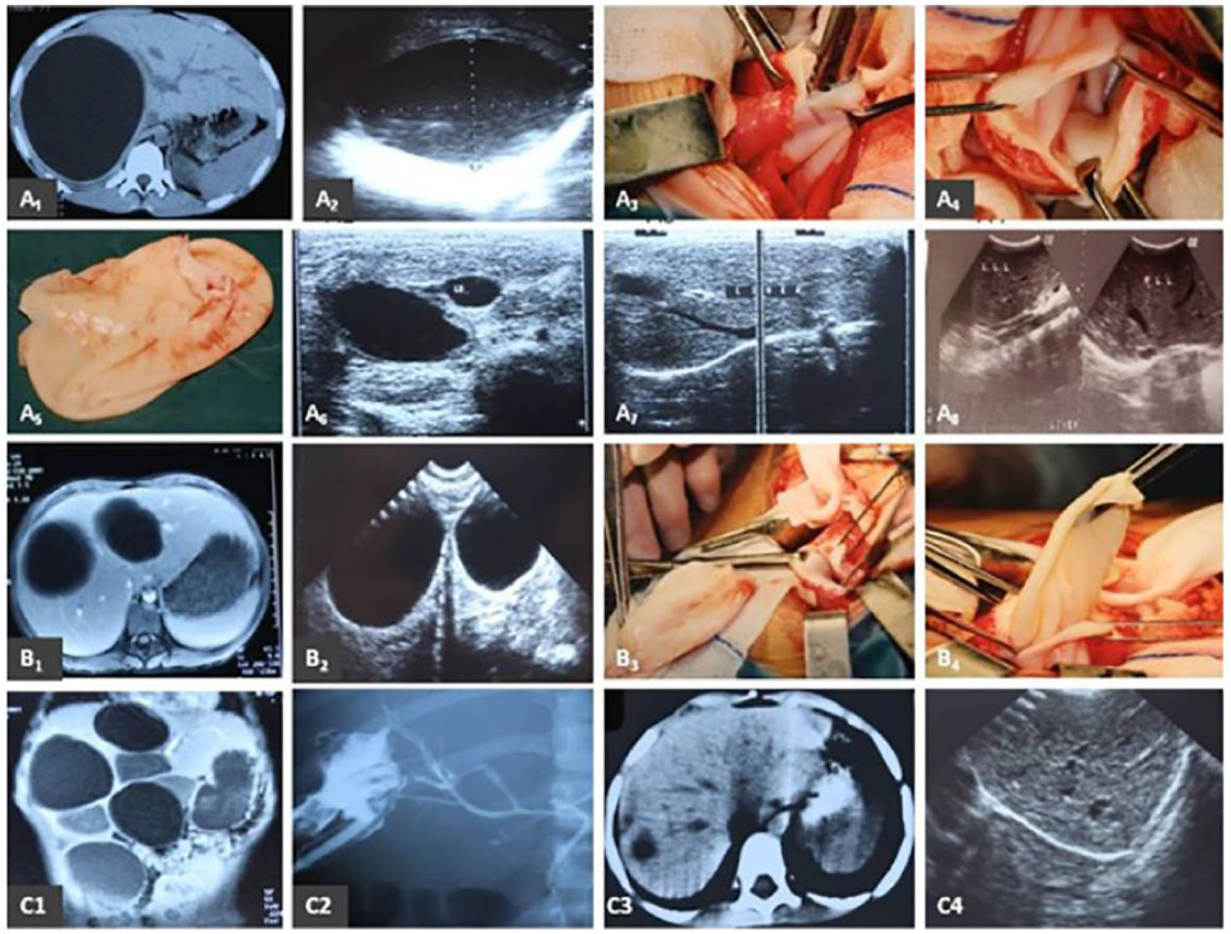
Liver echinococcosis of solitary, double and multiple localization. A1, A2: Computed tomography (CT) and ultrasound (US) appearance of enormous hydatid cyst, which occupies almost the whole right hepatic lobe. A3, A4: Intraoperative imaging of echinococcal pericystic cavity after cystic removal. A5: Echinococcal cystic surgical specimen. A6, A7, A8: Consecutive US images reveal gradual postoperative disappearance of the residual cavity. Simultaneously, there is a remarkable restoration of the liver parenchyma. B1, B2: CT and US preoperative imaging of right lobe echinococcal cyst with double localization. B3, B4: Intraoperative appearance of double cystectomy after cystic aspiration and deroofing. C1: CT appearance of multiple liver echinococcosis. C2: Surgical removal of the cysts at the same time. Postoperative bile leak and fistula are imaged in a fistulogram via the drain. Second operation managed to close the fistula. C3, C4: Postoperative CT and US imaging notifies complete restoration of liver parenchyma.
Other locations
(a) Diaphragm: This location is very rare and occurred in only 1 of our cases. To deal with it, low left thoracotomy was performed, and the Echinococcus cyst was removed with a small incision of the diaphragmatic surface (en bloc). There was a reconstruction of the diaphragm, with special sutures and Büllow placement (Figure 5).
(b) Peritoneum: There was 1 patient with multiple cysts that were completely excised by 1 laparotomy, and multiple cystectomies were performed. 25
(c) Bone: This was a case of Echinococcus located in the bones of the right upper limb, where complete cystic removal (en bloc) was performed without opening it.
(d) Spleen: This site was present in 2 cases, which were surgically treated with laparotomy. In the first case, cystectomy (puncture etc) and closure of the reactive capsule were performed, whereas in the second case, splenectomy was performed because of the large cystic size.
(e) Kidneys: This site was the most common and involved 4 cases. The operation was performed with a lumbar incision to access the retroperitoneal space. Then, in 2 cases, complete cystic excision (en bloc) was performed with partial nephrectomy, whereas in 2 other cases, cystectomy (puncture etc) and closure of the residual cavity were performed.
(f) Bladder: It included a case where laparotomy was performed for complete (en bloc) cystic excision.
(g) Multiple locations: They involved a total of 3 cases, of which 2 had synchronous localizations in the lung, liver, and peritoneum. In these cases, our patients underwent simultaneously thoracotomy (Barret technique), laparotomy with cystectomy and partial pericystectomy of the hepatic Echinococcus, and finally, multiple cystectomies of the peritoneal cavity. In the third case, there was multiple localization in the lung, liver, and spleen. The young patient underwent simultaneously a combined thoracotomy (Barret technique) and laparotomy, for hepatic cystectomy, and partial pericystectomy and splenectomy, for splenic echinococcosis (Table 2).
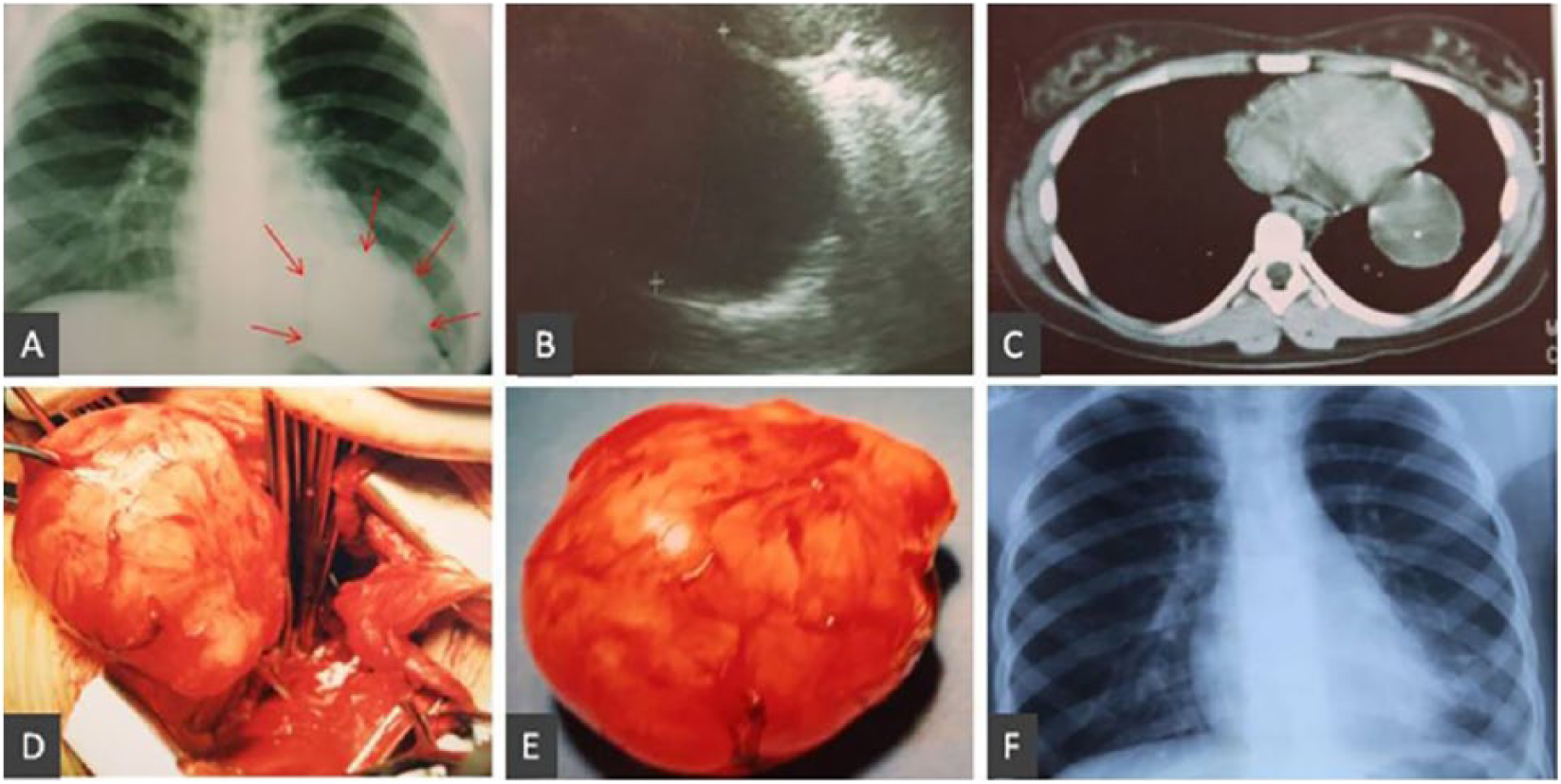
Diaphragmatic localization of echinococcal cyst. A. Preoperative X-ray imaging of echinococcal cyst localized in the left diaphragmatic crus. B. Ultrasound imaging of echinococcal cyst. C. Computed tomography appearance of the diaphragmatic cyst. D. Intraoperative image of cystic mobilization and excision. E. Surgical specimen of the cyst. F. Postoperative X-ray imaging with complete pulmonary reconstruction.
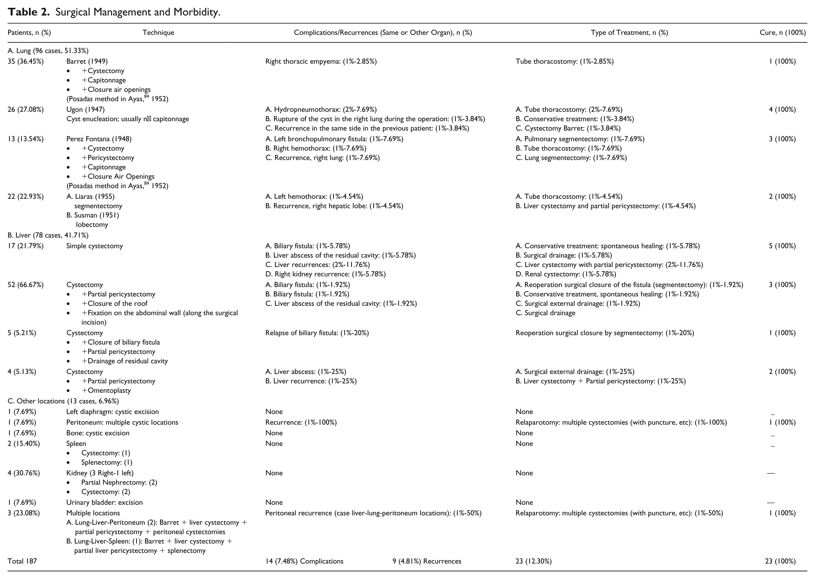
Surgical Management and Morbidity.
Statistical Analysis
Statistical analysis was performed for morbidity related to the different surgical methods to compare their efficacy in our young patients. The χ2 Monte Carlo test was used for categorical variables consisting of complications and recurrences for surgical methods that were used for hepatic and pulmonary echinococcosis. SPSS 23 (Statistical Package for the Social Sciences, version 23.0, SPSS Inc, Chicago, IL) was used for statistical analysis, and P <.05 was considered significant.
Results
The overall results of our cases are discussed in what follows.
Laboratory Findings
For the diagnosis of echinococcosis, chronologically, the following laboratory methods were used:
The initially used laboratory tests were Casoni’s dermoreaction and Weinberg reaction. These were positive in our cases, with percentages of 65.9% and 69.4%, respectively. However, because of the large number of false positive and false negative results, we have not used them in the past 2 decades.
Other newer methods, the indirect hemagglutination assay and the LAT, were positive, respectively, in 74.7% and 77.3% of our cases. Nevertheless, there were instances when they were associated with misleading diagnoses.
Subsequently modern diagnostic methods, such as ELISA and PCR, had high sensitivity and specificity rates: >90% of our cases. ELISA was positive in 91.5%, in a group of our cases, whereas PCR was applied to a small number of patients and was positive in about 100%.
The blood test for eosinophilia was found to be approximately 12% in our cases.
Radiological findings: The initial use of simple chest X-ray or abdominal chest X-ray imaging was valuable in the early diagnosis of echinococcosis. However, in the past 4 decades, US, CT, and more recently, MRI scanning have been highly specific and sensitive examinations, with diagnostic accuracy in approximately 90% of our young patients.
Therapeutic Results
The early postoperative results were completely satisfactory. Of the 187 cases, 173 (92.52%) were uneventful. However, 14 cases (7.48%) presented with various complications during the immediate postoperative period and were successfully treated.
Complications
Analytically, in the group of 14 children, the following complications were registered: 3 patients were surgically treated with various techniques; 1 bronchopulmonary fistula was excised by segmentectomy; and 2 with biliary fistula were treated by hepatic segmentectomies. Also, 8 cases underwent minimal intervention, with 5 thoracostomies (Büllow) and 3 liver abscess drainages. Ultimately, 3 patients (2 with biliary fistula and 1 with pulmonary spontaneous cystic rupture) were treated conservatively and did not require surgery. However, it should be clarified that in 1 case with spontaneous rupture of the echinococcal cyst during the operation, it was treated conservatively, by flushing the thoracic cavity with a solution of NaCl (15%). Later this patient presented with recurrent echinococcosis in the same lung.
The 3 cases with liver abscess were treated with minimally invasive methods by external drainage of the abscess. This procedure was easily performed because of our previous intraoperative technique of fixation of the cystic roof on the peritoneal surface along the abdominal incision. The complications and type of surgery used are outlined in Table 2.
Follow-up
Our patients were followed postoperatively for a period ranging from 1 to 12 years, with an average of 7.25 years. Overall, 9 (4.81%) cases developed recurrence of echinococcosis in the same or another organ. Ultimately, there was complete cure after recurrence management in 178 of our cases (95.19%).
Recurrences
The main recurrences of echinococcosis that developed in our cases are discussed here. Analytically, relapse developed in the same organ in 7 cases—that is, 3 cases in the liver, 2 cases in the lung, and 2 cases in the peritoneum. These relapses occurred within a 3-year follow-up period.
Recurrence of the disease in another organ occurred in 2 cases. Of these, in 1 case with an initial location of the echinococcal cyst in the liver, it reappeared after 2 years in the right kidney. In 1 case with an initial location in the lung, it reappeared in the liver after 3 years. All the recurrences of echinococcosis were successfully treated with a choice of analogous surgical techniques. Thus, the young patients fully recovered and were permanently relieved of hydatidosis (Table 2).
Antiechinococcal Agents
The systematic administration of antiechinococcal agents based on special protocols after the 1980s has contributed to a significant reduction in immediate postoperative complications and in particular recurrences.
Surgical Techniques
A total of 210 types of surgical operations were required to treat 187 cases of E granulosus along with overall complications, recurrences, and multiple locations. The types of operations were as follows: 96 thoracotomies, 78 laparotomies for liver location, 10 for other locations, and 3 cases with multiple locations, which required combined 3 thoracotomies and 3 laparotomies. For the treatment of the complications, a total of 11 surgical interventions were required: 1 was for the lung (thoracotomy), 2 were for the liver (laparotomy), and 8 were for minimal surgical interventions (5 Büllow thoracostomies and 3 drainages of liver abscesses). For 9 recurrences, 4 laparotomies for the liver, 2 thoracotomies for the lung, 2 laparotomies for the peritoneal locations, and 1 lumbar incision for the kidney were required.
Also, the choice of suitable surgical techniques at different intervals had contributed to an even greater reduction in the complications and recurrences of hydatid disease. The results of the surgical techniques in the various organs are as follows:
Lungs
(a) Barrett method: This was applied in 35 cases (36.45%). Postoperative complications were reduced (2.85%), and there were no recurrences.
(b) Ugon method: This was applied to 26 cases (27.08%), with associated high rates of complications in 3 cases (11.53%) and recurrence 1 case (3.84%).
(c) Perez-Fontana method: This was applied in 13 cases (13.54%), with increased complication rates in 2 cases (15.38%) and recurrence in 1 case (7.69%).
(d) Segmentectomy-lobectomy: This was applied in 22 cases (22.93%), with complication in 1 case (4.54%) and hepatic recurrence in 1 case (4.54%).
There was no statistically significant correlation between complications and surgical treatment of pulmonary echinococcosis. Regarding recurrences, the Barret method was superior to other methods, but this was not statistically significant (P <0.529 >0.05 95% CI 95%) Figure 6).
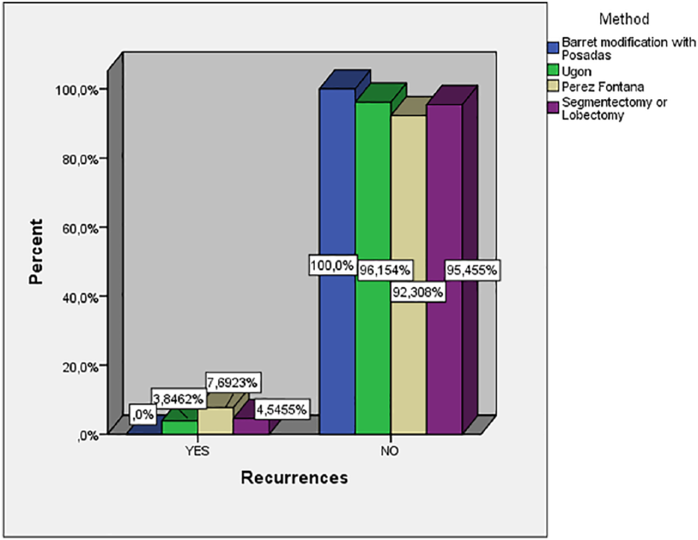
Surgical treatment of pulmonary echinococcosis, recurrences.
There was no statistically significant correlation between complications and surgical treatment of pulmonary echinococcosis. We also need to mention that the Barret technique is the only method without recurrences, whereas Perez-Fontana had the highest rate of recurrences 7.69% (P < 0.507, CI 95%).
Liver
(a) Simple cystectomy + Peritoneal fixation: This was applied in 17 cases (29.79%). This technique showed high rates of postoperative complications in 2 cases (11.76%) and relapses in 3 cases (17.54%).
(b) Cystectomy + Partial pericystectomy + Peritoneal fixation: This method was the most popular and was applied in 52 cases (66.67%). The rate of postoperative complications was 5.76% without any recurrence.
(c) Cystectomy + Partial pericystectomy + Peritoneal fixation + Cavity drainage: This technique was applied in 5 cases (6.41%), all associated with obscure biliary fistula. Four of the cases with bile leak were treated conservatively. In 1 case (20%), because of persistent bile leak, relaparotomy and closure of the biliary fistula were performed, with a small segmentectomy.
(d) Cystectomy + Partial pericystectomy + Epiplooplasty: This technique was applied in 4 cases (5.13%). One (25%) of these cases developed a hepatic abscess, and 1 (25%) developed a liver recurrence.
Ιn liver echinococcosis, there was a significance related to surgical treatment and recurrence, with a small superiority of the (b) and (c) categories, which had low recurrence, whereas high recurrence was seen in the (d) category (25%; P < 0.033 <0.05, 95% CI). Nevertheless, the correlation of biliary fistula and the (c) category was remarkable (P <0.414, 95% CI; Figure 7).
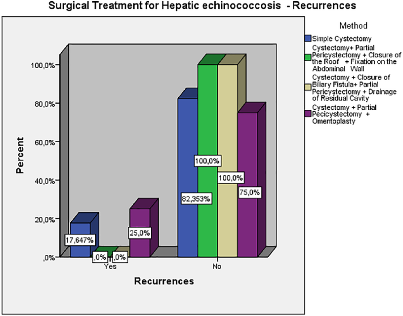
Surgical treatment for hepatic echinococcosis recurrences.
Other Locations
Of the 13 cases, 10 had solitary localization and 3 had 3 synchronous localizations in different organs. Thus, the total number of affected organs was 19. Of the 19 locations, 12 were treated with cystectomies (puncture, etc) and suturing of the reactive capsule. These different locations were in the following sites: 3 in the liver, 3 in the lungs, 3 in the peritoneum, 2 in the kidneys, and 1 in the spleen. The remaining 7 cases of partial or total resection of the affected organs were as follows: 2 splenectomies, 2 heminephrectomies, and 3 complete removal of the cysts (en bloc) from the diaphragm, bone, and bladder (Table 2).
Outcome and Mortality
The overall morbidity in our 178 cases was 12.29%. The mortality among our cases for the period 1970 to 2009 was zero.26,27
Discussion
E granulosus is colonized in the small intestine of dogs and other carnivores. These animals are the main final hosts, which do not develop echinococcosis. In contrast, herbivores are the intermediate hosts, which develop the disease. Human beings are the accidental intermediate hosts who develop the disease.
Transmission of hydatidosis occurs in the manner described below. The embryonated eggs of the parasite originally spread in the primary host’s digestive tract and are then excreted into the environment through the feces. Subsequently, they are recruited through the flora of the various herbivores. Following consumption, the embryonated eggs, under the influence of gastric juices, are transformed into hexacanth embryos or oncospheres. The hexacanth embryos are then transferred into the small intestine and adhere to the enteric mucosa, which they penetrate. They then enter the circulation through the capillaries and are transported and disseminated in the various organs of children, mainly in the lungs and secondarily in the liver, whereas the opposite is the case in adults. Rarely, there is a parasitic spread in other peripheral organs, such as the spleen, peritoneum, kidney, diaphragm, heart, bones, brain, and so on. After migration to the various organs, they are transformed into mature metacestode parasites (echinococcal cysts) in a period of several months or years. 28
The tissues of the host’s affected organs during the formation of the echinococcal cyst undergo direct pressure and displacement while at the same time being accompanied by a granulomatous reaction involving the development of fibrous and stromal tissue, particularly peripherally of the echinococcal cyst, and sometimes present with necrotizing regions. 28 There is also a remarkable immunological and histological response to echinococcosis. Thus, in most patients with hydatidosis, there is activation of interferon γ, interleukins (IL-4, IL-5, IL-6, and IL-10), and T helper (Th) cells. Particularly Th1 cells have a protective role against parasites and Th2 lymphocytes in the susceptibility of the host against hydatid disease.28,29
The wall of the Echinococcal cyst consists of the following 3 layers:
(a) a thin germinal epithelial layer, which, after the germinal epithelium is folded, initially induces germinal cysts or brood capsules containing protoscoleces or heads of the tapeworms;
(b) a thick multilayer intermediated laminate (laminar layer or membrane) located outside the germ; this layer does not contain cells but consists of multiple layer laminates rich in amino acids and carbohydrates that form the outer wall of the parasite; and
(c) a thick fibrous reactive capsule or adventitial or pericystic layer, which is a local reaction of the physiological tissues of the affected organ in a defensive effort to reduce the echinococcal cyst.30,31
Usually, the prevalence of echinococcosis in endemic areas is 50 cases per 100 000 population/year. In particular, in Peru, Argentina, East Africa, Central Asia, and China, echinococcosis can increase by a percentage of 5% to 10%. 32
In the Mediterranean Basin, annual incidence rates for human cystic echinococcosis of 4 to 8 per 100 000 have been recorded. Analytically, the incidence rate in France is <0.28 per 100 000, Spain 1.1 to 3 per 100 000, Morocco 4.5 per 100 000, and Algeria 3.6 to 4.6 per 100 000 population. 4 Specifically, the prevalence of echinococcal cyst in Greece 50 years ago was a serious problem because the incidence rates in the population were very high, in proportion to the level of echinococcosis worldwide. Thus, between 1967 and 1971, the incidence of echinococcosis was 14.8 cases per 100 000 population. However, since 1984, after severe preventive measures, the incidence of echinococcosis in Greece has reduced more than 100 times, according to the statistics of the Hellenic Ministry of Health (2011). 33 Analytically, the incidence rate between 2000 and 2009 varied from 0.05 to 0.37 cases per 100 000 population, whereas the average incidence of echinococcosis was 0.14 cases per 100 000 population. This percentage was 0.08 per 100 000 population for children <14 years old (Figure 6).33-35
It is important to note the following important preventive measures, which have shown promising results:
(a) sheep vaccination with the recombinant vaccine EG 95 offers 99% elimination of the viable parasitic cysts (MORO) 5 ;
(b) vaccination of dogs with soluble native proteins isolated from protoscoleces of E granulosus using recombinant proteins derived from a developmentally regulated gene family (egM) because 3 genes—egM 4, egM 9, and egM 123—associated with a very high level of protection (97%-100%) interfere in worm growth by suppressing egg development and embryogenesis 36 ; and
(c) an alternative effective preventive model against echinococcosis is a combination of sheep vaccination and dog anthelmintic treatment offering 96% to 100% protection against E granulosus (MORO). 5
Our data of 187 cases in comparison with international literature are as follows:
Gender: Among the overall cases, 54% were boys and 46% were girls. According to the international literature, there are controversial data, but usually, the parasite appears more often in boys.17,22,37
Age: Occurrence of echinococcosis ranged from 2 to 14 years, with an average of 9.2 years. The most common was between 7 and 13 years of age, with a large number of cases (41 cases, 22%) at the age of 10 years. Our youngest patient was 23 months of age, whereas the Greek bibliography reported an echinococcal liver cyst in a 17-month-old infant. Our previously mentioned data are consistent with the international literature.17,22,38,39
Localization: Regarding the location of the parasite in the various organs, in our cases, the most frequent location was the lungs in 96 cases (51.33%), followed by the liver in 78 cases (41.71%), and other rare locations in 13 cases (6.96%). In adults, hepatic location is dominant. According to the literature, the primary location of the echinococcal cyst in children is in the lung, at a rate of 47% to 65%. 17
Affected Lung
The most frequently affected lung was the right one, at 58.33%, and the most common lobe was the lower right, in 41 cases (73.21%). These findings are consistent with the data of the medical literature (Table 1).39-41
Combined Localization
This group had 6 cases (6.25%), and there was combined localization in the liver and lungs. This percentage is comparatively lower than that in other studies—16% in children, whereas adult rates are usually <4%.42-46
Unilateral localization: The unilateral placement in the lungs was very high, at 96.88%, whereas in the international literature, it is about 86%.47,48
Bilateral localization: Bilateral pulmonary localization was recorded in 3 cases (3.12%). There are bibliographic reports in children where the rates are disproportionately higher: 15% or 18%.41,49
Explanation of Frequent Location
To explain the more frequent location of Echinococcus in the lungs, compared with the liver, in children, several theories have been developed over the years: In one hypothesis it is reported that, the hexacanth embryo is transferred directly into the lung, through the middle and lower hemorrhoid venus plexus, which directly drains to the vena cava. Another prevalent point of view is that the large space of the arteriovenous sinuses of the liver in children allows easier passage of the hexacanth embryo toward other sites and particularly the lungs.21,22
Size of Echinococcal Cyst
Echinococcal cysts in the lung are usually of a larger size than those in the liver in children. A possible explanation for this is the high elasticity and low lung compliance in these studies, which justifies the more rapid development of Echinococcus cyst in the lungs compared with the liver. So this rapid growth rate of pulmonary hydatidosis in children is 5 times more than in hepatic echinococcal disease.21-23,39-41
In detail, the locations of the 78 cases of hepatic Echinococcus are the following:
Right lobe: 48 cases (61.55%)
Left lobe: 24 cases (30.77%)
Bilateral lungs: 2 cases (2.56%)
Multiple: 4 cases (5.12%; Table 1)
Other rarer locations are as follows:
Main Symptoms
In our cases, echinococcal cysts were asymptomatic: 51 cases (53.12%) with lung locations, 33 cases (42.3%) with liver cyst location, and 4 cases (30.77%) at other sites. The symptoms of a noncomplicated echinococcal cyst are usually mild or nonexistent.
The residual symptoms of lung echinococcosis were the following: cough occurred in 23 cases (23.95%), chest pain in 14 cases (14.58%), fever in 11 cases (11.46%), dyspnea in 6 cases (6.25%), and hemoptysis in 3 cases (3.12%); hydatidemesia occurred in 1 case (1.04%) and hydrothorax and thoracic empyema in 2 cases (2.08%; Figure 2A).
In contrast, complicated cysts usually have more pronounced symptoms. In our cases, complicated cysts were few (3%-1.6%), with much more severe symptoms. Symptoms are consistent with the location of the cysts. Chest pain is reported in 57% of cases of pulmonary localization, whereas in other studies phlegm and dyspnea have the highest prevalence.47,48,50
Symptoms, such as fever, dry or productive cough, chest pain, dyspnea, and hemoptysis occur in complicated cysts. Vomiting a large amount of clear fluid, rich in NaCl (hydatidemesia), or fragments of echinococcal cystic wall (“grape skins”) characterize a complicated tapeworm cyst, which is usually accompanied by bronchial erosion and communication. The above symptoms are accompanied by open bronchial communication, which is considered to be a precocious echinococcal cyst. In the literature, high rates, of up to 30%, of spontaneous rupture of the Echinococcus cysts of the lung are reported, which was not confirmed in our series. 47
Localization of the parasite in liver echinococcosis is usually in the right liver lobe. 17 Thus, in our 78 cases, 48 cases (61.55%) were located in the right lobe and 24 (30.77%) in the left lobe. Also, 2 cases (2.56%) of Echinococcus cysts were located in both lobes, and in 4 cases (5.12%), multiple echinococcal liver cysts were found. To our knowledge, there is no bibliographic report with simultaneous bilateral (2 locations) or multiple (3-6) liver localizations. 39
A rare complication of the echinococcal cyst has to be mentioned. This consists of erosion of the bronchial tree and causes a spontaneous rupture inside the lung, resulting in the formation of the biliobronchial fistula. This is characterized by the abnormal excretion of bile (bilioptysis) in the bronchial tree, although in our series, such a case did not occur. This complication is observed in the cysts found in the dome of the liver; both the pressure and the irritant effect of bile are responsible for septal rupture and bilioptysis.47,48
In the frequency range, the main symptoms of the echinococcal cyst in our 78 cases were the following: 43 cases (55.13%) had a palpable mass or swelling of the liver (the main symptom), 7 cases (8.97%) had fever, 5 cases (6.41%) had intense abdominal pain, and 1 (1.28%) was jaundiced. Abdominal pain in 73% of some studies and palpable mass in other recent studies are main symptoms of liver localization (Figure 2B).47,48,50
The other sites of the echinococcal cyst were rare and consisted of 6.96% (13 cases) of our 187 cases. These symptoms were varied, depending on the infected organ. Overall, the main symptoms of the 13 cases, single and multiple sites, were the following:
Asymptomatic: 4 cases (30.77%; 1 diaphragmatic single, 1 pulmonary multiple location, and 2 renal single sites)
Peritoneum: 3 cases (23.08%; 1 single and 1 multiple—ascites 3 and abdominal distension 3)
Liver: 3 cases (23.08%; multiple localization—liver swelling 2, liver mass 1, abdominal pain 1)
Spleen: 3 cases (23.08%; 2 single sites and 1 multiplex splenic)
Kidney: 2 cases (15.38%; 2 single—hematuria 1, pain 1, and swelling 1)
Lungs: 2 cases (15.38%; 2 multiple locations—cough 2, fever 1)
Bladder: 1 case (7.69%; 1 single—swollen lower abdomen)
Bone: 1 case (7.69%; 1 single—swollen soft tissue of upper extremity; Figure 2C)
Generally, to exhibit symptoms from the various organs, an echinococcal cyst should be >5 cm. But children are usually treated with cysts of >10 cm in diameter. In our cases, the echinococcal cysts were large, and their diameter ranged from 7 to 38 cm. Of these, 19 (10.16%) had a diameter of <10 cm, whereas the usual diameter was >15 cm. 47
Laboratory Diagnosis
Previously, laboratory examination of echinococcosis, was based on Casoni’s dermoreaction and Weinberg reaction. These, although positive, were 65.9% and 69.4% in our cases, with high rates of divergence—18% to 45% and 28%, respectively. For this reason, they are currently not used. In addition, the indirect hemagglutination test was positive in 74.7% of our cases, and the LAT was positive in 77.3% of our cases. The aforementioned examinations show lower rates of false results, corresponding, respectively, to 1% to 2% and 5% of the cases; the sensitivity of these methods has been determined to range from 66% to 100%.
A WHO study (2002) reported that the sensitivity of the LATEX method was 58% for the lungs, 80% for the liver, 88% for the combination liver/lungs, and 57% for the other sites. However, for the indirect hemagglutination assay, sensitivity ranges from 80% for the liver, 61% for the lungs, 90% for the combination liver/lungs, and 57% for the other sites. 31 However, nowadays, ELISA is widely used and has a sensitivity of 80% to 99% and a specificity of >60%. This method was positive in 91.5% of our cases. 51 The WHO (2002) reports that the IgG ELISA sensitivity for the liver is 93%, for the lungs 83%, for the liver and lungs 96%, and for the other sites 93%. Whereas other authors report that immunoelectrophoresis has a sensitivity of >70% and 100% specificity. 51
The double diffusion test (DD5) is considered a method that is most susceptible to immune electrophoresis. A specific confirmation can be obtained by demonstrating echinococcal antigens by immunodiffusion (arc 5) procedures or immunoblot assays (8-, 21-kD bands).52,53
However, today’s most recent and sensitive method of diagnosis of echinococcosis is the PCR test, which is the most reliable diagnostic method with 100% sensitivity and specificity. In our cases, PCR was applied to a relatively low number of small patients, with sensitivity in 100% of cases. 54
However, ELISA-specific antibodies cannot be detected in 20% of the liver and 40% of lung echinococcal cysts because of partial or completely calcified cysts that combined with a low level or absence of such antibodies.26,27,55 The ELISA assay diagnosis is based on the determination of specific antiechinococcal antibodies (IgG1, IgG4, Arc5, and Ag5).31,56
As regards a comparison of the 2 methods, these antibodies may be few, especially in young children with echinococcosis, whereas in difficult conditions, PCR can elucidate the parasitic factor.26,27,57
Echinococcosis is usually accompanied by eosinophilia (>5%-8%) of leucocytes in 20% to 34% of cases. In our cases, presence of eosinophilia was 12%. While finding leukocytosis, with elevated neutrophil polymorphonuclear leukocytes, is usually indicative of the echinococcal cyst infection. 58
Radiological studies of echinococcosis were previously based on simple X-rays, mainly of the chest and, second, the abdomen. Especially for the chest, simple X-rays can show several cysts resulting from echinococcosis. These cysts are usually large, single, or multiple and are located in one or both lungs. Usually, the wall of these cysts is smooth, and in some cases, it is possible to find calcifications of the wall of the cysts. All these bulges should be differentiated from other cystic or solid formations of the lungs. 59
The main pathognomonic signs of cracked or complicated Echinococcus cysts of the lung in children are the following: notch, water lily, combo (point of 2 layers of air) or onion peel, annular solar eclipse, meniscus, or air crescent (point of the meniscus or half crescent). The above radiographic images are usually found in complicated, cracked echinococcal lung cysts. 60
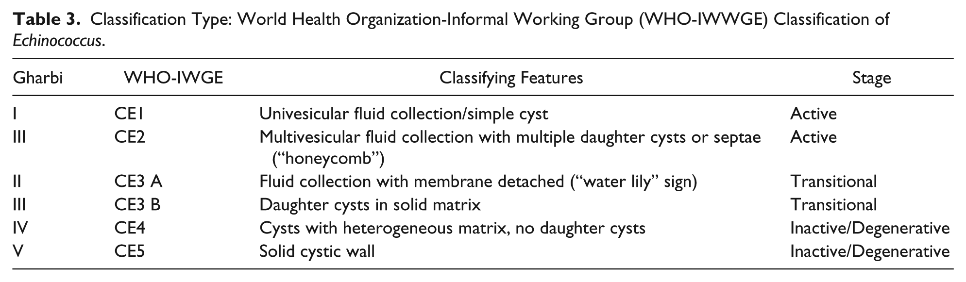
Abdominal radiography can also reveal large swelling of the liver, compression of the intra-abdominal viscera, deformation of the right crura, calcifications, and so on. However, after ultrasonography of the upper and lower abdomen was used, the early diagnosis of echinococcosis was easier, and this technique yielded a lot of data on parasitosis. The WHO (2001) classified the US liver echinococcosis into 5 categories, depending on the imaging findings (Table 3). 61
Classification Type: World Health Organization-Informal Working Group (WHO-IWWGE) Classification of Echinococcus.
Another comparative study between imaging modalities reveals that US and especially microbubble contrast-enhanced ultrasound (CEUS), has a 90% specificity and is superior to CT and MRI. In addition, CEUS is a valuable preoperative diagnostic tool when used in conjunction with CT scan. 62 CT liver imaging appears as usual with large hydatid cysts, averaging 5 cm in size, and may possibly grow 2 to 3 cm per year. These lesions may appear as simple cysts, but sometimes they may contain intracystic membranes or calcifications. The latter reflect the immune reaction of the host against the echinococcal hepatic invasion. In addition, CT of the lung reveals an area with circular peripheral halo in pulmonary echinococcosis. 63 Because complete calcification or disappearance of the cysts can reveal parasite death, CT is superior to MRI as a follow-up imaging modality. 64
According to the literature, indications for surgical treatment are analogous to symptoms and cystic characteristics. The main symptoms in children are abdominal pain, shortness of breath, fever, jaundice, or heart failure. According to the WHO, the cystic clinical features requiring surgical treatment are large hepatic cysts with internal multiple daughter cysts, contaminated cysts, and cysts with bile communication.65-67
Conservative treatment of echinococcosis involves the administration of various antiechinococcal agents, preoperatively or even postoperatively. Mebendazole (MBZ-Vermox) has begun to be administered since 1970. But it is now the second choice antiparasitic medication for children; albendazole is the primary antiechinococcal medication. The mechanism of action consists of inhibiting glucose absorption through the parasitic wall, resulting in consumption of glycogen and in degenerative lesions in the mitochondria and the endoplasmic reticulum. Mebendazole dosage amounts to 40 to 50 mg/kg body weight/d and is administered continuously orally, 1 week preoperatively and 4 to 6 weeks postoperatively, whereas in some cases, it is continued for 3 to 6 months.
The absorption of mebendazole from the digestive system is less than that of albendazole. For this reason, it should be administered for a longer period of time. The side effects are the following: digestive disorders, temporary alopecia, leucopenia, albuminuria, allergic reaction, fever, and elevation of liver transaminases. Contraindications are chronic liver disease, immunosuppression, extraordinarily large cysts, and dehydrated cysts.20,53
Albendazole (Zentel, Eskazole) was discovered in 1972 and, today, is the medicine of choice for the conservative treatment of childhood echinococcosis. Albendazole typically is more easily absorbed by a small patient’s digestive system, which results in high plasma levels of the drug. The mechanism of action of this agent is to reduce the production of ATP in the cytoplasm of the parasite, resulting in great energy loss, immobilization, and death. Albendazole is usually administered to children at a dose of 10 to 15 mg/kg B.W./d in 2 divided doses. The drug should be given simultaneously with fatty meals, and the total 24-hour dose should not exceed 800 mg. The duration of treatment is 4 weeks and is followed by an interval of 2 weeks. Then, it can be repeated to a minimum for 3 cycles and maximum for 6 cycles, usually applied to older children and adults. Undesirable effects and contraindications are similar to those of membendazole.5,20,53,68-70
The 2 aforementioned antiechinococcal agents belong to the benzimidazoles family. Treatment with benzimidazoles is preferred, particularly in patients with multiple locations of Echinococcus cysts in various organs, and small and multiple cysts, and can produce positive results in one-third of cases. Whereas it was reported that in up to 50% of cases, there was a significant reduction in the size of the cysts and symptoms, in 20% to 40% of cases, there was absolutely no response. These cases corresponded to complicated or very large echinococcal cysts or those with daughter cysts or others that had a thick or stratified wall. 53
In cases where albendazole monotherapy fails and particularly in the multiple localization of echinococcosis, the combination of albendazole and Praziquantel (PZQ, Biltricide) is recommended as an alternative treatment. The latter substance is the third antiechinococcal drug, which chemically belongs to isoquinolones and was discovered in the mid-1970s. This is used in difficult cases of echinococcosis during the preoperative period as well as during surgery, when rupture of the Echinococcus cyst occurs in the chest, abdomen, or other cavities. The usual dosage of the drug is 40 mg/kg B.W./1 week or 2 weeks.20,67,71
In reports of adults with solitary cysts, treatment duration never exceeded 1 year because of the possible adverse effects of albendazole. 72 In another study, Demirbilek et al 73 reported a 27% healing rate, with 3 months of treatment with albendazole prior to surgery. Franchi et al 74 reported that after treatment with albendazole, cyst calcification (reflects complete cure) appears between 6 months and 3 years.
A study about the predictive value of localization of hydatid disease in children supports the different responses to treatment of multiple cysts. Thus, peritoneal, pulmonary, and lung echinococcosis have better response to therapy with albendazole followed by surgical treatment, whereas hepatic echinococcosis has the worst treatment response. 64 Finally, administration of albendazole is indicated for children with multiple cysts <5 cm and with locations in multiple organs.62,75
Also, nitazoxanide, discovered in 1980 at the Pasteur Institute, is a thiazolide, which has antisaccharide properties and is used as an alternative in severe cases of echinococcosis as a rescue therapy in combination with albendazole and praziquantel (PZQ, Biltricide). 69
Conservative invasive methods include the percutaneous puncture-aspiration-injection-reaspiration (PAIR) technique, not used in any of our surgical cases. This method includes closed cystic puncture with ultrasonic guidance, aspiration of echinococcal fluid, injection of scolicidal agents, and reaspiration. PAIR could be accompanied by complications such as anaphylactic shock, parasite dispersion (treatment with synchronous albendazole administration), sclerosing cholangitis in the case of bile communication, and systemic metabolic toxicity from hypertonic sodium chloride solution.5,25,51,62
PAIR was first used in 1986, by Tynesian surgeons, as a supplemental therapy associated with albendazole and mebendazole. Nowadays, this method should be followed by treatment with albendazole, 10 days before and 30 days after surgery. 70 This method is used most often for liver cysts, but not for the lungs, heart, brain, and spine. Also, many cases are not reported in children but in adults. 16 Ιndications for PAIR are the following:
A. Anechoic cystic areas ≥5 cm diameter
B. Cysts plus daughter cysts associated with detachment from pericyst
C. Multiple cysts that are suitable for puncture
D. Contaminated cysts
E. Children >3 years old
F. Failure of conservative therapy with antiparasitic agents
G. Surgery contraindicated because of associated diseases
H. Recurrence after surgery
I. Patient’s noncooperation
Subsequently, indications for PAIR include WHO-informal working group on echinococcosis (IWGE) classification CE1 and CE3a cysts (single compartment cysts) <5 cm that have not responded well to medical therapy and in combination with medical therapy for cysts >5 cm (Table 3).26,27
Contraindications for PAIR are the risk of spillage, pulmonary cysts, and inactive or calcified cystic lesions. 20 Other contraindications for PAIR include percutaneously inaccessible cysts; multiple cysts in Guinaud sections I, II, and III; large cysts >5 cm; type I, II, III (by J. H. Charbi WHO classification) nonalveolar cysts; patients who do not wish to undergo surgery; relapse after surgery or medication; cysts communicating with biliary structures; inactive cysts; and complex multiseptated cysts.26,27
In a study by Goktay et al, 76 in 34 children with 51 echinococcal liver cysts, PAIR and albendazole combination were successful, and no recurrence was reported in 97% of cases over a 3-year period.
Surgery is the main treatment for cystic echinococcal cysts. Nevertheless, the administration of antiechinococcal agents in combination with surgery has been used as an effective treatment for cystic echinococcosis. Surgical indications vary with cyst characteristics, including cyst type, location, size, and complications. Surgery may be the best treatment for liver cysts that are secondarily infected or cysts located in the brain, lungs, or kidney. Liver cysts larger than 7.5 cm are likely to have biliary communication; surgery may be the best option for these cysts. 53
In all our cases, both liver and lung cystic puncture were used, with aspiration of the cystic content and infusion of sodium chloride into the cystic cavity. 25 Intraoperatively, large cysts (≥4 cm) can be treated by injection of scolicidal chemical solutions into the cyst, followed by evacuation, prior to further manipulations and extirpation of the cyst. Several scolicidal agents are used during cystectomy (puncture, solution infusion, reabsorption, cystic removal). The most common solution is the NaCl solution, in densities of 5% to 30%. According to our experience, the use of the 15% solution is very important; this causes the least electrolyte disturbances in children. This solution has high osmotic pressure and affects the outer membrane of the protocell, causing it to rupture.26,27,70,77
In our lung cases, between 1970 and 1982, more conservative surgical methods were used in 61 cases (63.54%). After 1982, 35 cases (36.46%) were subjected to more radical surgeries.
The Barrett method, according to the statistical analysis of our cases, has been quite popular in our department for a number of years because it has shown very low rates of postoperative complications and recurrence. The superiority of this method is reflected in the low morbidity and recurrence, even though this was not statistically significant (P < .507; Figure 6).
Thus, the Barret technique refers to a pulmonary hydatid cyst and was initially applied by Barret in 1949. It includes cystectomy and reduction of the residual cavity with special internal absorbable sutures (capitonnage). Moreover using capitonnage accelerates the healing process and causes shrinkage of the residual cavity, preventing empyema and air leak at the same time.
In the appearance of open bronchi, we applied Posada’s technique, by closing the communication with absorbable sutures. The above method was our most popular surgical technique and is considered a more radical method compared with the Ugon technique. We performed this technique in about 37% of our cases, with excellent results and no recurrence rate. Nevertheless, we recorded a complication of thoracic empyema (2.85%), which we treated with a second thoracostomy tube. In our selective cases of pulmonary echinococcosis, the combination of Barret and Posada techniques (along with capitonnage) when air leak is present, is still dominant, with excellent postoperative results.78-80
The reduction of the recurrence according to Nicks is consistent with stringent precautions against spillage and implantation associated with careful bronchial suture and capsular obliteration. 79
The Ugon technique was first applied in 1947 and defined as a technique with pulmonary cystic removal, under positive end-expiratory pressure and without removing the reactive pericyst. Another techniques used as a second priority was the Ugon technique, in which surgeon’s experience and surgical skills are required as well as very good cooperation during the critical phase of cystic delivery, with an anesthesiologist in case of serious risk of an automatic cystic rupture (Table 2). 81
With this technique, there is a risk of rupture of the cyst, resulting in local inoculation of the echinococcal cyst, but is a rare occurrence. Rupture usually occurs during enucleation (in toto) of the cyst when half of it is outside the reactive capsule and there is high endocystic pressure. To select our Ugon series (15.38%), in order to shrink a large residual cavity, we used special internal absorbable sutures (capitonnage). This technique is associated with increased frequency of cystic rupture and subsequent recurrence (3.84% of our cases). We also have recorded 2 (7.69%) cases with hydropneumothorax. Even though there is bibliographic report of delay in lung recovery after the Ugon method, we did not notice that in our cases. 82
The Perez Fontana technique (1948) consists of the Barret plus pericystectomy and occlusion of bronchial orifices. The excision of the pericyst is unnecessary nowadays because the latter is part of the host and not of the parasite. On the other hand, the Perez Fontana technique is associated with a high rate of complications (15%), which can be justified by pulmonary pericystectomy. This method was applied to a smaller number of patients (13.54%), but it was a much more radical surgery compared with the previous ones to treat the echinococcal cyst. Its initial disadvantage was the relatively high rates of postoperative complications and recurrence rate, as also in our cases (7.69%, P < .507; Figure 6).82,83
Segmentectomy or lobectomy to the lung are 2 radical techniques for complicated cysts and substantially improve the treatment of young patients, with low morbidity (4.54%), recurrence (4.54%), and hospital stay and with a better outcome. According to the literature, there is debate about the effect of radical surgery on a child’s development. In our experience, there was no effect of radical lung surgery on development20,51(Figure 3).
The overall complication rate following pulmonary hydatidosis in our cases were hydropneumothorax, hydrothorax, bronchopleural fistula, and thoracic empyema was 7.29% (7 cases). Additionally, the overall recurrence rate was 3.12% (3 cases). Bibliographic morbidity in pulmonary echinococcosis varies from 2.19% to 2.54%. This morbidity was recorded in the majority of the adult population in contrast to our study, which involved children. 48
Hepatic echinococcosis, which includes cystectomy and partial pericystectomy with abdominal wall fixation (in total 61 our cases), was the mainstay of our technique, with very good final results. It is noteworthy that omentoplasty, performed as complementary surgery in hydatid liver disease in adults, is not so popular in childhood echinococcosis because of its effects on childhood physical development (Table 2).23,26,27
In our cases of liver echinococcosis, neither segmental nor liver lobectomy was performed because they both have increased morbidity and have the same clinical effects as the above-mentioned conservative surgical technique (Table 2). With regard to liver cysts, large size is more common, and a radical treatment such as cystectomy and partial pericystectomy is more appropriate for their elimination. Nevertheless, as regards the correlation with biliary fistula, it perhaps could be explained by the surgical component of pericystectomy, which increases the morbidity by opening fistulous tracts from the cut liver surface (P < .414, 95% CI). Moreover, anchoring the cystic wall on the peritoneal surface is the additional technical step that was introduced by our department in order to control postoperative bile leak or fluid collection22,24,26,27,38,79,82,84 (Figure 4).
Liver echinococcosis treatment by total cystectomy was accompanied by high morbidity(12%) in the literature. 38 Our records suggested that the most prevalent hepatic surgical technique is partial pericystectomy and abdominal wall fixation, which is associated with high therapeutic efficacy and less morbidity (in 61 cases, 6.6%; Figure 7). Additionally, our overall recurrence rate was approximately 4.81%(the last 2 decades were zero), whereas bibliographic recurrence reports were 2% 38 .
It should also be noted that removal of part of the liver is not considered necessary, because the results are positive in almost all cases. Also, our reported technique can be applied in cases with multiple echinococcal liver cysts at 1 year, which has been successfully applied. Based on our experience, we provide our proposed algorithm for the diagnosis and treatment of pulmonary and hepatic echinococcosis in children (Figure 8).
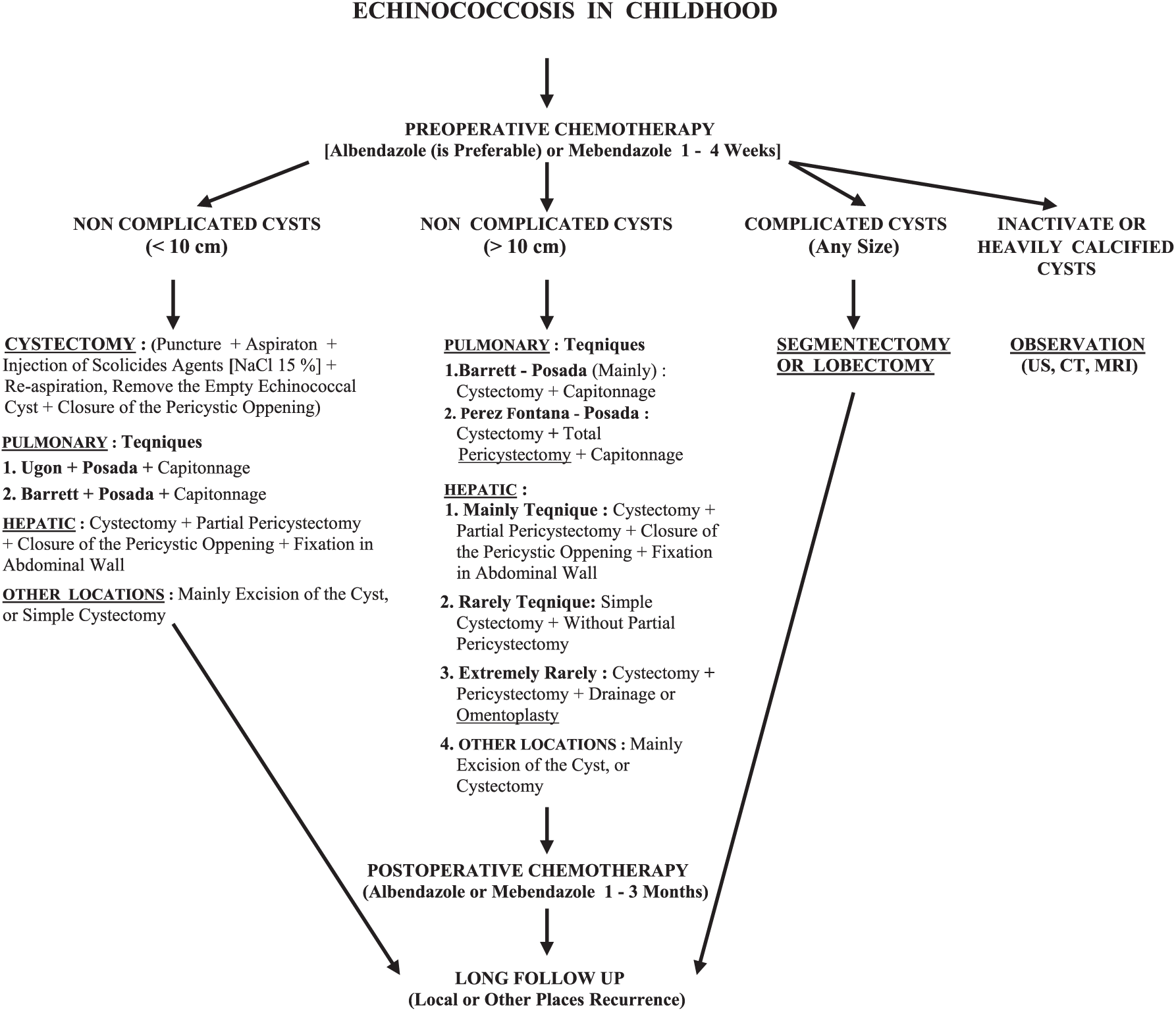
In this algorithm, we recommend that appropriate surgical techniques be chosen according to the cystic size. For example, the Barret technique can be used for cysts smaller than 10 cm because rupture and dissemination is more likely in larger cysts. We believe that the Barret technique has great cystic elimination efficiency with a cystic diameter >10 cm but is less satisfactory in cysts <10 cm. As regards the latter, Barret with Posadas modification is our preferable method. In addition, the Ugon technique is more applicable to cysts <10 cm. For these small cysts, enucleation is technically feasible, whereas in larger cysts, the likelihood of rupture and anaphylactic shock increases. On the other hand, Perez Fontana is more radical than the 2 previous methods because it includes a total pericystectomy and closure of the bronchial orifices. Perez Fontana is indicated for large cysts >10 cm but is associated with more complications. Because pericyst is a result of host reaction, its removal is sometimes responsible for these complications.
Finally, regarding other locations, in 5 cases, we performed cystic excisions (without puncture etc) en bloc with the affected organ. Moreover in 1 case, complete organ excision (splenectomy) was performed. Additionally, in 7 cases, our patients were treated with cystectomy (puncture, etc). Overall, all the aforementioned cases were associated with cure. Especially, for the patient with multiple echinococcosis of the peritoneal cavity, multiple cystectomies (puncture, etc) were required. However, 14 months later, this patient presented with recurrence and was treated successfully with surgery.
Our overall mortality rate was zero. According to bibliographic reports, overall operative mortality in pulmonary echinococcosis is 1% to 2% and liver echinococcosis is 1%. 38
Conclusions
It is very important to mention the significant reduction in the incidence of echinococcosis in Greece, due to special preventive measures as it is recorded from the Hellenic Ministry of Health-Center for Disease Control & Prevention (H.C.D.C.P) 2011 (Figure 9). 34
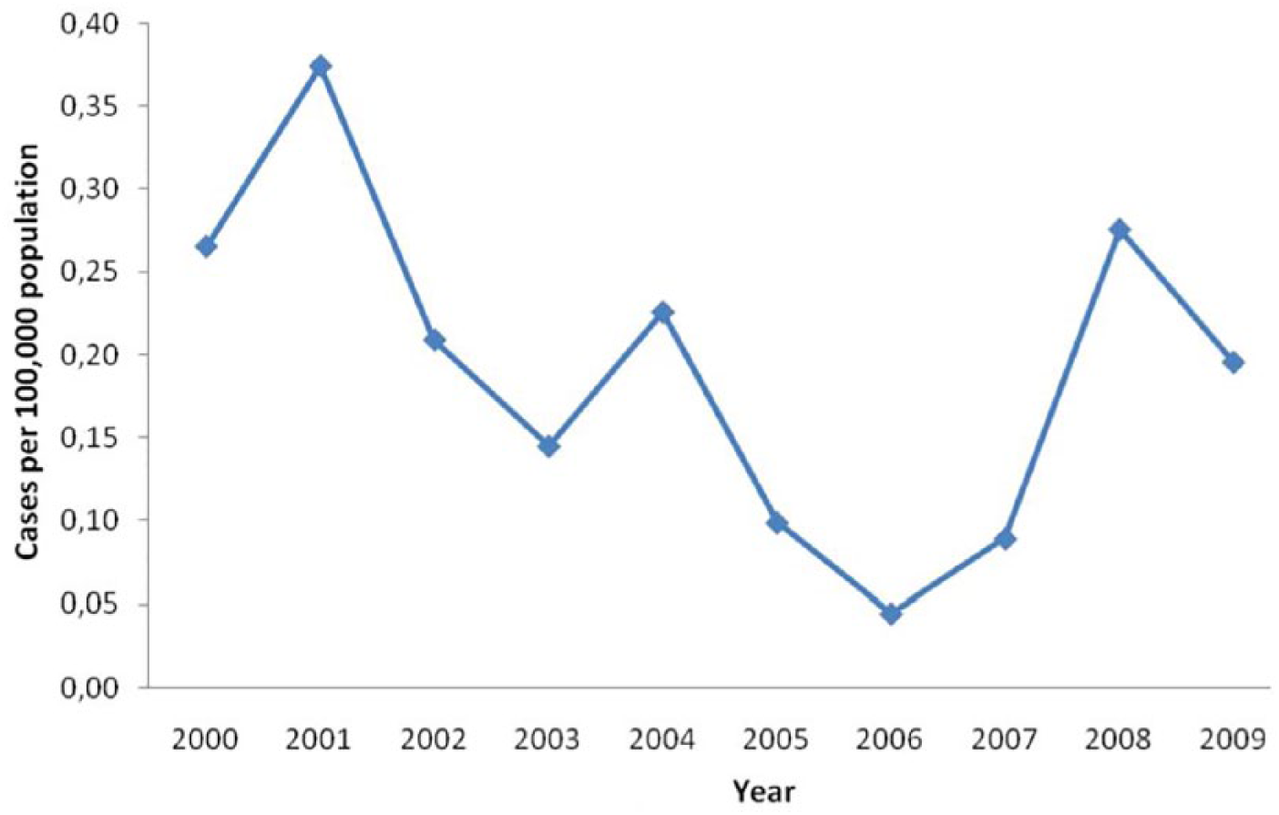
The overall incidence of echinococcosis for all ages, in Greece, per 100.000 Population, during the years 2000-2009, has decreased recently [Hellenic Ministry of Health - Center for Disease Control & Prevention (H.C.D.C.P), 2011]. 34
It is remarkable that there was no mortality recorded in all our aforementioned cases. The overall complications and recurrences were managed with additional minor surgical interventions, with ultimately successful results.
The Barret technique, according to our experience, is the procedure with a higher therapeutic efficacy, whereas Ugon demands anesthesiologist’s cooperation and is suitable for small-sized echinococcal cysts. On the other hand, avoidance of total pericystectomy (Perez-Fontana) minimizes possible complications. More radical operative techniques such as segmentectomy and lobectomy are associated with enhanced healing process, and they are recommended for complicated cysts, according to our experience.
For liver hydatid disease in children, the suggested method is partial deroofing of the cyst, closure of biliary openings, and anchoring of the residual cystic wall on the peritoneal wall for postoperative control of bile leakage or management of contamination (liver abscess or bile leak). This technique was introduced by the Greek pioneering pediatric surgeon in 1961 (V. Petropoulos); we applied it in our cases with some modifications.26,27,85
Ultimately, regarding other cystic localizations, en bloc excisions with part of the affected organ or complete organ resection were performed. More recently, video-assisted thoracoscopic surgery seems to be a good alternative therapeutic method to thoracotomy for pulmonary hydatid disease treatment in children, for selected small patients. 86
Finally, in our experience, perioperative use of albendazole improves the surgical effects of the above techniques, prevents recurrence, and decreases the possibility of serious allergic reactions from hydatid dissemination.
Author Contributions
GAC wrote the initial draft,collected data,conducted references and helped in the final modification of the manuscript. ASP was the main operator of surgical cases, collected data and with the cooperation of GAC modified and completed the final manuscript.
Footnotes
Declaration of Conflicting Interests
The author(s) declared no potential conflicts of interest with respect to the research, authorship, and/or publication of this article.
Funding
The author(s) received no financial support for the research, authorship, and/or publication of this article.