
Abstract
Starches can be used to form edible or biodegradable films, and recently modified starches have been used to form self-supporting films by casting from aqueous solution. In this work, we aimed to propose a novel starch-based composite biomaterial matrix for use in biomedical applications, especially tissue engineering. The goal of the study was to evaluate the cytocompatibility of composite hydrogels of methylated starch and chitosan, using glutaraldehyde as the cross-linker. Commercial cassava starch with high purity (96.69%) was methylated with dimethyl sulfate in order to obtain a rigid material that could possibly render stronger mechanical properties to chitosan hydrogels. Therefore, methylated starch was mixed with a solution of chitosan and the cross-linking was induced by the addition of glutaraldehyde, allowing the formation of hydrogel films which were visualized under scanning electron microscopy. The method of fabrication was optimized based on the capacity of the cells to attach to the material and proliferate. After thorough washes with ethanol and saline solution, human fibroblasts were seeded on top of the gels and allowed to grow for 3 to 5 days. Cell viability was measured using an (3-(4,5-dimethylthiazol-2-yl)-2,5-diphenyltetrazolium bromide) MMT assay, and cell morphology was visualized by light microscopy. It was found that cells were viable at every time point, with their metabolic activity comparable to the controls (tissue culture plastic and chitosan alone), as well as clear cell–matrix interactions. Moreover, an increase in the metabolic activity over time indicated the capacity of the material to support cell proliferation. The proposed methylated starch–chitosan system is an excellent matrix that allows cell adhesion and could thereby be further assessed as a scaffold for tissue engineering.
Introduction
Chitosan hydrogels have been widely utilized in the engineering of different tissues due to the materials’ high cytocompatibility, ease of modification with different bioactive molecules, and antimicrobial properties, without forgetting its availability and relatively low cost. These features make chitosan an attractive candidate for practical tissue engineering applications. The versatility of this bio-polymerhas been demonstrated through its use in bone, cartilage, heart muscle, among other tissues, supporting cell growth and differentiation into different tissue-specific lineages.16–22 Despite such beneficial features, these hydrogels lack the appropriate mechanical properties necessary for the regeneration of tissues that are subjected to significant mechanical loads.23,24 The ideal tissue engineering scaffold must be able to withstand the mechanical demands of the damaged zone while inducing and conducting the regeneration process.25,31 It is therefore imperative to find materials that could confer better mechanical properties to chitosan hydrogels.
In the last decades, chemical modification methods have been applied on starches, from different botanical sources, in order to achieve the development of new products which require that the starch complies with specific properties and functions.1,2 Thus, modified starches are used in a wide array of industrial applications.4,6,8,27 The chemical modification of interest in the present work was methylation. Starch is easily modified using iodide methyl or dimethyl sulfate (DMSO4), and, when dried, it forms rigid, plastic-like films. Numerous methylation procedures are available; however, the choice of the procedure shall depend on the desired degree of substitution. On the other hand, the hydrophilic nature of native starches gives a brittle feature to the films elaborated with the methylated product, caused by the high intermolecular forces of the amylaceous components.7 Consequently, it is believed that methylated starch could improve the mechanical properties of chitosan hydrogels at the same time that the chitosan could help overcome brittleness. At the same time, chitosan could act as a plasticizer of the methylated starch, reducing the intermolecular forces through the increase of intermolecular spaces.39 We hypothesize that the appropriate combination of these two materials could yield three-dimensional matrices that could be employed in the engineering of different tissues.
Starch has already been employed in tissue engineering and other biomedical applications.28,29 Biocompatible polymers have been modified with maize starch, and scaffolds for tissue engineering have been fabricated with them. These matrices not only support cell adhesion and proliferation but also the differentiation of mesenchymal stem cells into specific lineages such as the osteogenic phenotype. Nevertheless, up to date, methylated starch is yet to be tested in this field.
The use of starches has the aggregated value of low cost, since they can be obtained from local produce by efficient, economical industrial processes that have been standardized. One of the main sources of starch in Venezuela is cassava, Manihot esculenta C., a tubercle of great importance in the Venezuelan diet and locally produced in large amounts. When dried, the methylated starch forms rigid structures that could potentially render better mechanical properties to chitosan scaffolds. Thus, the objective of this work is to assess the cytocompatibility of chitosan hydrogels modified with methylated cassava starch and the potential of the composite matrix as scaffolding for tissue engineering.
Materials and methods
Raw materials
Cassava starch
Five kilograms of Brazilian native cassava starch (M. esculenta C.) with purity of 96.68% were donated by Alfonzo Rivas & Cia., edo. Aragua, Venezuela. Chemical modification of the commercial native starch was carried out at the laboratory.
Human dermal fibroblasts (HDFs)
Primary cells (HDFs) were provided by the Laboratory of Tissue Culture and Tumor Biology (Instituto de Biología Experimental, Facultad de Ciencias, Universidad Central de Venezuela). The HDFs were obtained through the enzymatic treatment of human skin samples, as reported by Arvelo. 3 HDFs were expanded in Dulbecco’s Modified Essential Medium (DMEM), supplemented with 10% fetal bovine serum (FBS), 100 U/ml penicillin, and 100 U/ml streptomycin, and incubated at 37℃ in a controlled atmosphere of 5% CO2 and 95% relative humidity.
Methods
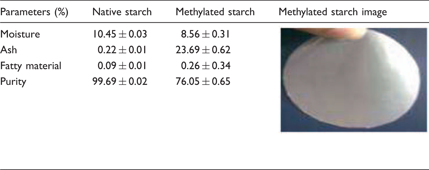
Purity of the native cassava starch
Native and modified starches were analyzed for moisture, ash, crude protein (N × 6.25%), and fatty material contents as a percentage (w/w), following the methodology described in AACC (2003) and Whistler (1964).40,41 Purity was calculated from the difference between 100 and percent of moisture, crude protein, fatty material, and ash content following the equation

Starch methylation
The methylated starch was obtained by using the Whistler and Paschall 4 procedure with some modification as follows: Aseptically, 6 g of starch was mixed with 8 ml of distilled water at 28℃. Subsequently, a barium solution (3.3 g Ba(OH)2 in 8 ml H2O) was added. The slurry was heated to boil (98℃) and 1 ml of DMSO4 was slowly added dropwise. Once the boiling point was reached, the slurry was kept in a water bath at 45℃ for 24 h. The slurry was resuspended in distilled water at 55℃ and centrifuged at 2500 r/min, for 15 min several times, in order to discard the excess BaSO4. The BaSO4-free slurry was neutralized (pH = 7) with H2SO4 0.5 N and centrifuged at 2500 r/min for 15 min. Finally, a portion of the obtained slurry was stored at 4℃ for posterior cytocompatibility assays, and the other one was dried at 45℃ for 24 h or to completion, for composition analysis.
Polarized light microscopy (PLM)
Granular shape and Maltese cross were evaluated by optical microscopy using a polarized light filter. Starch was sprinkled onto a glass slide, 1–2 drops of distilled water were added, and the sample was kept for 5 min. After that time, the slide was covered with a cover slip, and it was kept for 2 more minutes, examined, and photographed in a Nikon Optiphot-2 microscope.
Matrix preparation
In order to prepare the matrix, chitosan was dissolved in a solution of acetic acid to 1% v/v, to produce a final concentration of 1% w/v. The chitosan solution was mixed with the stored methylated starch slurry at a 1:1 ratio (50/50). A total of 200 µl of the mixture were poured in sterile 24 well-plates. Chitosan cross-linking was induced by adding and mixing 50 µl of 2.5% glutaraldehyde into the chitosan–methylated starch suspension, and the hydrogels were allowed to form for 15 min. After that, the hydrogels were sterilized with 70% ethanol, followed by several thorough rinses with a phosphate buffered saline (PBS) solution. Chitosan hydrogels, without starch, were prepared in the same manner.
SEM
The prepared matrices, chitosan, and chitosan–methylated starch composites were observed by SEM. For these, the samples were frozen at –80℃ and lyophilized for 24 h. Once in the SEM facility, the lyophilized matrices were placed in an oven at 50℃ for 8 h, followed by drying in a vacuum chamber for 24 h. The samples were then gold–palladium coated using a sputter coater (EMS 950 turbo evaporator, Electron Microscopy Science), followed by observation in a field emission SEM operated at 10 keV.
Culture of HDFs on the matrices
Expanded HDFs were cultured on the surfaces of the already prepared hydrogels. Briefly, cells were detached from the culture plate by incubating them in 2.5% trypsin–EDTA at 37℃ for 5 min. The detached cells were then counted, using a hemocytometer and trypan blue to account for dead cells, and centrifuged at 400 g for 5 min. The supernatant was discarded, and the pellet was resuspended in supplemented (Dulbecco's modified essential medium) DMEN. HDFs were seeded on the surface of the wells where the hydrogels were prepared at a density of 20 × 103 cells/cm2. Culture over the substrates was carried out for 3 and 5 days, using supplemented DMEM (previously mentioned), at 37℃ in a controlled atmosphere of 5% CO2 and 95% relative humidity. At each time point, cellular proliferation and morphology were evaluated as described later. Culture conditions were defined as follows:
C1: tissue culture plate (TCP), without hydrogel C2: chitosan hydrogel, without starch C3: methylated starch suspended in DMEM, at a concentration equivalent to that present in the hydrogels (M) M: chitosan–methylated starch hydrogels at 1:1 ratio.
Light microscopy
Cytocompatibility can be qualitatively assessed by the observation of adherent cells and their morphology, which was evaluated by light microscopy, using a Nikon microscope, Type 120.
Metabolic activity by MTT assay
A quantitative measure of cytocompatibility is through the determination of cellular metabolic activity, done by the MTT assay, described by Nam et al. 5 This assay relies on the ability of viable cells to reduce a yellow tetrazolium salt (MTT; Sigma) metabolically to a purple formazan product. This reaction takes place when mitochondrial reductases are active [16]. Cultured cells, in the different experimental conditions, were incubated in the presence of MTT solution in PBS (200 µg/ml, 250 µl per well) for 2 h at 37℃, 5% of CO2, and 95% relative humidity. The MTT was discarded and the formation of purple formazan crystals was carried out by adding 250 µL of dimethyl sulphoxide. Absorbance was measured using an ELISA plate reader, TECAN infinite M200, at a wavelength of 570 nm. The metabolic activity is directly proportional to the absorbance. The greater the metabolic activity, the more viable the cells are, and thereby a greater cytocompatibility is implied.
Statistical analysis
Mean and standard deviation were calculated. It was also applied a two-way Analysis of variance (ANOVA) test, and multiple pair-wise comparisons were carried out using the Tukey-HSD method with a 95% confidence level (p ≤ 0.05).
Results and discussion
Percent of purity of the native and methylated cassava starches
Proximate composition (db) and percent purity of the native cassava starch.
PLM
The photomicrograph with PLM of the native starch reveals the average characteristic size of 20–40 µm for cassava starch.
6
The polarized microscopy allowed the observation of the granule's well-defined edges, a central hilum and Maltese cross. Furthermore, granules had round and oval-truncated shapes and were grouped in small conglomerates. (Figure 1). In contrast, the photomicrograph of the modified granule displays an increment in its size, ranging between 47 and 72 µm. This augmented size of the methylated starch could be due to the inclusion of the methyl groups into the granules, which should generate forces of repulsion that are increasing the inter- and intramolecular spaces, allowing the inclusion of a greater number of water molecules.26 Sívoli et al.
7
have noted that the morphological characteristics, crystalline structure, and susceptibility to hydrolysis were dramatically affected by the process of methylation in maize and potato starches; similar results were reported by Sitohy and Ramadan.
8
Polarized light microscopy photomicrography of Native cassava starch (100×).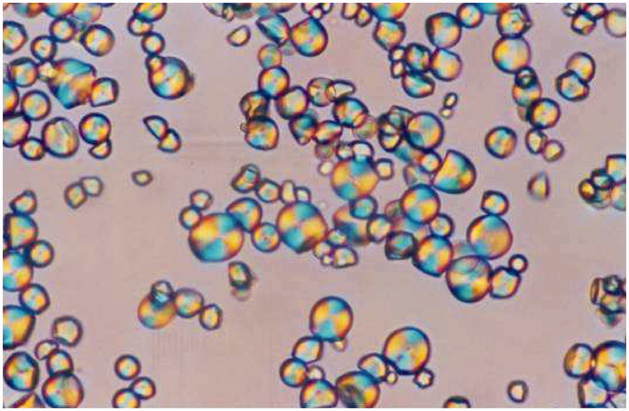
Matrix morphology
To define the composition of the composite hydrogels, several chitosan/starch ratios were previously prepared (100/0, 50/50, and 75/25). Hydrogels at these compositions were made by the addition of glutaraldehyde (as explained in “Materials and methods” section) and these were incubated for a defined period of time to assess their stability. Initially, all hydrogels formed well (Figure 2), with some showing a yellow to brown coloration which was observed as signs of significant cross-linking.23,24 However, when incubated for 24 h, the 75/25 showed significant signs of degradation, while the other gels were able to maintain their integrity (Figure 2). The chitosan gel displayed a more translucent appearance than that observed in the composite hydrogels. It was also observed that the pure chitosan gel was brittle, when manipulated with a spatula, fracturing easily, while the chitosan–starch scaffold (50/50) conserved its integrity under the same conditions of manipulation. It is possible that in the hydrogels with 75% methylated starch, the amount of starch did not allow the proper formation of the chitosan cross-linked matrix, thereby inducing its degradation over time. For all other hydrogel conditions, no degradation was observed, even during cell culture.
Composite hydrogels made in a 24 well-plate at 15 min and incubated for 24 h under agitation. Composition is depicted as % methylated starch/% chitosan.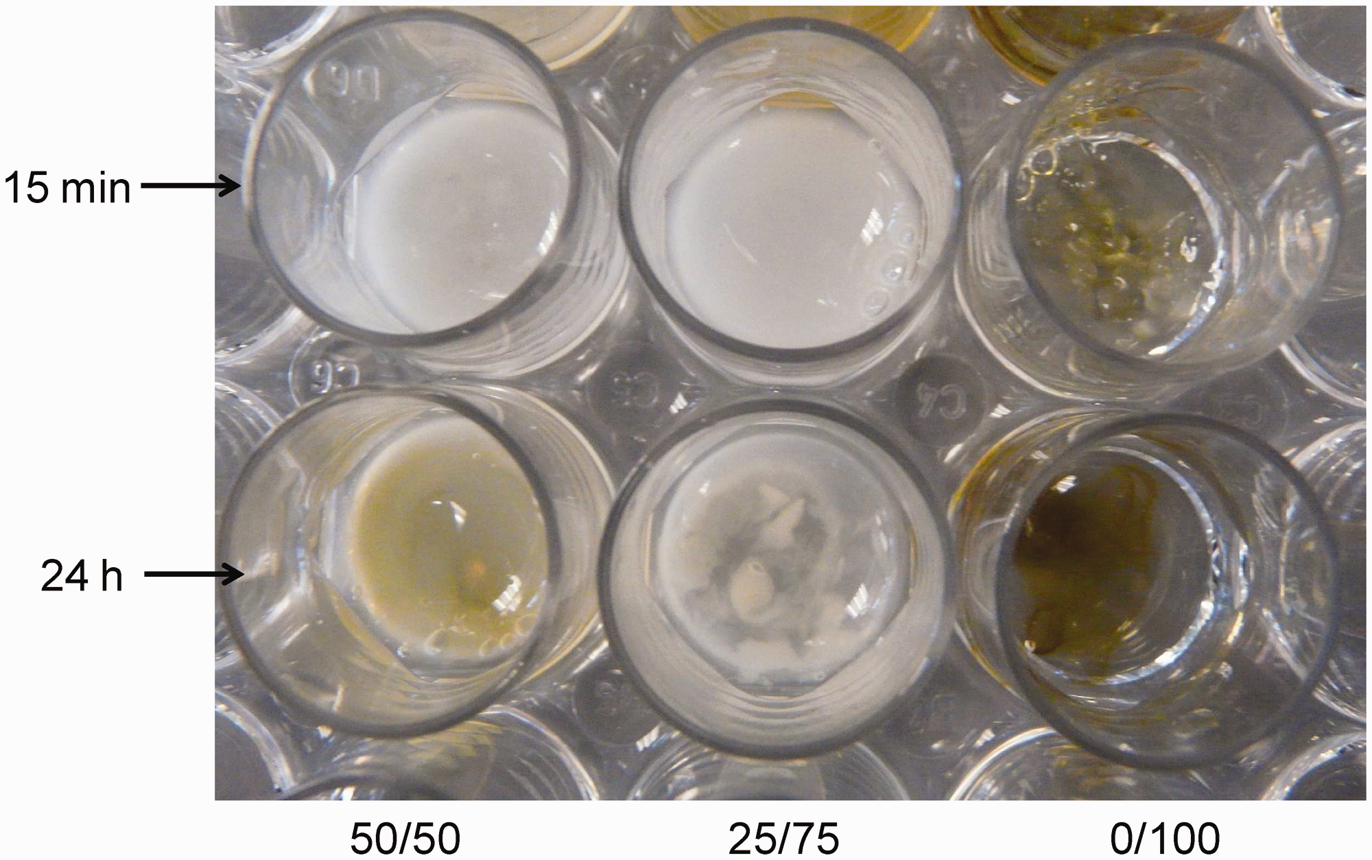
Matrix morphology was evaluated by SEM. In Figure 3, the porous structure of lyophilized chitosan and composite hydrogels is shown. It can be observed that the chitosan matrix (Figure 3(a) and (c)) presented very thin walls (<1 µm) and a very poorly preserved porous structure. In fact, it is clear from the micrographs that the pores tend to collapse by showing folded walls and a brittle appearance. The composite matrices (Figure 3(b) and (d)), on the other hand, appear to display better preserved pores, when compared to the chitosan gels; this was observed during the SEM session, in which images of chitosan structures were difficult as a result of their collapse due to the SEM beam. This indicates that there might be an improvement of the mechanical integrity of the matrix provided by the presence of the methylated starch. However, further analysis would be needed to determine the mechanical properties of the matrices. Another important feature of the composite matrices is the appearance of superficial rugosity by what appears to be entrapped starch granules within the cross-linked chitosan. It has been previously reported in the literature that the presence of micro-rugosity is important in processes of cell adhesion and activity; thus, providing a more appropriate surface to modulate cellfunction.12,32,35
Field emission scanning electron micrographs of chitosan hydrogels ((a) and (c)) and composite, chitosan-methylated starch hydrogels ((b) and (d)).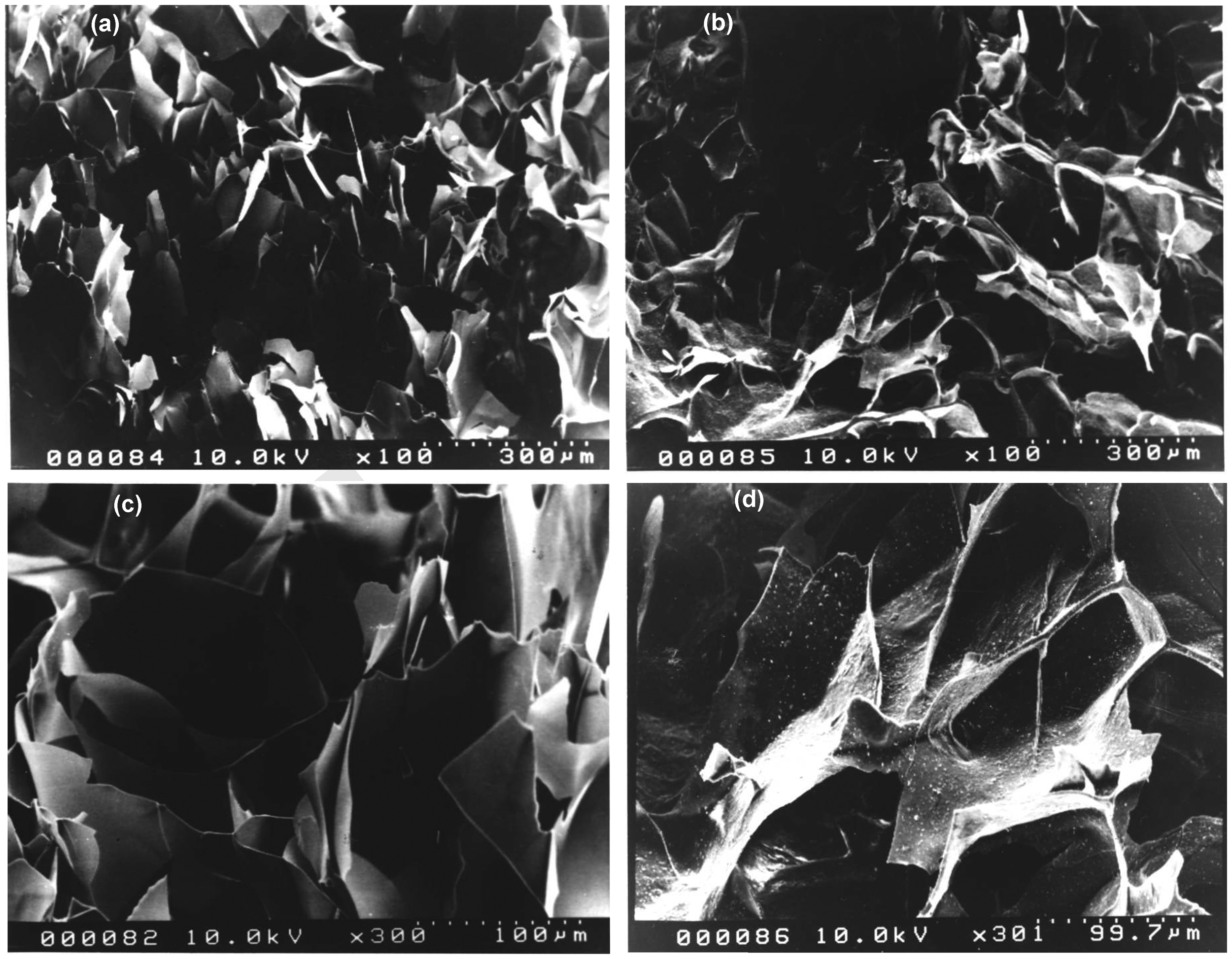
Biocompatibility of the hydrogels
HDFs were grown in medium DMEN supplemented with 10% FBS, 1% glutamine, and 1% antibiotic (penicillin Confluent HDF cultures at 10 × 35 (a) and 20× (b) magnification. The HDFs were obtained from enzymatic digestion of healthy human skin and cultured with supplemented DMEM at 37℃, 5% CO2, and 95% relative humidity following standard protocols.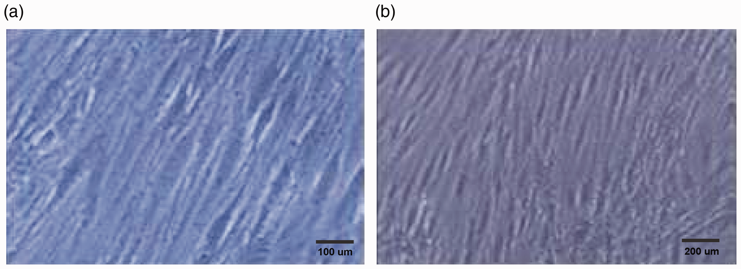
HDFs are commonly used as an in vitro model to test biomaterial biocompatibility. In this study, it was important to evaluate not only the biocompatibility of the material but also that of the fabrication process. Glutaraldehyde is inherently cytotoxic and thereby it was of crucial importance to develop a hydrogel fabrication protocol that would not be cytotoxic.13,14 Initially, trials were carried out to discard most of the glutaraldehyde so as to have nontoxic levels within matrix. After assaying the metabolic activity of the cells via MTT, it was determined that the protocol described previously (“Materials and methods” section) was the most appropriate for cell culture, with thorough washes under shaking to get rid of the unreacted cross-linker.
Figure 5 shows the micrographs of HDFs cultured for 5 days on chitosan hydrogels subjected to ethanol sterilization and only two washes with PBS. The cells presented a rounded morphology atypical of healthy fibroblasts, as shown in Figure 4, indicating poor cell adhesion and thereby cell–matrix interactions. Clusters of cells floating free in the culture medium were abundant, along with an acidification of the medium evidenced by a change in color from red to yellow. All these observations, along with a metabolic activity close to zero (data not shown), were a clear indication that the material was cytotoxic, and this could be due to the material itself or to the lack of sufficient washes to discard the unreacted glutaraldehyde, as suggested previously,
9
and ethanol from the sterilization step. At this point, it is important to point out that ethanol sterilization was chosen since (Ultraviolet) UV and autoclave sterilizations reportedly degrade the polymers.
Micrographs of HDF cultures on a chitosan–methylated starch hydrogel after 3 days. Arrows indicate the presence of round shaped and detached fibroblasts.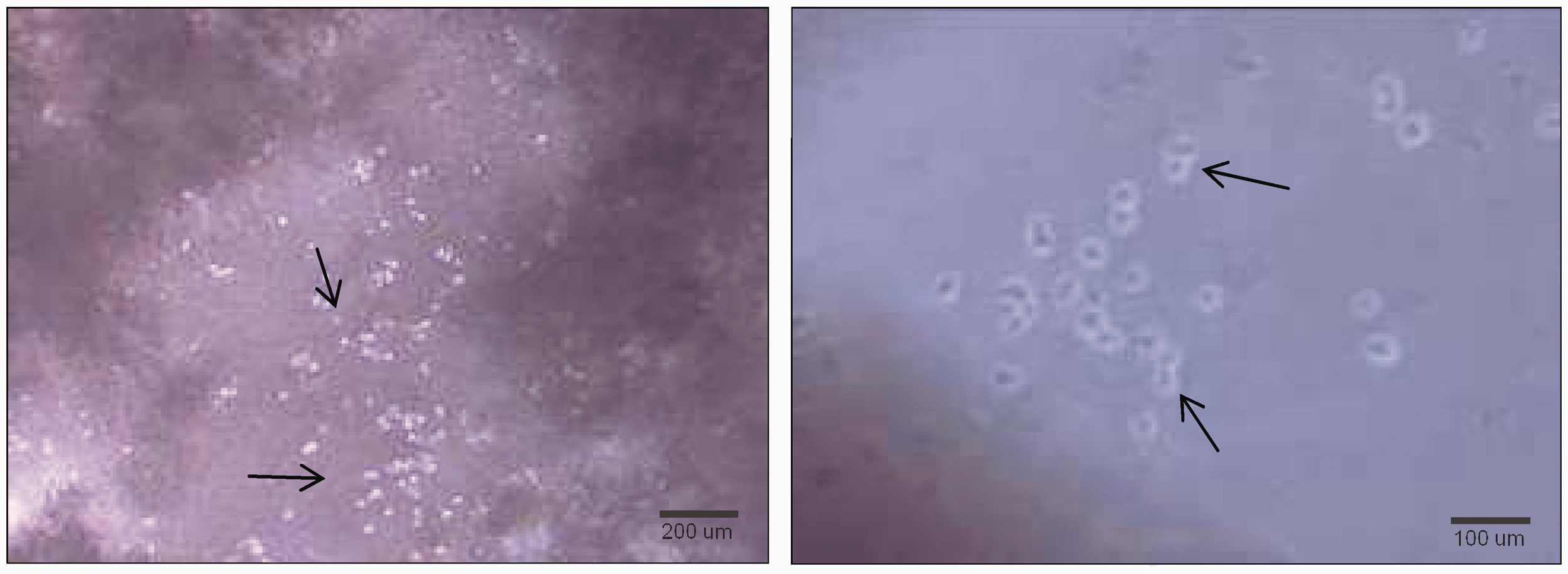
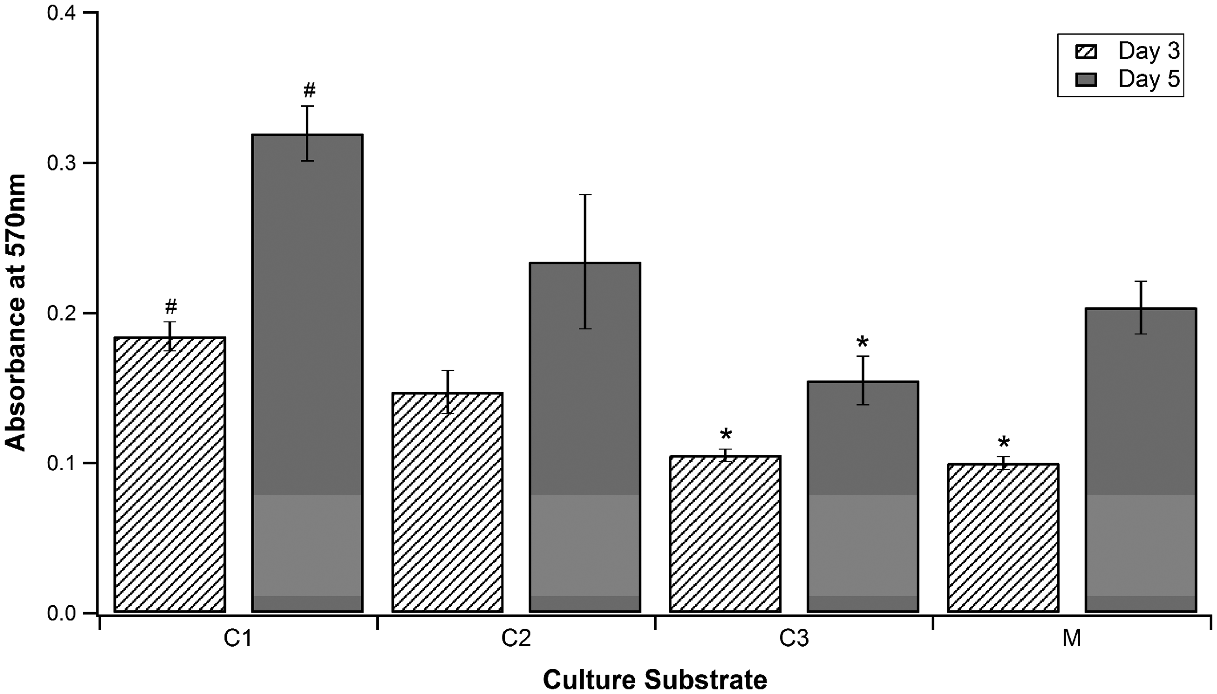
After five washes with PBS, the cells adhered well to the material, as shown in Figure 6(b) (cells cultured on chitosan hydrogels). Here, the cells presented an elongated morphology similar to the one observed in Figure 4. When the starch was incorporated, however, the optical properties of the material changed, and it became difficult to observe the cells under simple light microscopy. A few cells could be appreciated, encircled; these cells also displayed a typical, healthy fibroblast shape. Nevertheless, the MTT assay revealed significant metabolic activity under all culturing conditions (Figure 7). Metabolic activity increased from day 3 to day 5 in all culture conditions, indicating the ability of the cells to adhere and proliferate, which translates into good cytocompatibility of the material.
Photomicrography of the HDF cultured for 5 days on (a) C3 (methylated starch suspended in DMEM), (b) C2 (chitosan hydrogel), and (c) M (chitosan–methylated starch hydrogel). Metabolic activity of HDF cultured on different substrates. C1: TCP; C2: chitosan gel; C3: methylated starch suspended in DMEM; and M: chitosan–methylated starch hydrogel. # and * represent the highest and lowest metabolic activity at a given time point, respectively. Metabolic activity at day 5 was significantly higher than at day 3 for all substrates (p < 0.05).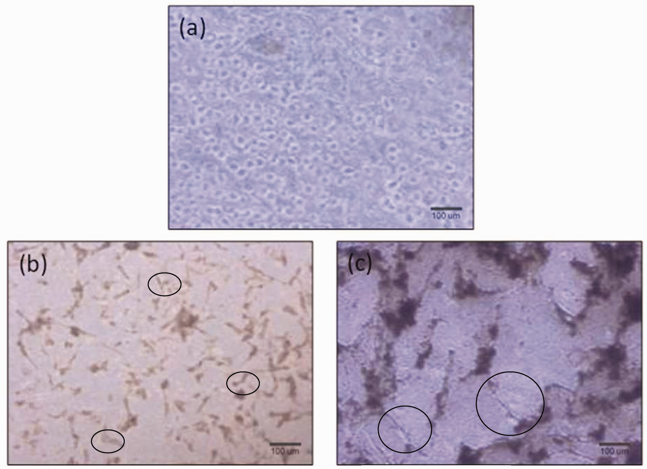
Generally, cells initially adhere to the surface through the interactions of the matrix with the cell membrane receptors. Amino groups from chitosan represent substrate clutches that are ideal for cell adhesion.16–19,22,25 Subsequently, fibroblast cells produce type I collagen, which is characteristic of the connective tissue, allowing them to begin the process of cellular division,33 evident at day 5 of culture. Once the cells have established their extracellular matrix, a more pronounced cellular proliferation takes place to begin with the exponential growth on the substrate used. Gutshe et al. 10 had indicated that after 24 h of cultivation, fibroblasts have produced sufficient extracellular matrix proteins to continue their proliferation.
TCP surface has been specially treated by the manufacturer to make it an ideal substrate for cell growth; therefore, it is expected to observe greater cell adhesion and growth in this group when compared to the hydrogels. After 5 days of culture, chitosan hydrogels show significant metabolic activity, corroborating chitosan’s ability to support cell adhesion and expansion, reported by numerous authors. Wang and Hon 11 performed in vitro biocompatibility assays using different types of cells on chitosan, demonstrating its excellent cytocompatibility. Cells can adhere strongly to this material and proliferate properly. Chitosan also can support and modulate growth and proliferation of vascular, neural cells, fibroblasts, epithelial cells or keratinocytes, osteoblasts, and chondrocytes.12,13
The composite hydrogel used in this study showed a metabolic activity very similar to that obtained with the chitosan matrix, with no statistically significant differences among these two cultures, thus indicating a high level of cytocompatibility. A significantly lower metabolic activity resulted from the cultures with methylated starch suspended in medium. This could be due to the fact that the starch precipitated on top of the cell layer creating a barrier for nutrient and oxygen transport that could slow down cell growth. However, it is evident, from the increment in metabolic activity in time, that the starch was not cytotoxic. From these findings, it is possible to say that the hydrogels and their components are cytocompatible, supporting cell adhesion in proliferation, and that the composite hydrogels of chitosan and methylated starch are suitable candidates for tissue engineering applications.
Conclusions
From the obtained results, it can be concluded that composite hydrogels, made out of methylated cassava starch and chitosan cross-linked with glutaraldehyde, are cytocompatible and support cell adhesion and proliferation. Furthermore, each of the matrices components was proven noncytotoxic; thus, indicating that methylated starch could be used in other polymeric systems for biomedical applications. The proposed matrix is a promising candidate for tissue engineering, and its potential for such applications can be further assessed.
Footnotes
Funding Acknowledgment
This work was funded by the National Fund for Science, Technology and Innovation (Fondo Nacional de Ciencia, Tecnología e Innovación, FONACIT) of the Bolivarian Republic of Venezuela.