
Abstract
A novel antimicrobial dental composite resin has been developed and evaluated. Alumina filler particles were covalently coated with an antibacterial resin and blended into a composite resin formulation. Surface hardness and bacterial viability were used to evaluate the modified composite resin. Results showed that almost all the modified composite resins exhibited higher antibacterial activity along with improved surface hardness, as compared to unmodified one. Effects of antibacterial moiety content on the modified fillers, modified alumina particle size and loading, and total filler content were investigated. It was found that increasing antibacterial moiety content, particle size and loading of the modified filler, and total filler content generally increased surface hardness. Increasing antibacterial moiety, filler loading and total filler content increased antibacterial activity. On the other hand, increasing particle size showed a negative impact on antibacterial activity. The leaching tests indicate that the modified experimental composite resin showed no leachable antibacterial component to bacteria and 3T3 mouse fibroblasts.
Introduction
Long-lasting dental restoratives are attractive to both dental clinics and scientific community, because they can reduce patients’ dental office visit and resultant physical pains as well as expenses. 1 Clinically attractive dental filling restoratives should not only have mechanical and physical properties that are comparable to natural teeth but also exhibit antibacterial property that can prevent teeth from forming secondary caries.2–4 Secondary caries is a type of caries that is generated by oral bacteria after dental restorations. It is a tooth demineralization produced by plaque bacteria such as Streptococcus mutans in the presence of fermentable carbohydrates. The process occurs at the interface between the cavity preparation and the restoration. 3 Secondary caries has been reported to be the main reason to the dental restoration failure.1–4 Therefore, prevention of secondary caries is very important in restorative dentistry. So far, two main strategies have been used to reduce or prevent secondary caries caused by bacteria. One is to incorporate low molecular weight antibacterial compounds into dental restorative formulations. The antibacterial mechanism is based upon release or slow-release of these low molecular weight compounds. Such compounds include but are not limited to various antibiotics, chlorhexidine, zinc ion, silver ion and iodine.5–8 However, release or slow-release of compounds can suffer from a mechanical property reduction of restoratives over time, short-term effectiveness but long-term run-out of the releasing compound, possible toxicity of the compound to surrounding tissues, and an enhanced chance for antibiotic-resistant bacteria formation due to decreasing concentration of the released compound.5–8 The other strategy is to incorporate high molecular weight antibacterial polymers or covalently link antibacterial compound to restoratives or devices.9–18 The antibacterial mechanism of this strategy is based on “kill by contact”. 12 This strategy seems a more effective strategy, as compared to release or slow release. One of the typical examples is to incorporate organic quaternary ammonium salts into restoratives.9–14 The quaternary ammonium salt-containing materials have been found to show a broad spectrum of antimicrobials and also be able to kill or inhibit bacteria that are resistant to other types of cationic antibacterial compounds. 19 The examples of using the quaternary ammonium salt derivatives for dental restoratives include applying polymerizable methacryloyloxydodecyl pyridinium bromide in composite resins, 12 using curable methacryloxylethyl cetyl ammonium chloride in antibacterial bonding agents, 20 , 21 adding polyethylenimine quaternary ammonium nanoparticles to composite resins, 13 and incorporating polymerizable quaternary ammonium bromide derivatives with different chain lengths into glass-ionomer cements. 14 The results showed that all the above quaternary ammonium salt-modified dental restoratives did exhibit significant antibacterial activities. Another example of using this strategy is to incorporate furanone-derivatized compounds into restoratives. The furanone derivatives have shown strong antitumor 22 , 23 and antibacterial functions. 15 Recently these derivatives were incorporated into dental glass-ionomer cements 16 and dental composite resins, 17 resulting in the promising outcomes. 16 , 17 The formulated cements and composite resins showed a significant bacterial inhibition that is comparable to those formulated with the quaternary ammonium salt derivatives. 14 Unfortunately, our unpublished lab results have shown that by in situ polymerization the cured composite resins showed leachable due to incomplete monomer-to-polymer conversion. 8 , 24 Therefore, in this study, we proposed to use alumina particles as a delivery vehicle to deliver antibacterial agent by covalently coating a cured antibacterial polymer on alumina particle surface, to formulate an antibacterial composite resin with an enhanced antibacterial function and improved mechanical hardness without leaching antibacterial moieties.
The purpose of this study was to covalently coat an antibacterial furanone derivative onto crystalline alumina particles, use these coated particles as a delivery vehicle to formulate a novel antibacterial dental composite resin for improved antibacterial activity and surface hardness, and evaluate the surface hardness and antibacterial property of the formed composite resin.
Experimental
Materials
Bisphenol A glycidyl methacrylate, triethylene glycol dimethacrylate, acrylic acid, 2-hydroxyethyl acrylate, p-toluenesulfonic acid monohydrate, 3,4-dichloromalealdehydic acid, toluene, sodium bicarbonate, γ-(trimethoxysilyl)propyl methacrylate, potassium persulfate, camphoroquinone, and 2-(dimethylamino)ethyl methacrylate, and alumina particles with different sizes were received from Sigma-Aldrich Co. (Milwaukee, WI) and used without further purifications. The Herculite-XRV (particle = 0.7 microns, untreated) glass fillers were received as a gift from Kavo Kerr Dental Specialties (Orange, CA).
Synthesis and characterization
Synthesis of 3,4-dichloromalealdehydic acid-hydroxyethyl acrylate (DAHA) is shown below. To a solution containing 3,4-dichloromalealdehydic acid (DA, 0.1 mol), toluene and p-toluenesulfonic acid monohydrate (1% by mole), 2-hydroxyethyl acrylate (HA, 0.11 mol) in toluene was added. 25 After the mixture was run at 90–100°C for 4 h, toluene was removed using a rotary evaporator. The formed DAHA was purified by washing with sodium bicarbonate and distilled water, followed by freeze-drying. The synthesis scheme is shown in Figure 1.
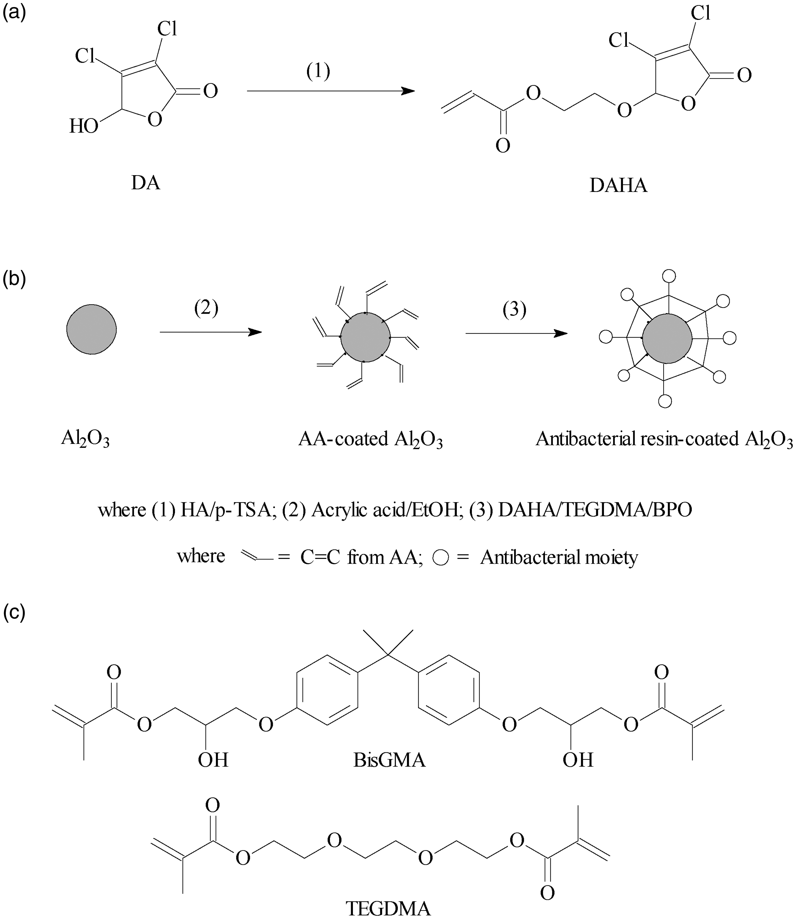
Schematic diagrams for synthesis of DAHA and antibacterial resin-coated alumina particles as well as oligomer structures: (a) DAHA synthesis; (b) Antibacterial resin-coated alumina particle preparation; (c) BisGMA and TEGDMA structures.
Surface coating of antibacterial resin onto alumina particles is shown below. Surface coating was accomplished with the following three steps: (1) Surface activation with acrylic acid. Briefly alumina particles (0.5 g) were dispersed in acrylic acid (30 ml) with ultrasonic vibration for 10 min, 26 followed by heating at 70 °C overnight, washing with methanol and filtering. (2) Fixation of antibacterial agent on particle surface. This process was conducted by immersing acrylic acid-activated alumina particles in a mixture of DAHA (20% by weight) and triethylene glycol dimethacrylate (5%) in methanol, followed by removing methanol with a rotary evaporator. (3) Covalently coating antibacterial agent on the particle surfaces. This process was completed by dispersing the particles in distilled water containing potassium persulfate, followed by heating at 70 °C for 3 h, washing, filtering and freeze-drying. The coating scheme is shown in Figure 1.
Characterization
The alumina particle surfaces were characterized with Fourier transform-infrared (FT-IR) and thermal gravity analysis (TGA). FT-IR spectra were acquired on a FT-IR spectrometer (Mattson Research Series FT/IR 1000, Madison, WI). The thermal decomposition history of selected alumina particles was determined on a thermogravimetric analyzer (Mettler Toledo, Columbus, OH) at a heating rate of 10 °C/min under nitrogen.
Specimen preparation for evaluations
The experimental composite resins were formulated with a two-component (powder and liquid) system. 24 The glass powders (silicon dioxide, Herculite XRV) were treated with γ-(trimethoxysilyl)propyl methacrylate, following the published protocol. 24 The treated glass powders were then blended with the antibacterial resin-coated alumina particles using a vortex mixer. The liquid portion was formulated with bisphenol A glycidyl methacrylate (BisGMA, 50% wt/wt), triethylene glycol dimethacrylate (TEGDMA, 50%), camphoroquinone (photoinitiator, 1%), and 2-(dimethylamino)ethyl methacrylate (activator, 2%), following the protocol elsewhere. 24 The composite resin without any antibacterial resin-coated alumina filler addition is named as “Kerr”. A glass filler content at 75% (wt/wt) was applied throughout the study unless specified.
Specimens were prepared by mixing the liquid with the glass fillers thoroughly at room temperature, according to the published protocol. 24 Briefly, the cylindrical specimens were made in a glass ring with dimensions of 4 mm in diameter x 2 mm in thickness, having a transparent microscope glass slide on each side, for surface hardness, bacterial viability and cell viability tests. All the prepared specimens were illuminated with a blue light device (EXAKT 520 Blue Light Polymerization Unit, EXAKT Technologies, Inc., Oklahoma City, OK) for 2 min, removed from the mold, and conditioned in distilled water at 37 °C for 24 h prior to testing.
Hardness test
The hardness test was performed on a micro-hardness tester (LM-100, LECO Corporation, MI) using a diamond indenter with 25 g load and 30 s dwell time. Knoop hardness number (KHN) was averaged from six readings for each sample.
Bacterial viability test
The bacterial viability test was carried out based on the protocol elsewhere. 18 In short, bacterial colonies were suspended in 5 mL of tryptic soy broth, supplemented with 1% sucrose, to form a suspension with 108 CFU/mL of bacteria and incubated for 24 h. Four bacteria species including Streptococcus mutans (S. mutans), Staphylococcus aureus (S. aureus), Pseudomonas aeruginosa (P. aeruginosa) and Escherichia coli (E. coli) were assessed. The disk specimen was sterilized with 70% ethanol for 10 s and incubated with the bacterial suspension in tryptic soy broth at 37 °C for 48 h under 5% CO2. To 1 mL of the above bacterial suspension, 3 μL of a green/red (1:1 v/v) dye mixture (LIVE/DEAD BacLight bacterial viability kit L7007, Molecular Probes, Inc., Eugene, OR, USA) was added, followed by vortexing for 10 s, sonicating for 10 s, vortexing for another 10 s and keeping in dark for about 15 min before analysis. Then 20 μL of the stained bacterial suspension was added onto a glass slide and viable (green) and dead (red) bacteria were imaged with an inverted fluorescence microscope (EVOS FL, AMG, Mill Creek, WA, USA). A bacterial suspension without disks was used as control and viable bacteria counts from the suspension were used as 100%. The viability was analyzed by counting from the recorded images. Triplicate samples were used to obtain a mean value for each material in each test.
The specimen elute test was conducted based on the following protocol. Briefly, the disc specimen was sterilized with 70% ethanol and sterile phosphate buffer saline (PBS), followed by immersing in a 96-well plate containing tryptic soy broth at 37 °C for 48 h under 5% CO2. After the specimen was removed, the bacterial suspension in tryptic soy broth was added into each well and incubated at 37 °C for 48 h under 5% CO2. Then the viability was analyzed by counting from the recorded images.
3T3 Mouse fibroblast viability test
The 3T3 mouse fibroblast viability test was conducted based on the protocol described elsewhere. 27 Briefly, three steps were followed as below: (1) Culturing cells: 3T3 cells were cultured at 37 °C for 48 h in an air atmosphere containing 5% CO2 and 95% relative humidity, with Dulbecco’s modified Eagle’s medium (DMEM, Hyclone Laboratories, Inc. Logan, UT) containing low glucose, supplemented with 10% heat-inactivated fetal bovine serum (Hyclone Laboratories), 4 mM L-glutamine (Hyclone Laboratories), 100 U/ml penicillin (Sigma-Aldrich, St. Louis, MO), 50 μg/ml gentamicin (Invitrogen Life Technologies, Carlsbad, CA) and 2.5 μg/ml amphotericin B fungizone (Lonza, Walkersville, MD). (2) Elute preparation of the test materials: The disc specimen was sterilized with 70% ethanol and sterile phosphate buffer saline (PBS), followed by immersing in a 48-well plate containing 300 μl serum minus DMEM for 48 h. (3) The water soluble tetrazolium salt-1 (WST-1) test: The cells were plated in a 96-well plate at 2 x 10⁴ cells per well in 100 μl of DMEM supplemented with 10% FBS, 100 U/ml penicillin and 100 μg/ml streptomycin. After incubation at 37°C overnight, the medium was replaced with 100 μl of the fresh medium containing different concentrations of eluate (50%). The cells were then incubated for 48 h before the WST testing. The positive control was serum minus DMEM with untreated cells and the negative control was serum minus DMEM without cells. The WST-1 test was carried out by adding 10 µl of WST-1 reagent (Roche Diagnostics, Indianapolis, IN) and 90 μl of serum minus DMEM into a well and then incubating the plate at 37 °C for 2 h. The absorbance of the solution was measured at 450 nm using a microplate reader (Perkin Elmer 1420 Multilabel Counter, Victor 3, Akron, OH). Cell viability (%) was obtained by the equation: cell viability (%) = (absorbance of the sample eluate - absorbance of the negative control)/(absorbance of the positive control - absorbance of the negative control) × 100. Triplicate samples were used to obtain a mean value for each material.
Statistical analysis
One-way analysis of variance (ANOVA) with the post hoc Tukey-Kramer multiple-range test was used to determine significant differences of the measured properties among the materials or formulations in each group. A level of α = 0.05 was used for statistical significance.
Results and discussion
Characterization
Figure 2 shows the TGA weight-loss curves for alumina, acrylic acid (AA)-coated alumina and antibacterial resin-coated alumina particles. The quantitative transition points for weight-loss are: (a) alumina: 2.5% weight loss due to absorbed moisture or water. (b) AA-coated alumina: 2.5% weight loss from 30 to 150 °C (absorbed water) and 11% loss from 150 to 500 °C (AA coating). (c) Antibacterial resin-coated alumina: 2.5% weight loss from 30 to 200 °C (absorbed water), 11% loss from 200 to 350 °C (AA coating), and 12% loss from 350 to 500 °C (cross-linked antibacterial resin coating).
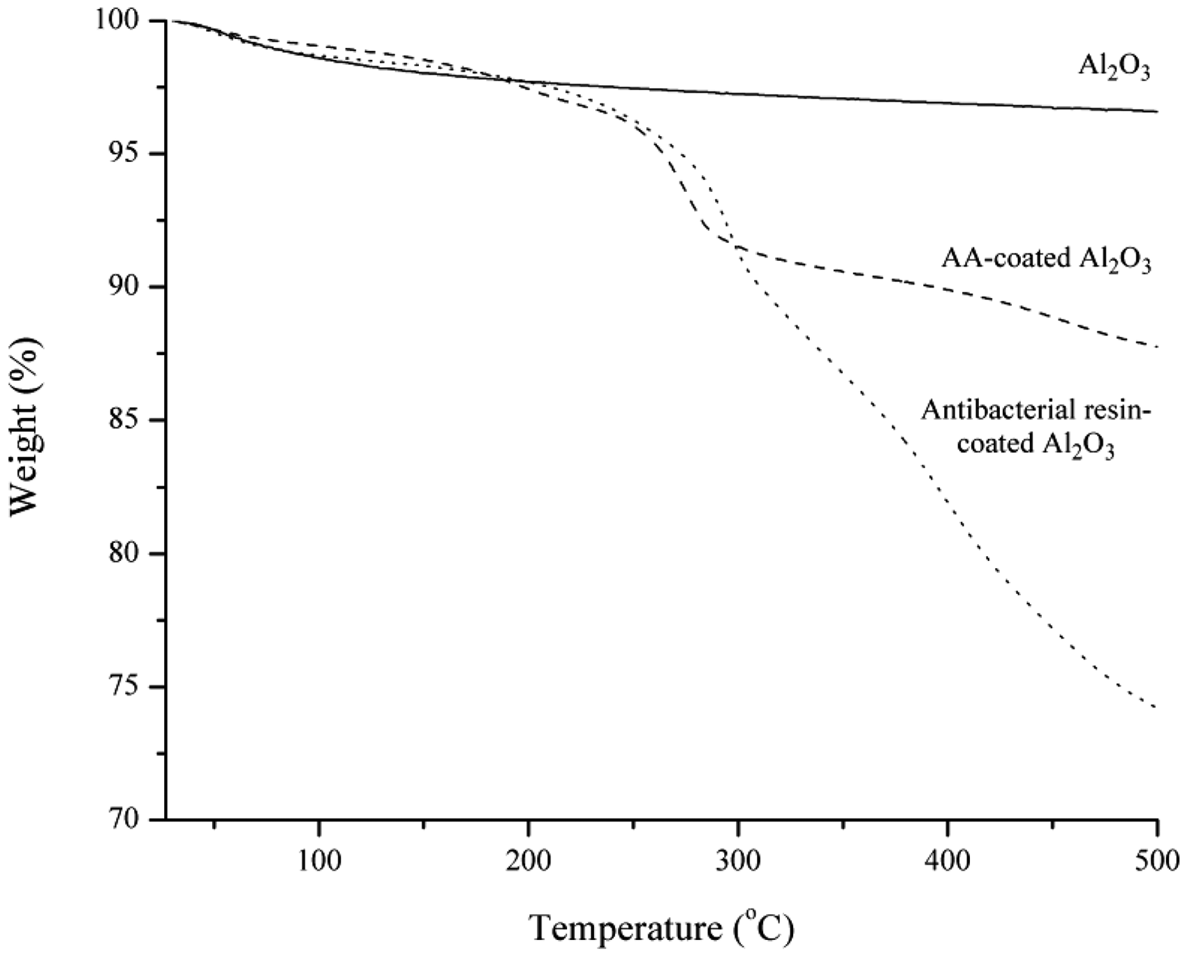
TGA of surface-coated and non-coated particles.
Figure 3 shows a set of FT-IR spectra for alumina (a), AA (b), AA-coated alumina (c), DAHA (d), and antibacterial resin-coated alumina (e). Spectrum a (alumina) shows peaks at 3415 and 1643 for hydroxyl groups from adsorbed water on alumina particles. 28 It has been reported that this type of water is generally present on any manufactured ceramic particles and difficult to exclude. 29 Spectrum b (acrylic acid) shows a broad peak between 3600 and 2400 for carboxyl group (-COOH), a strong peak at 1724 for carbonyl group and two peaks at 1636 and 1618 for carbon-carbon double bonds. In comparison with spectra a, b and c (AA-coated alumina), the appearance of strong peaks at 1728 for carbonyl group and at 1654 for carbon-carbon double bond on spectrum c confirmed successful coating of AA on alumina surface. Furthermore, lack of broad carboxyl peak in spectrum c is also consistent with carboxylic groups intra-structurally linked to alumina particles. Spectrum d (DAHA) shows strong peaks at 1790 for intra ester group on DA, at 1727 for ester group on HA, and 1626 (small) and 1639 (large) for carbon-carbon double bonds. In comparison with spectra c, d and e, except for the peaks at 3415 and 1643 for hydroxyl groups from adsorbed water on alumina particles (spectrum a and c), the appearance of a peak at 1790 for an intra-ester group on DA, and a peak at 1727 for an ester group on HA on spectrum e confirmed that both DA and HA were successfully coated on AA-coated alumina particle surfaces.
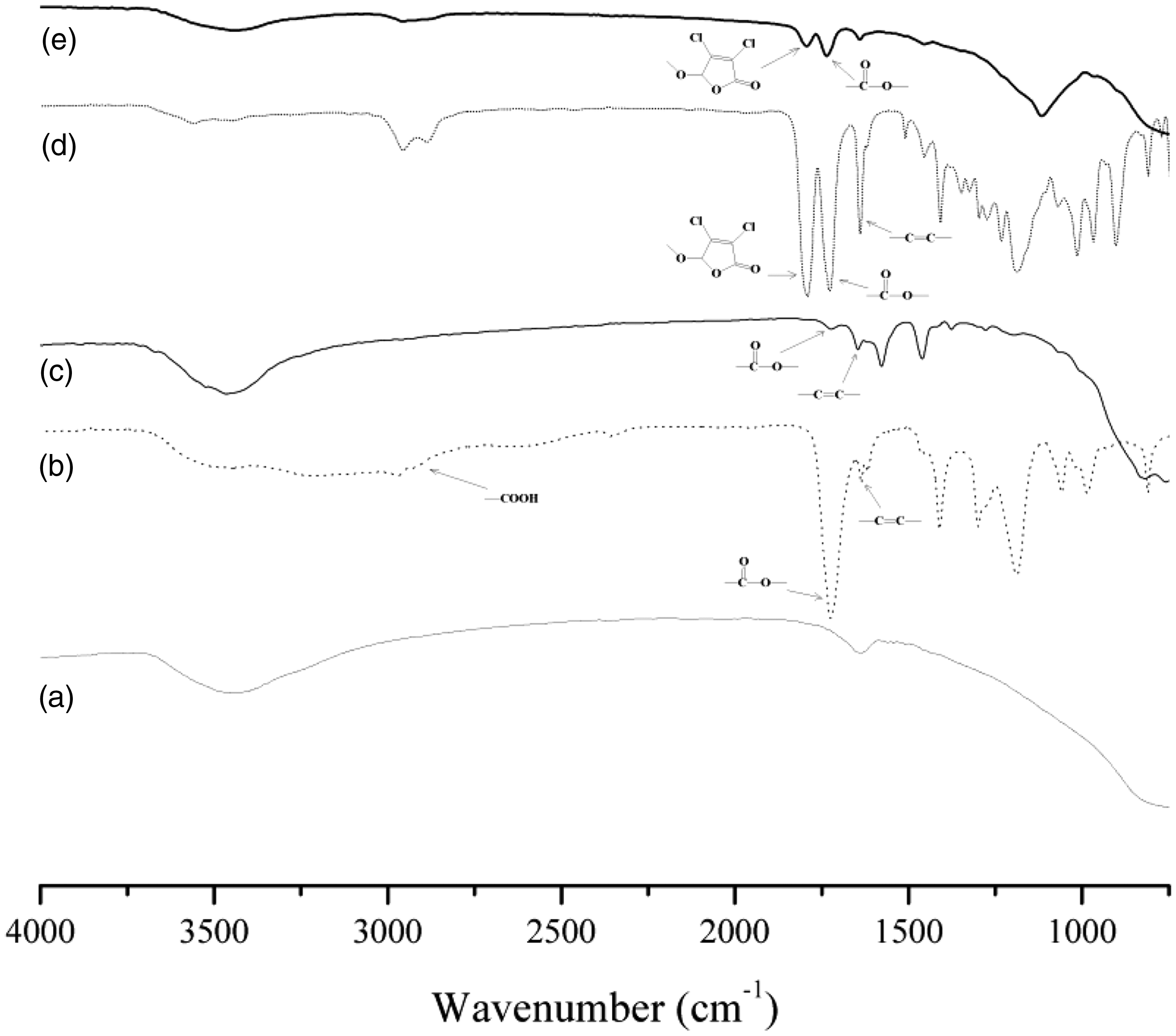
FT-IR spectra of surface-coated and non-coated particles.
Figure 4 shows a photograph describing the dispersion characteristics of unmodified and modified alumina particles in water and monomer. The unmodified alumina particles (left) were found to be well-dispersed in water layer but not in methyl methacrylate layer at all due to the hydroxyl groups on alumina particle surfaces. On the other hand, either AA-coated (middle) or antibacterial resin-coated alumina particles (right) were found to be well-dispersed in organic methyl methacrylate layer but not in water layer at all. This can be attributed to the hydrophobic nature of acrylate groups and/or cross-linked hydrophobic antibacterial resin coatings on the modified alumina particles. This photograph also indicates that surface coating was successful.
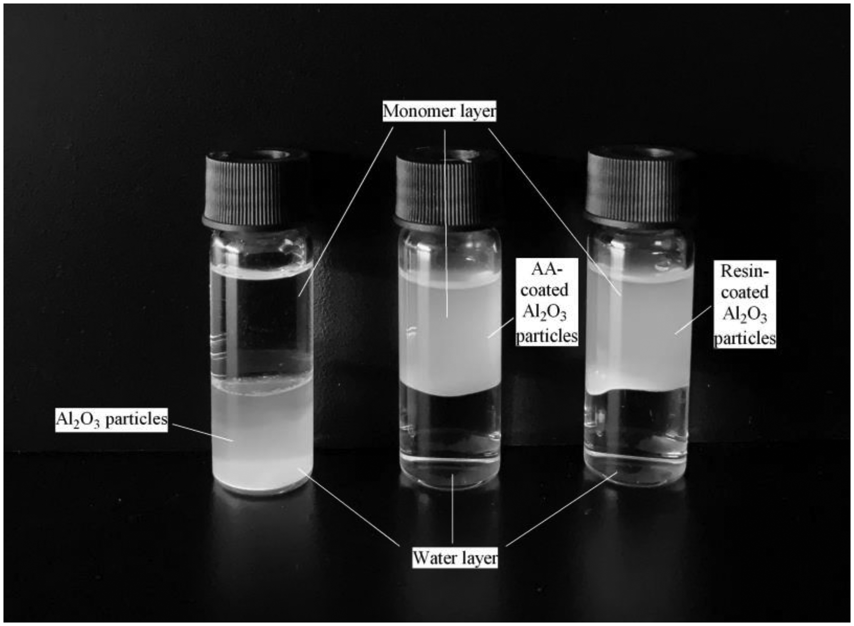
Photograph describing non-coated, AA-coated and antibacterial resin-coated alumina particles in water and methyl methacrylate monomer.
Evaluation
Figure 5 shows the effects of antibacterial moiety content on Knoop hardness number (KHN) and S. mutans viability. The mean KHN was in the decreasing order of 30% = 25% > 20% > 15% > 10% = 5% > Kerr, where no significant differences were found between 5% and 10% and between 25% and 30% (p > 0.05). The mean S. mutans viability was in the decreasing order of Kerr > 5% > 10% > 15% > 20% > 25% > 30%. Apparently, increasing antibacterial moiety content significantly increased KHN and reduced bacterial viability or in other words increased antibacterial activity. Regarding KHN, antibacterial resin-coated alumina particles increase KHN, which may partially be attributed to stiffer and harder DAHA ring structure (see Figure 1). On the other hand, addition of DAHA moiety does exhibit a significantly strong antibacterial function.
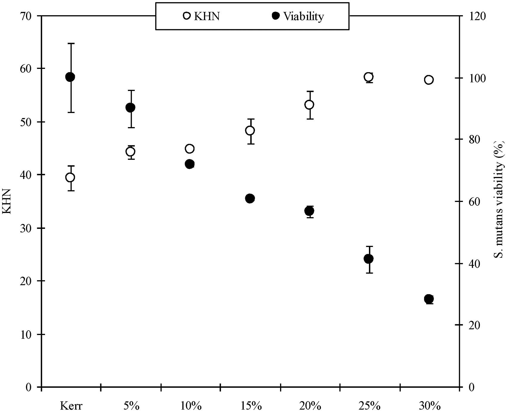
Effect of DAHA moiety content on KHN and S. mutans viability.
Figure 6 shows the effects of modified alumina particle size on KHN and S. mutans viability. The mean KHN was in the decreasing order of 162 μm > 94 μm = 63 μm > 10 μm > Kerr, where no significant differences were found between 94 μm and 63 μm and between 10 μm and Kerr. The mean S. mutans viability was in the decreasing order of Kerr > 162 μm > 94 μm > 63 μm = 10 μm, where no significant difference was found between 10 μm and 63 μm. It appears that increasing antibacterial alumina particle size slightly increased KHN but did not significantly affect bacterial viability, although the incorporated antibacterial alumina fillers did significantly improve the antibacterial activity as compared to Kerr. The largest particles (162 μm) showed the 2nd highest bacterial viability to Kerr, indicating that under the same weight smaller particles can deliver more antibacterial residues to the composite resin due to their larger surface areas, as compared to larger ones.
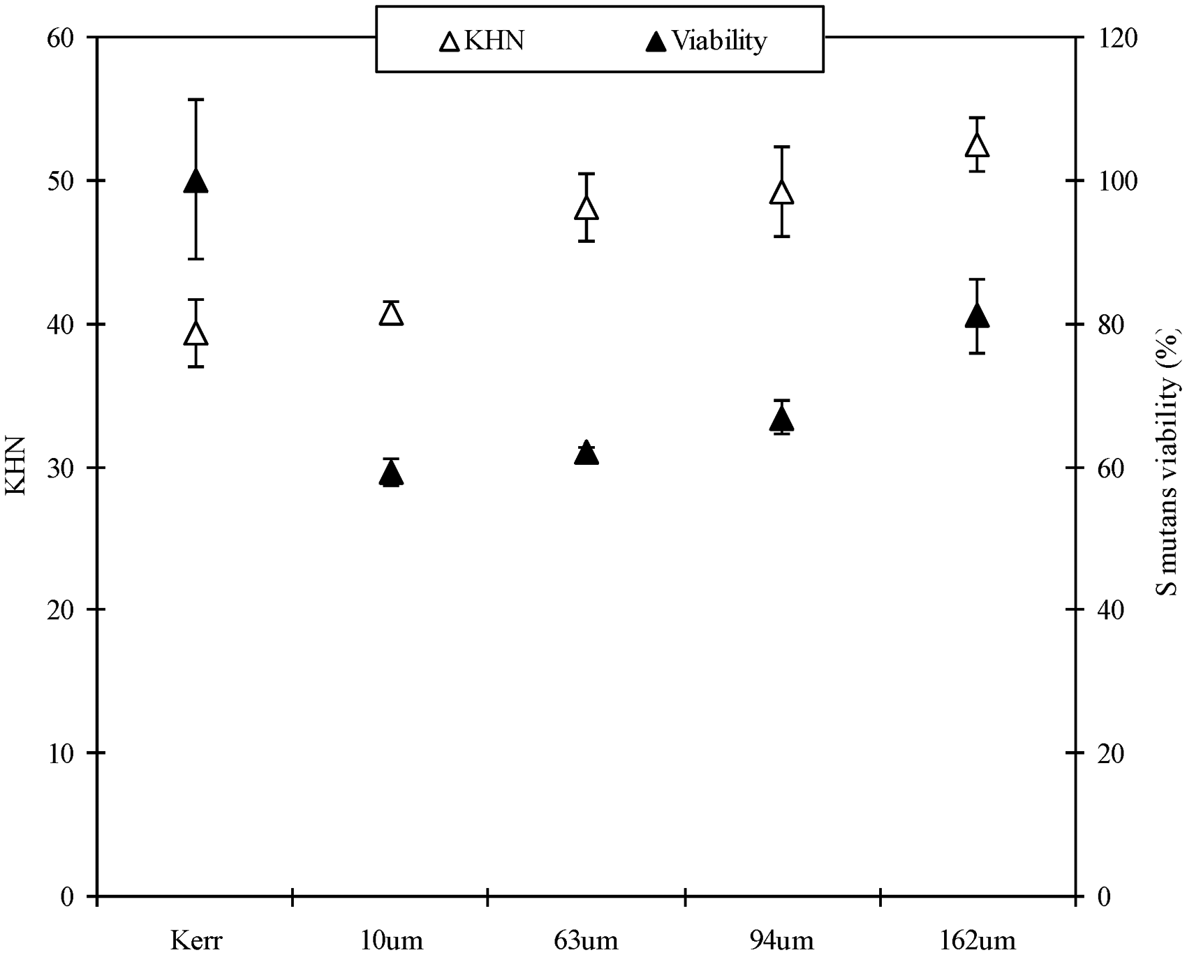
Effect of antibacterial resin-coated particle size on KHN and S. mutans viability.
Figure 7 shows the effects of antibacterial alumina filler loading on KHN and S. mutans viability. The mean KHN was in the decreasing order of 15% = 20% = 25% > 10% > Kerr, where no significant difference was found among 15%, 20% and 25%. The mean S. mutans viability was in the decreasing order of Kerr > 10% > 15% > 20% > 25%. Increasing antibacterial filler loading increased KHN up to 15% and then nearly no change is observed. On the other hand, increasing filler loading decreased bacterial viability. This can be easily explained as that at the same particle size increasing antibacterial filler loading increases antibacterial moiety contents, thus enhancing the antibacterial activity of the composite resin.
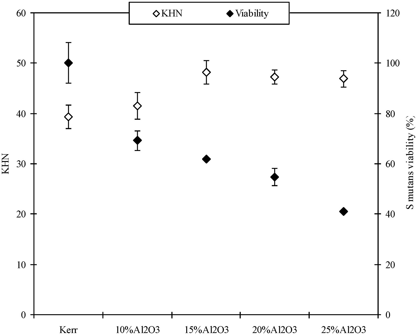
Effect of antibacterial resin-coated filler content on KHN and S. mutans viability.
Figure 8 shows the effects of total filler content on KHN and S. mutans viability. The mean KHN was in the decreasing order of 79% > 77% > 75% > 73% by weight. The mean S. mutans viability was in the decreasing order of 73% > 75% > 77% > 79%. Increasing total filler content significantly increased KHN but reduced bacterial viability. In composite resin formulations, total filler content determines hardness and other properties. 8 , 30 The higher the filler loading, the higher the hardness the composite resins are anticipated. 8 , 30 Highly filled composite resins often show the properties that are closer to natural teeth, because teeth are highly mineralized tissue. 8 , 30 Therefore, higher filler contents are favored in composite resin formulations. However, the problem that is often faced during composite resin preparations is hard to incorporate more filler particles into composite resins due to interfacial incompatibility between inorganic fillers and organic resin. 31 Fortunately, in this study, since we were able to well coat the alumina fillers with antibacterial resins, the interfacial compatibility was found to be significantly improved. Thus the total filler loading was significantly increased, resulting in the fact that not only the total filler content was added up to 79% but also more antibacterial fillers were incorporated. That is why higher total filler loading showed increased hardness and antibacterial activity.
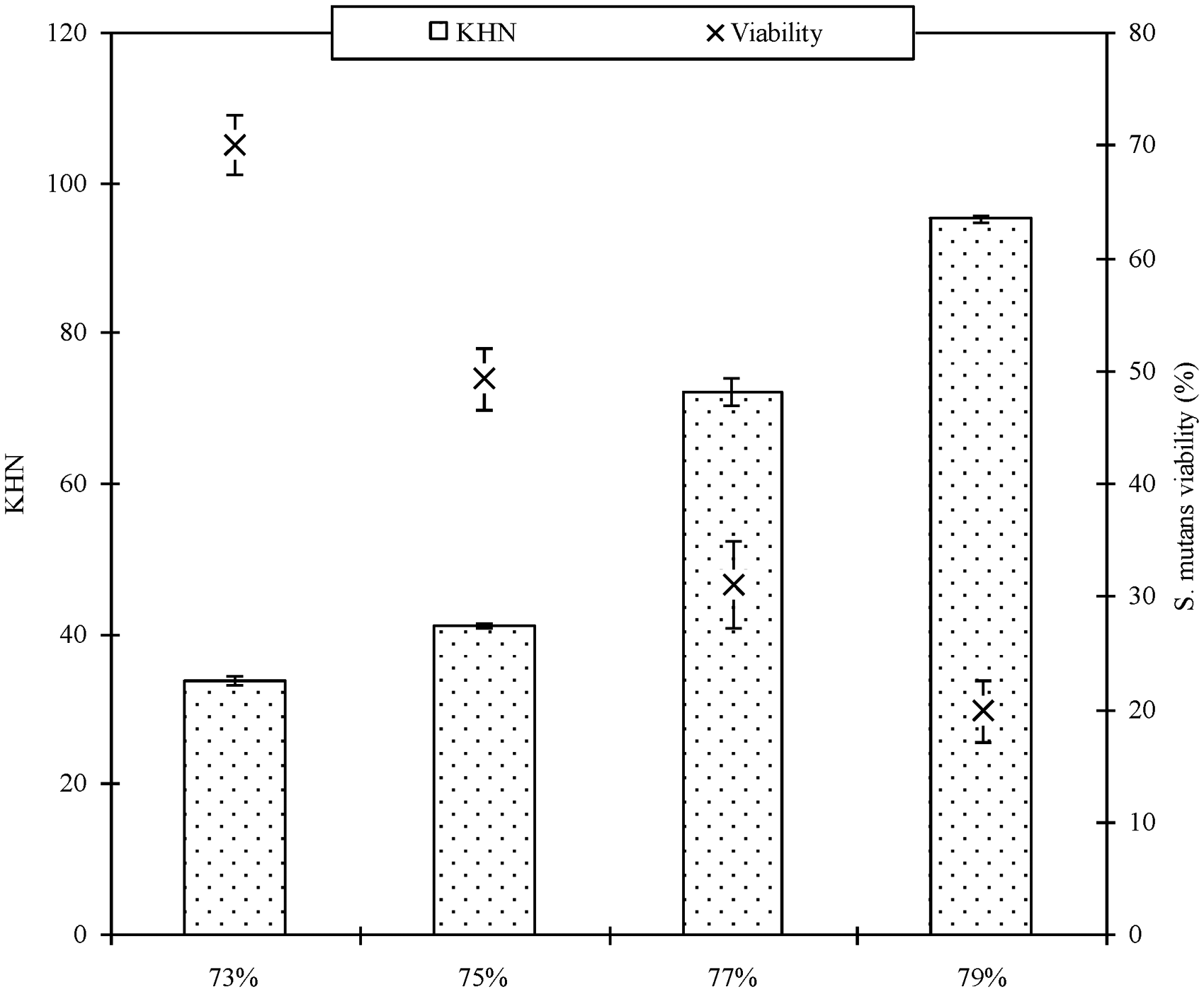
Effect of total filler content on KHN and S. mutans viability.
Table 1 shows the effect of the modified composite resins with different filler loading on the viability of four bacterial species. From the results, it is clear that increasing total filler loading decreased bacterial viability. Different bacterial species showed different responses to antibacterial composite resins. From 73% to 79% total filler loading, S. mutans, S. aureus, P. aeruginosa and E. coli showed 72%, 62%, 74% and 80% decrease in bacterial viability, respectively. This indicates that the responses of different bacterial species to the antibacterial compound are different. S. mutans, P. aeruginosa and E. coli are more vulnerable to the antibacterial compound than S. aureus.
Effect of antibacterial fillers on viability of four bacteria (%). a
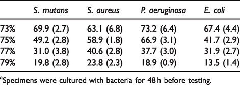
aSpecimens were cultured with bacteria for 48 h before testing.
Figure 9 shows a set of photomicrographs of bacterial viability after incubating bacteria with the composite resins, with green fluorescence indicating live bacteria in the culture and red fluorescence indicating dead bacteria. The images depict (a) S. mutans with Kerr (live), (b) S. mutans with Kerr (dead), (c) S. mutans with experimental antibacterial composite resin (live), (d) S. mutans with experimental composite resin (dead), (e) S. aureus with experimental composite resin (live), (f) S. aureus with experimental composite resin (dead), (g) E. coli with experimental composite resin (live), and (h) E. coli with experimental composite resin (dead). Figure 9(a) shows many green (live) bacteria but (b) shows no red (dead) bacteria. In contrast, Figure 9(c) shows significantly lower number of live bacteria whereas (d) exhibits dead bacteria. Figure 9(e) and (g) show lower number of live S. aureus and E. coli but (f) and (h) exhibit dead S. aureus and E. coli. Apparently, antibacterial composite resin showed significant antibacterial activity by not only inhibiting bacterial growth but also killing bacteria.
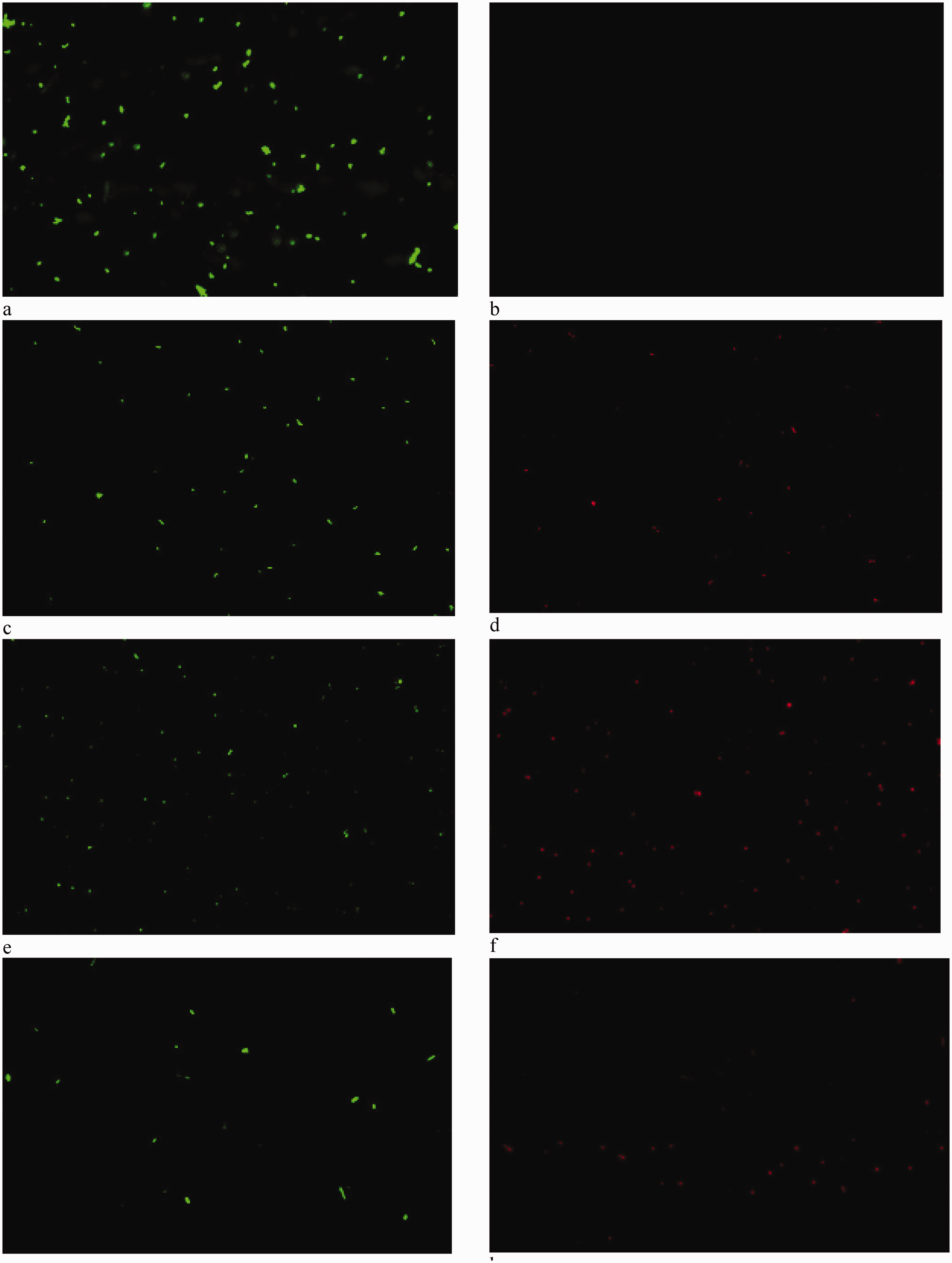
Bacterial images after incubating with antibacterial composite resin vs. Kerr for 48 h. Bacteria were stained with a fluorescence viability stain, with green fluorescence indicating live cells in the culture and red fluorescence indicating dead cells. (a) S. mutans with Kerr (live), (b) S. mutans with Kerr (dead), (c) S. mutans with antibacterial composite resin (live), (d) S. mutans with antibacterial composite resin (dead), (e) S. aureus with antibacterial composite resin (live), (f) S. aureus with antibacterial composite resin (dead), (g) E. coli with antibacterial composite resin (live), and (h) E. coli with antibacterial composite resin (dead).
Table 2 shows the results from the leaching tests. To test whether antibacterial components from the experimental antibacterial composite resin would leach out, three experiments - aging, extractable to S. mutans viability and extractable to 3T3 cell viability, were conducted. Theoretically speaking, if there were no changes in bacterial viability during specimen aging, it means that the composite resin would have no leaching. If there were no changes in bacterial number after S. mutans were cultured with elute, it means that the composite resin would have no leaching. Regarding 3T3 fibroblasts, if there were no changes in cell viability after culturing with elute from the composite resin, it means that the composite resin would have no leaching. From Table 2, it is clear that no significant change in bacterial viability was found between 1 day and 7 day aging in bacterial broth. Furthermore, no significant change in bacterial number between the antibacterial composite resin and Kerr was found after culturing with the 48-h elute. For 3T3 viability test, no significant difference in 3T3 viability was found between Kerr and the experimental composite resin. The results indicate that the experimental composite resin is a non-leaching antibacterial composite resin, which eliminates the concern on potential cytotoxicity due to attached antibacterial residues. It also confirms that this novel composite resin inhibits or kills bacteria by contact but not by the released leachable.
Leaching tests.
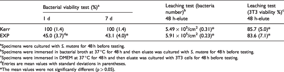
aSpecimens were cultured with S. mutans for 48 h before testing.
bSpecimens were immersed in bacterial broth at 37 °C for 48 h and then eluate was cultured with S. mutans for 48 h before testing.
cSpecimens were immersed in DMEM at 37 °C for 48 h and then eluate was cultured with 3T3 cells for 48 h before testing.
dEntries are mean values with standard deviations in parentheses.
*The mean values were not significantly different (p > 0.05).
Conclusions
In this study, a novel antimicrobial dental composite resin has been developed and evaluated. Alumina filler particles were covalently coated with antibacterial resin and blended into a composite resin formulation. Results showed that almost all the modified composite resins exhibited higher antibacterial activity along with improved surface hardness, as compared to the unmodified one. Increasing antibacterial moiety content of the added fillers, particle size and loading of the modified fillers, and total filler content generally increased surface hardness. Increasing antibacterial moiety, filler loading and total filler content increased antibacterial activity. On the other hand, increasing particle size showed a negative impact on antibacterial activity. The leaching tests indicate that the experimental antibacterial composite resin showed no leachable antibacterial component to bacteria and 3T3 mouse fibroblasts.
Footnotes
Declaration of Conflicting Interests
The author(s) declared no potential conflicts of interest with respect to the research, authorship, and/or publication of this article.
Funding
The author(s) disclosed receipt of the following financial support for the research, authorship, and/or publication of this article: This study was partially sponsored by IUPUI Research Support Funds Grant.