
Abstract
Type I collagen, the major organic component of human dentin, plays an important role in regulating the mechanical strength of dentin. Collagen in dentin can be strengthened by heating. We hypothesized that UV irradiation could produce similar strengthening effects and might maintain the strength of dentin after rehydration. Beam-shaped dentin specimens from the crowns of human third molars were subjected to flexural testing. Flexural strengths were two and three times greater than those in the control group after 5 minutes’ UV irradiation and heating to 140°C, respectively. After 30 days of rehydration, the heated specimens reverted to their original strength, whereas the UV specimens were 69% stronger than the original. Raman spectra of dental collagen were unchanged after heating, whereas several peaks, including a C-C bond in a proline ring, were amplified by UV irradiation. It is concluded that dentin strengthened by UV irradiation retains strength after rehydration because of chemical changes in collagen.
INTRODUCTION
In our previous research (Hayashi et al., 2008), we discovered that the flexural and microtensile strengths of human dentin were from 2 to 2.4 times greater after heating between 110°C and 140°C for 60 min. The stress intensity factors at fracture also increased after heating, while the modulus of elasticity was unchanged. X-ray diffraction analyses indicated that shrinking of the lateral packing of the collagen triple-helices from 14 Å to 11 Å is the probable cause of the increased strength of heated dentin. However, the question remained as to whether such strength produced by heating would be retained in hydrated conditions, because hydration is the normal oral environment.
In human bone, which has a composition comparable with that of dentin, the collagen network plays an important role in its toughness, and the toughness and strength of bone decrease with increasing collagen denaturation (Wang et al., 2001). The strength of collagen is affected by cross-linking, which may increase its mechanical properties and molecular stability (Oxlund et al., 1995, 1996; Banse et al., 2002; Wang et al., 2002; Miguez et al., 2004). A previous study demonstrated that the strength of reconstituted tendon collagen could be improved by varying the time and temperature of dehydrothermal cross-linking (Wang et al., 1994). Chemical agents (such as formaldehyde and glutaraldehyde) and ultraviolet (UV) irradiation are among the basic strategies which introduce stable and covalent intermolecular cross-links within collagen fibrils (Cooper and Davidson, 1965, 1966; Weadock et al., 1995; Jorge-Herrero et al., 1999; Bedran-Russo et al., 2008). Since the organic matrix of human dentin is 30% by volume, mainly type I collagen (Nanci, 2003), we intended to investigate whether heating or UV irradiation could significantly strengthen the mechanical properties of human dentin by promoting chemical changes in collagen and then retain them.
The purpose of this study was to investigate the effects of heating and UV irradiation on the mechanical properties of human dentin by focusing on their changes when dehydrated and rehydrated. The null hypothesis tested was that the mechanical properties of human dentin were not affected by heating or UV irradiation, whether under dehydrated or rehydrated conditions.
MATERIALS & METHODS
Preparation of Beam-shaped Dentin Specimens
Human third molars free of caries were stored in Hanks’ balanced salt solution (HBSS) at 4°C and used within 3 mos of extraction. Protocols were approved by the ethics committee of Osaka University. Informed consent was obtained from all donors for collecting the teeth. Beam-shaped dentin specimens, measuring approximately 1.7 x 0.9 x 8.0 mm, were obtained from coronal central portions of the molars. Dentinal tubules in the specimens were organized to run parallel to the loading surface along the specimen length. In our previous study, we had confirmed that dentin specimens with this orientation were most strengthened by heating (Hayashi et al., 2008).
Heating, UV Irradiation, and Rehydration Treatment
Heating for Various Times
Beam-shaped specimens were heated according to the following protocols: Control (wet) specimens were soaked in HBSS; heated specimens were heated in an oven to 110 or 140°C and then removed immediately on reaching the targeted temperature or when heated to those temperatures for 10 to 60 min. The heating rate was 10°C/min. Our previous extensive research had confirmed that the mechanical properties of human dentin were most improved by heating to 110 or 140°C (Hayashi et al., 2008).
UV Irradiation
Other sets of specimens were irradiated with UV light for 5, 15, 30, or 60 min at 356 nm wavelength with a power of 3200 mW/cm2 in a UV LED irradiation unit (Omron ZUV-C30H, Kyoto, Japan). The distance from the UV light source to the specimen was approximately 10 mm. The moist specimens became dehydrated after UV irradiation.
Rehydration after Heating and UV Irradiation
We wanted to examine whether the dentin strengthened by heating or UV irradiation would retain the strength after rehydration. The specimens heated at 110°C for 10 min or 140°C, or UV-irradiated for 5 or 15 min, were rehydrated in HBSS at ambient temperature for 3, 7, and 30 days.
Each experimental group included 10 specimens, each obtained from a different tooth. The sizes of the specimens before and after heating or UV irradiation and after rehydration were measured by a digital caliper (CD15, Mitutoyo, Kawasaki, Japan) with an accuracy of 0.001 mm, after which the percentages of linear shrinkage were calculated.
Flexural Fracture Testing
Flexural strengths of heated and UV-irradiated dentin with or without rehydration were measured as reported previously (Hayashi et al., 2008). Specimens subjected to flexural fracture testing were secured in a custom-made metallic holder with a cantilever beam geometry, and a flexural load was applied by means of a universal testing machine (Autograph AG-IS, Shimadzu Co., Kyoto Japan) with a crosshead speed of 0.1 mm/min. Fracture stress (sec, MPa) was calculated with the following equation:
where M (N) is the load at fracture, L (m) is the valid length in the cantilever geometry, and b (m) and d (m) are the width and thickness of a specimen, respectively.
The flexural strengths of specimens taken from different environments were compared by means of ANOVA and Scheffé’s F test at a 95% level of confidence.
Correlation between Numbers of Dentinal Tubules and Their Flexural Strength
After flexural testing, the fractured surfaces of all the specimens were observed with a scanning electron microscope (SEM) (JSM9-840A, JEOL, Tokyo, Japan) at a magnification of X200. The dentinal tubules were counted by the use of SEM images, and the correlations between the numbers of dentinal tubules and their flexural strength under the different conditions of heating and UV irradiation were analyzed mathematically by use of curve analysis software (Excel, Office 2004, Microsoft, Redmond, WA, USA).
Laser Raman Spectroscopic Analysis
A beam-shaped dentin specimen was demineralized in 0.5 M EDTA at a pH of 7.4 for 10 days. In our previous study (Hayashi et al., 2008), complete demineralization of human dentin with this method was confirmed by the disappearance of Bragg diffraction rings originating from hydroxyapatite crystals. The demineralized dentin was subjected to heating to the targeted 140°C or to UV irradiation for 15 min. The same specimens were tested before and after heating or UV irradiation.
Raman spectra were measured by laser Raman microscopy (RAMAN-11, Nanophoton, Osaka, Japan). The excitation source was a semiconductor laser operating at 785 nm with nominal power of 100 mW. The laser beam was focused and irradiated onto the specimen with an objective lens (x5, NA = 0.3). The area of irradiation was around 1.0 x 1.0 μm2, and the accumulation time was 180 sec. The excited Raman scattering light was collected with the same objective lens and guided to the spectrograph, which had a focal length of 500 mm. Raman light was then recorded with a thermoelectrically cooled CCD camera as the Raman spectrum, which contained chemical bond information.
The Raman spectra obtained were normalized for the comparison, since the intensity of the raw spectra changes depending on the surface condition of dentin, such as the roughness and density of the collagen molecules. The normalization protocol was as follows: The background intensity caused by fluorescence was reduced with the algorithm developed by methods previously reported (Lieber and Mahadevan-Jansen, 2003); then, the spectra were normalized with the peak intensity at 815 cm−1, which corresponded to the backbone vibration of collagen (Frushour and Koenig, 1975).
RESULTS
After dehydration, the greatest flexural strengths were found in specimens that had been heated to 110°C for 10 min or longer or to 140°C. After that, further heating did not increase the strength of the specimens (Table 1). The flexural strength reached a plateau of 234.3 to 253.5 MPa, or 3 times greater than that of the control specimens. Linear shrinkage was stabilized at 2.3 % after heating at 110°C for 10 min and to 140°C. Judging from the increased flexural strengths and stabilization of linear shrinkage, we identified that heating at 110°C for 10 min and heating to 140°C were the most favorable conditions to strengthen human dentin.
When the heated specimens were rehydrated, their strength diminished (Table 2). In those heated to 110°C for 10 min, rehydration led to reversal to the initial wet condition after 3 days (back to 101.9 MPa from 253.5 MPa). The linear shrinkage, which was 2.25% after heating, was reduced to 0.20% after 3 and 7 days of rehydration. Heating at 140°C increased the flexural strength to 234.3 MPa, which was then reduced to 130.1 MPa after 3 days of rehydration, still significantly stronger than in the wet condition. However, this strength after heating disappeared completely after 30 days of rehydration.
The effects of UV light irradiation for 5 and 15 min were found to increase flexural strengths to 157.3 and 145.9 MPa, respectively, and to produce linear shrinkage of 0.39% (Table 1). However, this strength dropped when UV irradiation continued for longer than 30 min. When the irradiated specimens were rehydrated, the flexural strength dropped. However, specimens treated with UV irradiation for 5 min were still 69% stronger than the control group, even after 30 days of rehydration.
The correlation between the number of dentinal tubules and their flexural strength after heating and UV irradiation is shown in Fig. 1. As previously reported (Inoue et al., 2003), the flexural strengths of specimens in the present study decreased as the numbers of dentinal tubules increased in wet conditions; the same tendency was observed in dentin subjected to UV irradiation. However, in the heated dentin, there was no correlation between the numbers of dentinal tubules and their flexural strength.
Raman spectra of collagen were found to be unchanged after heating (Fig. 2a), whereas a peak of 922 cm−1, which indicates a C-C bond in a proline ring (Frushour and Koenig, 1975), was amplified by UV irradiation (Fig. 2b). In addition, several other peaks of 1323, 1407, and 1453 cm−1, which indicate deformation and twisting of CH2 and CH3, were also amplified (Fig. 2b).
DISCUSSION
In our previous study, we reported that heating strengthens dentin, and that shrinkage of the lateral packing of the collagen triple-helices is the probable cause of the increased strength. The present study aimed to clarify whether heated dentin retained its strength when rehydrated, since several studies have reported that changes in the biomechanical properties of dentin after dehydration could be reversed by rehydration (Jameson et al., 1993; Nalla et al., 2005, 2006). We also examined whether any other dehydration process might produce strength that could be retained.
Loss of water during heating is the probable cause of the shrinkage of the collagen network, and the residual shrinkage after rehydration suggests the possibility of dehydrothermal cross-linking. If one hypothesizes that cross-linking by heating is one of the reasons for the shrinkage, other methods of generating collagen cross-linking, such as UV irradiation or chemical agents, could be expected to produce similar effects. In the present study, we investigated how heating and UV irradiation strengthen dentin, focusing on the chemical change in collagen detected by the Raman spectroscopic analysis. Chemical agents, which are commonly used to promote cross-links (Bedran-Russo et al., 2008), were not considered because of their toxicity.
We demonstrated in the present study that the strength gained by heating as a result of dehydration was reversed by rehydration. The water content of dentin is about 10 to 13% by weight (Burnett and Zenewitz, 1958), of which approximately 75% is in the dentinal tubules and the remainder in the mineralized matrix (van der Graaf and ten Bosch, 1990). Thus, the loss of water during heating is almost certainly the reason for the overall shrinkage. It is known that dehydrothermal cross-linking is dependent on exhaustive removal of bound water from collagen molecules, which results in condensation reactions between the carboxyl and amino groups on adjacent amino acid chains (Weadock et al., 1995). This reaction is accelerated in a vacuum environment. Judging from the results of flexural testing and linear shrinkage after rehydration, and the laser Raman spectroscopic analyses of collagen in the present study, the reason for the strengthening of dentin by heating is the shrinkage that occurs as the water is removed. This generated a higher-density structure without any detectable chemical change in the collagen. The likelihood is that this high-density structure can overcome the structurally weak configuration of dentin, and as a consequence, the increase in the dentinal tubules did not lead to negative effects on the flexural strength of heated dentin.
From the results of the Raman spectroscopic analysis, some chemical changes in the collagen subjected to UV irradiation were confirmed. If these chemical changes increased the hydrophobic characteristics of collagen, it would be reasonable to expect that the strengthening effects in dentin would remain after rehydration. Further detailed analysis of quantification of the cross-links in the dentin collagen after UV irradiation would be interesting to improve our understanding of the possible mechanisms of the chemical changes. This could best be investigated by high-performance liquid chromatography.
The possible clinical implication of this study is that UV—rather than heating—may be useful to strengthen dentin to prevent fracture in pulpless teeth, since the mechanical changes after UV irradiation were found to be stable even after rehydration, whereas the effects of heating were reversed.
It is concluded that human dentin can be strengthened by heating or UV irradiation. Dehydration may have generated higher-density structures, and UV caused chemical changes in collagen. Rehydration reversed the strengthening effect on heated dentin, but has less of a reversal effect on UV-irradiated dentin. Thus, the null hypothesis tested in this study was rejected.
Effects of Heating and UV Irradiation on Flexural Strength and Liner Shrinkage of Human Dentin
Effects of Rehydration on Mechanical Properties of Human Dentin with Heating and UV Irradiation
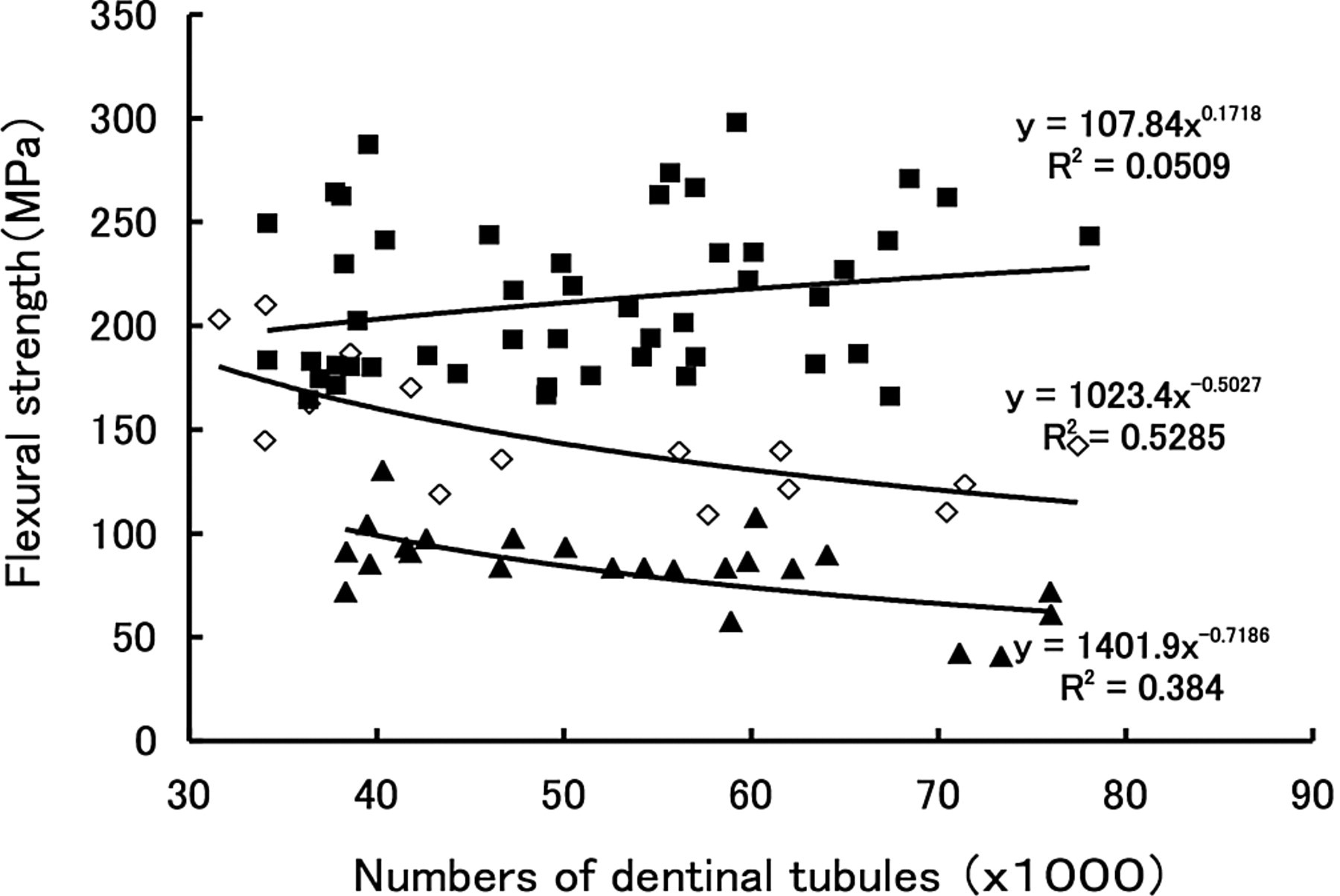
Correlation between the numbers of dentinal tubules and the flexural strength of heated and UV-irradiated human dentin. ▪ 140° C heating group, n = 48. ⋄ 5-minute UV irradiation group, n = 15. ▴control group, n = 24.
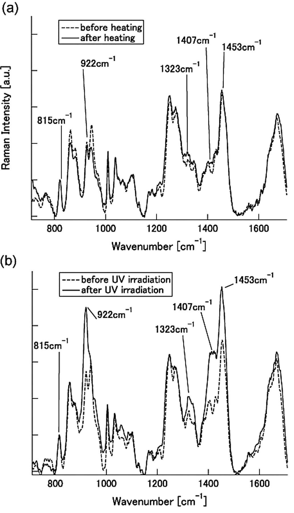
Laser Raman spectra of dentin collagen with heating up to 140°C
Footnotes
Notes
Acknowledgements
This study was supported by a Grant-in-Aid for Scientific Research (No. 19390482) from the Japan Society for the Promotion of Science.