
Abstract
Aspartate-serine-serine (DSS) repeats are abundant in naturally occurring proteins that are critical for tooth formation. We recently developed octuplet repeats of aspartate-serine-serine (8DSS) peptides to promote the nucleation of calcium phosphate carbonate from free ions. In this paper, we report a possible role of 8DSS in promoting mineral deposition onto human enamel. Human enamel specimens were demineralized, exposed briefly to 8DSS solution, and then exposed to simulated body fluid that favors mineral deposition. At various stages of treatment, nano-mechanical behaviors, namely, hardness and elastic modulus, were determined by nano-indentation. Nano-indentation results showed that 8DSS treatment effectively improved the mechanical and elastic properties of demineralized enamel. The values of hardness and elastic modulus for the 8DSS-treated enamel were significantly higher than those of surfaces without 8DSS treatment. Furthermore, 8DSS peptides promoted the uniform deposition of nano-crystalline calcium phosphate carbonate over demineralized enamel surfaces and reduced surface roughness of demineralized enamel.
Introduction
Human enamel is a highly mineralized extracellular matrix including 96% inorganic mineral and 4% organic material with water. The inorganic mineral is a crystalline hydroxyapatite (HA) organized in a highly complex three-dimensional micro- and nano-architecture that contributes to enamel’s remarkable hardness (Landis et al., 1993; Nanci, 2003).
Enamel is relatively stable in the healthy oral environment, where saliva and oral fluid continuously promote the balance between dissolution and deposition of minerals. However, if dissolution occurs at a rate kinetically greater than that of mineral deposition, demineralization or dental caries can result (Suga and Watabe, 1992; Cuy et al., 2002). Currently, the most common treatment for clinically detectable caries is to fill the decayed tissue with artificially restorative materials. While this method is effective, a preferred approach is to promote the remineralization of small, incipient demineralized lesions before they advance into full lesions (ten Cate and Featherstone, 1991). At a minimum, this latter approach is thought to reduce the likelihood of decay by slowing demineralization. Several strategies involving the use of fluorides and mineral precursors such as amorphous calcium phosphates and silicas are currently available (Tung and Eichmiller, 2004; Seidel-Bittke, 2005). Also, casein phosphopeptide-amorphous calcium phosphate (CPP-ACP) has been shown to promote mineral deposition and to increase enamel hardness (Panich and Poolthong, 2009). However, the effect of CPP-ACP remineralization is not universally reproduced (Lennon et al., 2006), and a recent review concluded that “the quantity and quality of clinical trial evidence are insufficient to make conclusions regarding the long-term effectiveness of casein derivatives, specifically CPP-ACP, in preventing caries in vivo and treating dentin hypersensitivity or dry mouth hardness” (Azarpazhooh and Limeback, 2008). Therefore, a need exists for alternative peptides that can promote mineral deposition onto dental tissues.
Dentin phosphoprotein (DPP) is the most abundant non-collagenous extracellular matrix component in dentin and is known to play a critical role in tooth mineralization (He and George, 2004). DPP is highly acidic (pI ~ 1.1), due in part to high concentrations of serine (45-50%) and aspartic acid (35-38%) (Jonsson and Fredriksson, 1978; Stetler-Stevenson and Veis, 1987). Furthermore, this highly anionic DPP is unusually specific to calcium ions (Kuboki et al., 1979).
Although intact DPPs have been evaluated for dental remineralization, DPP-derived peptides may offer several potential advantages over entire proteins. First, peptides can be synthesized in high purity, and can be formulated to optimize shelf life. Besides manufacturing efficiency, the synthetic route also avoids the common concerns regarding proteins derived from animal sources, such as allergies, immunogenicity, and potential disease transmissions. Finally, overexpression of DPP protein during growth and development is associated with abnormal enamel rod organization and weakened enamel (White et al., 2007). Hence, compact structure of the peptides may offer better conformational fit than intact DPP proteins on enamel, bone, and synthetic materials surfaces to favor mineralization where the intact proteins are less efficient.
Human DPP contains numerous repetitive nucleotide sequences of aspartate-serine-serine that are believed to promote the formation of hydroxyapatite (George et al., 1996). Several small peptides have been designed based on the sequence of the dentin phosphoprotein, and these peptides, consisting of multiple repeats of the tripeptide aspartate-serine-serine (DSS), bind with high affinity to calcium phosphate compounds. Moreover, these small peptides can recruit calcium phosphate to peptide-derived polystyrene beads (Yarbrough et al., 2010). Of these, the octuplet repeats of aspartate-serine-serine, 8DSS peptides, are the most active in the mediation of biologically directed mineral deposition (Sikes et al., 1991; He et al., 2003; Qiu et al., 2004; Bigi et al., 2006). However, the ability of 8DSS peptides to promote mineral deposition remains unknown. The objective of this study was to investigate the effects of 8DSS peptides on human enamel surface by evaluating surface microstructures and nano-mechanical behavior.
Materials & Methods
Materials Preparation
Fifteen human adult molars were collected from local dental clinics, cleaned of debris, and stored in distilled water with 0.5% thymol solution until use. Since nano-indentation requires smooth substrate surfaces, each whole tooth was embedded in long-cure epoxy resin (Leco, St. Joseph, MI, USA), and then the embedded teeth were sectioned along the parallel median plane with a diamond wheel (331-CA, Struers, Cleveland, OH, USA) on a precision cut-off machine (Accutom 50, Struers, Cleveland, OH, USA) under copious irrigation. Subsequently, the surfaces were polished sequentially with 800- and 1200-grit silicon carbide (SiC) sandpapers, followed by 3 µm, 1 µm, and 0.5 µm Al2O3, and 0.06 µm colloidal silica. All polished samples were individually sonicated in distilled water for 5 min to remove residual abrasives.
Demineralization and Surface Treatment of Enamel Surface
Enamel surfaces were demineralized with 35% phosphoric acid for 30 sec and then rinsed thoroughly with distilled water. The demineralized surfaces were exposed to either 0 or 12.5 µM 8DSS peptide dissolved in HEPES buffer solution for 30 min, followed by immersion in simulated body fluid (SBF) at 37°C for 24 hrs (pH 6.8) to promote the nucleation and growth of hydroxyapatite crystals (Murphy and Mooney, 2002; Papanearchou et al., 2005; Kokubo and Takadama, 2006).
Mechanical Characterization by Nano-indentation
Nano-indentation testing was carried out in an MTS XP nano-indenter (Agilent Technologies, Inc., Vista, CA, USA) with a three-sided Berkovich diamond tip. Both the diamond tip and the nano-indenter were calibrated with fused silica prior to testing. Each nano-indentation run consisted of two steps: loading and unloading segments. From the oscillation of the resulting depth signals, the contact stiffness was continuously measured so that the values of hardness and elastic modulus could be obtained along the various indent depths. The amplitude of the force oscillation was 0.1 µN, and 20 indentations for each sectioned enamel specimen were made on the dry surface after different treatments.
Due to the organic matrix phase, the mechanical behavior of human enamel is significantly sensitive to strain rate (Zhou and Hsiung, 2006). Efforts were made to ensure that the materials were loaded at a constant strain rate of 0.05 sec−1 in the loading segment for all tests.
Surface Characterization by SEM, AFM, and FIB/TEM
The microstructure of enamel was investigated with the use of a variable-pressure scanning electron microscope (VP-SEM) operated in secondary image mode without conductive coating on the top of the surface, which was beneficial for surface observation by avoiding carbon or gold coating artifacts. Additionally, the surface morphology was characterized by means of a Nanoscope IIIa atomic force microscope (AFM; Veeco Instruments, Santa Barbara, CA, USA) with tapping mode. The scan size and scan rate were 20 µm x 20 µm and 1 Hz, respectively. The average surface roughness and localized average roughness (Ra) were calculated from 3 µm x 3 µm and 1 µm x 1 µm squares, respectively, on an enamel rod for each sectioned enamel specimen.
Ultrathin cross-sectional specimens were prepared by focused ion beam (FIB; Nova 600 FIB, FEI Company, Hillsboro, OR, USA) (Engqvist et al., 2004), and then the microstructure of the outermost enamel surface was characterized by transmission electron microscopy (TEM; TF20 TEM, FEI Company, Hillsboro, OR, USA).
Results
Mechanical Characterization
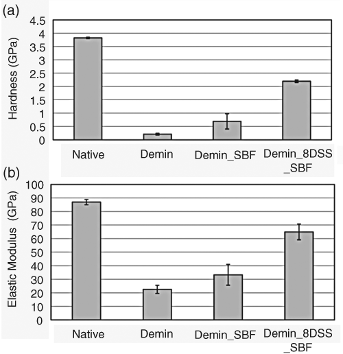
For evaluation of the ability of 8DSS peptide to promote mineral deposition, the native enamel specimens were acid-etched, rinsed, DSS-peptide-treated, and subjected to SBF treatment. The average surface hardness of enamel decreased from 3.83 to 0.21 GPa after demineralization (p < 0.0001) (Fig. 1a). The average hardness of demineralized enamel remineralized in SBF without 8DSS peptide treatment increased from 0.21 to 0.69 GPa (p < 0.01). In contrast, the average hardness increased to 2.20 GPa when the demineralized enamel was exposed to 12.5 µM 8DSS peptide and SBF (p < 0.0001).
Average surface hardness
Similarly, the average elastic modulus changed substantially, from 86.96 to 22.57 GPa, a 74% decrease (p < 0.0001), after demineralization (Fig. 1b). The average elastic modulus of demineralized enamel remineralized in SBF without 8DSS peptide treatment increased from 22.57 to 33.34 GPa (p < 0.05), while pre-exposure to 12.5 µM 8DSS peptide increased the elastic modulus to 64.93 GPa (p < 0.0001). These results indicated that treatment with 12.5 µM 8DSS peptide effectively increased both surface hardness and elastic modulus of demineralized enamel.
Surface Characterization
The new minerals may have been deposited into the interprismatic fissures and intercrystalline spaces; however, the demineralized enamel treated with SBF alone (Fig. 2e) produced prismatic and interprismatic surfaces similar to those observed in demineralized controls (Fig. 2c). The nanorods (black arrow in Fig. 2f) inside each enamel rod after SBF treatment became larger compared with those after the demineralization process (white arrow in Fig. 2d).
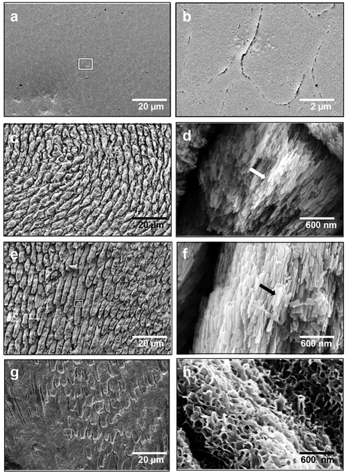
The scanning electron micrographs (SEM) of enamel upon different treatments.
Exposure to 12.5 µM 8DSS peptide prior to SBF treatment notably resulted in the mineral deposition onto enamel prisms and interprismatic spaces that appeared much smoother than demineralized controls (Figs. 2c, 2g). Most importantly, high-magnification SEM revealed a definitive change in surface morphology, from elongated hydroxyapatite nanorods in demineralized enamel to nanoscale flakes (Fig. 2h).
Based on AFM data, the average surface roughness of polished native enamel was 65 nm, and the localized average roughness was 33 nm (Fig. 3a). After surfaces were acid-etched, the average surface roughness increased from 65 to 2267 nm (p < 0.0001), and the localized average roughness was 457 nm (Fig. 3b). The overall average surface roughness and localized average roughness were 1829 nm and 246 nm, respectively (p < 0.0001; Fig. 3c), when demineralized enamel was treated with SBF alone. However, analysis of the AFM data for demineralized enamel treated with 12.5 µM 8DSS peptide prior to SBF immersion showed a reduction in average surface roughness from 2267 nm to 1734 nm, and the average localized roughness was reduced from 457 nm to 107 nm (p < 0.001; Fig. 3d).
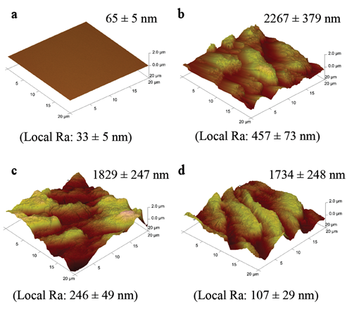
Atomic force micrographs for
These surface changes were further evaluated by TEM. A low-magnification TEM image of the polished native enamel prior to any surface treatment is shown in Fig. 4a, and the two insets are the high-magnification image and the corresponding electron diffraction patterns for the marked area, showing that the top surface of native enamel was flat and the hydroxyapatite within native enamel was highly crystalline. Although the low-magnification TEM image showed that the surface roughness increased to thousand-scale nanometers (Fig. 4b), the outermost remaining enamel maintained the periodicity and crystallinity (Fig. 4b insets) after demineralization. SBF treatment alone, without 8DSS peptide, promoted the deposition of carbonate-apatite [Ca10(PO4CO3OH)6(OH)2] that appeared to contain a mixture of crystalline and amorphous phases (white arrow in Fig. 4c left inset) on the enamel surface, while it produced crystalline calcium phosphate carbonate [Ca10(PO4)6CO3] minerals, forming a network on the top of the enamel surface with 8DSS peptide exposure prior to SBF treatment (Fig. 4d).
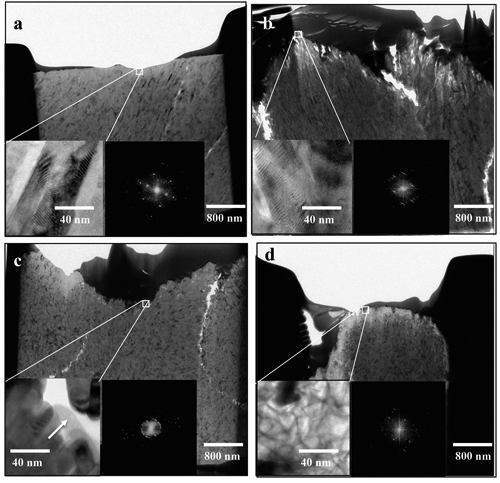
Transmission electron micrographs showed that
Discussion
The improvements in surface roughness, hardness, and elastic modulus were likely related to the formation of carbonate minerals on the surface of demineralized enamel. Furthermore, the values of hardness and elastic modulus depended on the amount and the microstructure of the newly grown mineral. Without applying 8DSS peptide, SBF treatment alone promoted the formation of carbonate-apatite [Ca10(PO4CO3OH)6(OH)2] on top of the demineralized enamel surface consisting of hydroxyapatite [Ca10(PO4)6(OH)2]. The surface mechanical and elastic properties had been increased, and the surface roughness had been reduced without significant changes in the morphology of demineralized enamel. In contrast, 8DSS peptide treatment promoted significant crystalline calcium phosphate carbonate deposition that appeared to cover all enamel prisms and also filled the spaces at enamel prism peripheries. This pattern of mineral deposition may be responsible for further reduction in localized surface roughness (from ~ 450 nm to ~ 100 nm). Therefore, 8DSS peptide exerted influence not only on the amount of mineral deposition, but also on the types and morphology of deposited minerals.
DSS peptides possess a high affinity to bind with the HA surface (Yarbrough et al., 2010) and a strong interaction with calcium and phosphate ions (George et al., 1996). The 8DSS peptides may play two roles in the remineralization process. First, they limit calcium and phosphate ions to depart from the demineralized enamel when 8DSS peptides bind with HA. Second, 8DSS peptides advance the capture of calcium and phosphate ions from SBF solution to form new mineral depositing on the demineralized enamel. As a result, the application of 8DSS peptides greatly improved the mechanical and elastic properties of demineralized enamel.
The fact that mineralization treatments did not return the mechanical properties to native enamel is likely due to the severity of this demineralization procedure (Hu and Featherstone, 2005), which uses a strong acid to etch enamel more aggressively (pH < 1) and more rapidly (30 sec) than other demineralization models that slowly create demineralized lesions with methylcellulose/lactic acid (pH 4.6) over 4 wks (ten Cate et al., 1996). Since TEM image and electron diffraction patterns showed that at least some of the residual enamel after demineralization treatment appeared to be intact, the decrease in mechanical property may be due to the loss of minerals and disruption of organic matrix. Although this demineralization model does not represent all enamel defects, the results here motivate additional evaluation of 8DSS effects in other demineralization models.
To the best of our knowledge, this study is the first demonstration of the use of 8DSS peptides to promote mineral deposition onto human enamel and improve the surface properties of demineralized enamel. The choice of octuplet repeats, the use of HEPES as a carrier, and the dose were selected based on extensive pilot experiments. However, much more work remains to make this approach more practical. For example, the choice of SBF was made due to its well-documented ability to produce apatites, facilitating the analysis of the effects of 8DSS. Future work is needed to document the effects of 8DSS in saliva and in other mineralization conditions. Pilot experiments show that 8DSS peptides can also promote the effects of commercially available remineralization agents (e.g., ACP) after only 60 sec of exposure to ACP (Tung and Eichmiller, 2004).
Footnotes
Acknowledgements
The authors express their gratitude to Dan Yarbrough and Jian He, School of Dentistry, University of California, Los Angeles, for providing 8DSS peptides. We are grateful for the financial support from C3 Jian Inc., the National Science Council (NSC 98-2218-E-035-002), and priority research fields plan grant (09G27101) from Feng Chia University, Taiwan, and for the collaboration of Center for Micro/Nano Science and Technology, National Cheng Kung University, Taiwan.