






Abstract
Lithium is an activator of β-catenin signaling, and β-catenin mediates bone acquisition in response to mechanical loading in the bone. We tested the hypothesis that lithium enhances new bone formation during midpalatal suture expansion. Forty-eight Wistar rats with or without suture expansion were gavage-fed daily with lithium chloride or sodium chloride. We labeled the rats with bromodeoxyuridine to evaluate the proliferation and differentiation of the osteoprogenitors. Lithium increased β-catenin expression and cell proliferation in expanding sutures. Initial delay in the differentiation of osteoprogenitors into mature osteoblasts by lithium treatment corresponded with expansion of pre-osteoblasts, which preceded the increase of new bone formation in the suture. These results suggested that β-catenin regulates proliferation of osteoprogenitors and maturation of osteoblasts during midpalatal suture expansion osteogenesis, and that lithium enhances bone regeneration by elevating β-catenin expression. Lithium treatment could be a pharmaceutical aid to improve the stability of orthodontic treatment like rapid palatal expansion.
Introduction
Mechanical strain across craniofacial sutures is well-recognized as a controlling stimulus for bone formation and remodeling in the skull and face (Alaqeel et al., 2006). This is the basis upon which growth modification of the craniofacial complex can be achieved through various orthopedic-orthodontic appliances. Stretching of the sutures, such as during rapid palatal expansion, induces a biologic chain of events that leads to new bone deposition in the midpalatal suture (Kobayashi et al., 1999). The clinical results are increased maxillary skeletal and dento-alveolar widths (Cameron et al., 2002). Although the midpalatal suture can be successfully opened, relapse of the posterior dentition width has been frequently reported (Lima et al., 2005; Gurel et al., 2010). Long-term stability of rapid palatal expansion is related to several factors, including resistance of the maxillary walls, soft tissue adaptation, and age (Baccetti et al., 2001; Lagravere et al., 2005). However, a major reason for early relapse is inadequate bone formation in the suture (Sarnas et al., 1992; Sannomiya et al., 2007). Therefore, enhancing bone formation in the midpalatal suture may improve the stability of maxillary expansion.
β-catenin, a central effector of canonical Wnt signaling pathway, participates in every stage of skeletogenesis in craniofacial bones, from the self-renewal and proliferation of skeletal stem cells to the specification of osteoprogenitors and the maturation of osteoblasts (Liu et al., 2007; Leucht et al., 2008). Wnts are secreted glycoproteins that bind to a receptor complex consisting of Frizzled proteins and low-density lipoprotein receptor-related protein 5/6 (LRP5/6) (Church and Francis-West, 2002). Bound ligands inhibit the activity of glycogen synthase kinase-3β (GSK-3β), followed by an increase in the amount of β-catenin in the cell. As a result, β-catenin is stabilized and translocates into the nucleus, where it binds to TCF/LEF transcription factors to regulate the expression of Wnt target genes (Church and Francis-West, 2002). Recently, one report showed that β-catenin functions as an important mechanotransduction mediator in osteoblasts (Case et al., 2008). Mechanical strain induces accumulation and nuclear transformation of active β-catenin in osteoblasts (Hens et al., 2005; Armstrong et al., 2007; Case et al., 2008). Moreover, conditions that increase β-catenin levels synergize the downstream effects of strain on bone (Robinson et al., 2006; Case et al., 2008). It is thus reasonable to hypothesize that activating β-catenin signaling will enhance bone regeneration in the midpalatal suture in response to mechanical expansion.
Lithium chloride (LiCl), a drug used for decades to treat bipolar disorder, increases β-catenin signaling by inhibiting GSK-3β (Hedgepeth et al., 1997; Schou, 2001). LiCl induces alkaline phosphatase expression in C3H10T1/2 pluripotent stem cells (Bain et al., 2003) and promotes osteocalcin mRNA expression and bone formation in calvarial osteoblast cultures (Kugimiya et al., 2007). In mice, lithium administration stimulates Wnt/β-catenin-mediated transcription in bone, leading to an increase in bone mass and an improvement in fracture healing (Clement-Lacroix et al., 2005; Chen et al., 2007). Thus, the purpose of our current study was to test whether activation of β-catenin by lithium enhances new bone formation in the expanding midpalatal suture.
Materials & Methods
Midpalatal Suture Expansion and Lithium Administration
Thirty male 4-week-old Wistar rats were subjected to rapid midpalatal suture expansion (Kobayashi et al., 1999). An expansion spring with 2 helices was fabricated with 0.018-inch orthodontic wire (TP Original Premier Wire, TP Orthodontic Appliance Co., Ltd., WuXi, China). The initial expansion force was calibrated to 100 ± 5 g. The spring was fitted between the upper right and left molars and was secured by Transbond™ LR light-cured resin (3M Unitek, Monrovia, CA, USA) (Fig. 1A). Animals were gavage-fed daily with 200 mg/kg body weight of LiCl (Sigma-Aldrich, St. Louis, MO, USA) in 300 µL de-ionized water or the same dose of NaCl solution. The rats were sacrificed after 3, 5, and 7 days (n = 5). Another 18 rats without suture expansion were also treated with LiCl or NaCl and sacrificed at the same time (n = 3) (Fig. 1B). The experiment was approved by Shanghai Jiao Tong University (Animal Ethics Approval No. SCXK 2008-0016).

Intra-oral view of the helix spring for midpalatal suture expansion in a rat
In vivo BrdU Labeling
Rats were labeled with bromodeoxyuridine (BrdU, Sigma-Aldrich) on experimental day 2, when cell proliferation was most active after midpalatal suture expansion (Kobayashi et al., 1999). For proliferation analysis, rats were given a single intraperitoneal injection of 5 mg/100 g body weight BrdU and sacrificed on day 3. For osteoblast differentiation analysis, animals were given 2 injections of 2.5 mg/100 g body weight BrdU spaced 8 hrs apart and sacrificed on day 5 (Erlebacher et al., 1998) (Fig. 1B).
Histochemical Staining
Paraffin sections were stained with 0.01% toluidine blue (Sigma-Aldrich) for histological evaluation. The BrdU signals and the expression of β-catenin were detected by immunohistochemical staining (Tang and Rabie, 2005). The mouse monoclonal β-catenin (E-5) primary antibody was purchased from Santa Cruz (Santa Cruz Biotechnology Inc., Santa Cruz, CA, USA) (Alvarez et al., 2004).
Quantitative and Statistical Analysis
Images were captured with a microscope (Nikon ECLIPSE E200, Nikon Instruments, Inc., Melville, NY, USA) and digital camera system (Pixera Penguin 600CL CCD, Pixera Corporation, San Jose, CA, USA). Semi-quantitative analysis was performed with computer-assisted image analysis software (Image-Pro Plus, Media Cybernetics, Inc., Bethesda, MD, USA). The amount of new bone formation was recorded as the percentage of new bone area relative to the total fibrosis area between the 2 sides of the suture cartilage when viewed with toluidine blue staining (Fig. 2). BrdU-labeled cells (at least 80 pixels) in the suture were identified and counted by the computer (Tang and Rabie, 2005). Data are presented as the number of labeled cells per 100,000 µm2 suture area. Labeled cells embedded in new bone matrix were recorded separately. All measurements were performed on triplicate sections for each animal, and the data were collected again 4 wks later by the same observer. The difference between LiCl- and NaCl-treated animals was statistically analyzed by unpaired t tests (SPSS for Windows, Version 11.0, SPSS, Inc., Chicago, IL, USA).
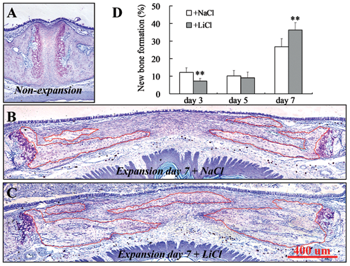
Toluidine blue staining showing an overview of a rat’s midpalatal suture (
Results
The midpalatal suture of the rat is a cartilaginous synchondrosis (Fig. 2A). Upon expansion, the 2 chondrocyte layers were forced apart laterally (Figs. 2B, 2C). Compared with NaCl-treated rats, quantitative analysis showed that new bone formation on day 3 was greatly retarded in LiCl-treated animals (Fig. 2D). On day 7, however, a significant increase in new bone volume was identified after LiCl administration (Figs. 2B-2D). β-catenin immunostaining was observed largely in pre-hypertrophic chondrocytes, with some staining in proliferative and mineralizing hypertrophic chondrocytes in suture cartilage (Fig. 3A). During suture expansion, β-catenin staining was detected in pre-osteoblasts lining the bone surface and in some mesenchymal cells close to the new bone (Figs. 3B-3E). A substantial increase in the expression level of β-catenin persisted in LiCl-treated rats (Figs. 3G-3J). In non-expansion sutures, no significant difference was observed between the LiCl- and NaCl-treated rats during the course of the experiment.
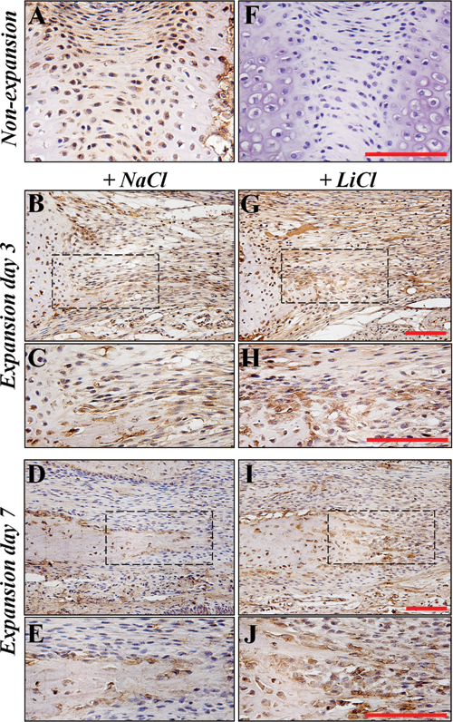
Immunohistochemical staining showing β-catenin expression in the midpalatal suture cartilage
When the rats were labeled with BrdU on day 2 and sacrificedon day 3, limited BrdU-positive cells were detected in non-expansion sutures (Fig. 4A). With expansion, many BrdU-labeled cells were seen approaching the new bone surface, and others were scattered in the suture (Figs. 4B, 4C). Few labeled cells were observed inside the bone. On expansion day 5, 38% of labeled cells were detected inside new bone matrix in NaCl-treated animals (Figs. 4D, 4F). In contrast, only 12% of the labeled cells were embedded in new bone in LiCl-treated rats (Figs. 4E, 4F). Nevertheless, the total number of labeled cells was significantly increased in LiCl-treated groups on both day 3 and day 5 (Fig. 4F).

BrdU labeling showing proliferating cells in the midpalatal suture cartilage
Discussion
The role of the Wnt/β-catenin pathway in controlling embryonic bone development and bone mass has been well-documented. In our current study, β-catenin proteins were observed in the synchondrosis of the non-expansion suture (Fig. 3A). A similar expression pattern has been reported in the growth plate, where β-catenin regulates chondrocyte differentiation and maturation (Tamamura et al., 2005). Wnt/β-catenin signaling may also be involved in the endochondral bone formation of the rat midpalatal suture during natural growth. Upon expansion, however, the stretching force induces an alternative bone formation mechanism consisting of intramembranous ossification in the midpalatal suture, where mesenchymal cells proliferate, differentiate into osteoblasts, and lay down bone matrix (Kobayashi et al., 1999). In our current study, we showed that β-catenin may actively participate in this process, and that oral administration of LiCl enhanced bone regeneration, possibly by elevating β-catenin expression.
We first identified β-catenin expression in mesenchymal cells (osteoprogenitors) and pre-osteoblasts around the new bone front on day 3 after midpalatal suture expansion (Figs. 3B, 3C). β-catenin promotes mitotic activity of osteoprogenitors in cranial sutures by stimulating cyclin D1, a key regulator of cell cycle entry (Liu et al., 2007). Coincidentally, an increase in the expression of active β-catenin and its target genes, including cyclin D1, was reported in osteoblasts subjected to mechanical loading (Robinson et al., 2006; Armstrong et al., 2007). Furthermore, elevating β-catenin signaling with a GSK-3β inhibitor, such as LiCl, enhances the anabolic effects of strain on bone (Robinson et al., 2006; Case et al., 2008). Thus, it is likely that mechanotransduction and osteoblast proliferation are connected through β-catenin. In our current study, stronger β-catenin staining was observed in expanding sutures in rats treated with LiCl (Fig. 3G, 3H). These results were not surprising, because the dosing schedule used in this study was previously demonstrated to activate β-catenin signaling in bone (Clement-Lacroix et al., 2005; Chen et al., 2007). An important finding of our current study is that the elevated expression of β-catenin in osteoprogenitors coincided with a significant increase in the number of proliferative cells in the expanding suture of LiCl-treated rats (Figs. 3, 4). Therefore, it is conceivable that midpalatal suture expansion creates a strain alignment that causes a deformation of the cytoskeleton of osteoprogenitor cells in the suture (Kobayashi et al., 1999), triggering β-catenin expression. β-catenin subsequently induces cell division, possibly by up-regulating its target gene cyclin D1 (Liu et al., 2007). Analysis of these data, taken
We have shown that a significant increase in cellular renewal occurred during expansion of the midpalatal suture in response to LiCl treatment, but we have not discussed how these osteoprogenitors become osteoblasts and lay down bone matrix. It has been estimated that 3 days are required for osteoprogenitor cells to divide and differentiate into osteoblasts (Turner et al., 1998). We thus traced the migration of the labeled cells by visualizing BrdU signals on day 5. Our current results showed that 38% of labeled cells were embedded in new bone matrix in NaCl-treated rats (Figs. 4D, 4F), meaning that these proliferating osteoprogenitors had differentiated into mature osteoblasts. In contrast, only 12% were inside new bone in LiCl-treated animals (Figs. 4E, 4F). This result suggested that LiCl inhibited the differentiation of the osteoprogenitors into mature osteoblasts. As a result, the amount of new bone formation may have been initially retarded on day 3 in the LiCl-treated rats (Figs. 2D, 4B, 4C). Our current results are in agreement with earlier in vitro findings that exogenous Wnt 3a, as seen with increased β-catenin nuclear localization, led to increased proliferation of adult human multipotential mesenchymal cells and suppressed osteogenic differentiation of these cells (Boland et al., 2004). Similarly, activation of the Wnt pathway resulting from a mutation in Lrp5 or stabilization of the gene encoding β-catenin causes a delay in injury-induced bone regeneration (Chen et al., 2007; Kim et al., 2007). In these mice, osteoprogenitor cells in the injury site remain in a proliferative state, and differentiation into osteoblasts is blocked (Chen et al., 2007; Kim et al., 2007). Nevertheless, β-catenin plays a different role in undifferentiated mesenchymal cells and cells committed to the osteoblast lineage (Chen et al., 2007). Transgenic mice expressing a constitutively active form of β-catenin in all cells show an absence of bone regeneration at fracture sites, whereas osteoblast-specific expression of stabilized β-catenin enhances bone healing (Chen et al., 2007). In our current investigation, it is important to note that LiCl led to a significant increase in the number of BrdU-labeled cells around the bone surfaces on day 5 (Fig. 4E), indicating that proliferating osteoprogenitors were committed osteoblasts (pre-osteoblasts). The elevated β-catenin signals in the large population of pre-osteoblasts in LiCl-treated animals thus led to a robust and significant increase in new bone formation by day 7 (Figs. 3I, 3J, 2D). These results do not conflict with those of Chen et al. (2007), who reported that LiCl interferes with bone healing when given 2 wks before the fracture. In their report, β-catenin was continuously activated, similar to the mutation resulting in constitutively active β-catenin, which leads to a block of osteoblast differentiation and a delay in bone healing (Chen et al., 2007). In our current study, LiCl was given on the day of suture expansion. The differentiation and maturation of pre-osteoblasts were initially inhibited, but not totally blocked. Thus, analysis of our data further supports the importance of β-catenin signaling in regulating the proliferation, differentiation, and function of osteoblasts during bone regeneration.
Growth modification by the use of mechanical force on the bony margins of many sutures in the craniofacial complex, including the midpalatal suture, has been successfully used to treat disorders of craniofacial development. Although many factors, such as the amount, direction, and duration of force applied, play an essential role in the success of such treatments, manipulating the key regulatory factors and signaling pathway through which biomechanical stimulations regulate bone formation and remodeling at the suture will be a novel approach to improving treatment. Our current study demonstrated that activating β-catenin signaling by oral administration of LiCl in rats enhanced new bone regeneration during midpalatal suture expansion. Oral lithium has been used widely and effectively in humans for over a half-century. Yet, the adverse effects of systemic lithium intake remain a major concern. Local administration of other β-catenin activators or clinical trials in humans receiving lithium therapy for bipolar disorder should help to confirm its potential use in daily orthopedic-orthodontic practice.
Footnotes
Acknowledgements
This research was supported by the Research Fund of the Science and Technology Committee of Shanghai (Grant No. 08DZ2271100 & 10QH1401600) and by the Shanghai Leading Academic Discipline Project (Grant No. S30206).