






Abstract
Previous studies have demonstrated that botulinum toxin type A (BoNT-A) attenuates orofacial nociception. However, there has been no evidence of the participation of the voltage-gated sodium channels (Navs) in the antinociceptive mechanisms of BoNT-A. This study investigated the cellular mechanisms underlying the antinociceptive effects of BoNT-A in a male Sprague-Dawley rat model of trigeminal neuropathic pain produced by malpositioned dental implants. The left mandibular second molar was extracted under anesthesia, followed by a miniature dental implant placement to induce injury to the inferior alveolar nerve. Mechanical allodynia was monitored after subcutaneous injection of BoNT-A at 3, 7, or 12 d after malpositioned dental implant surgery. Subcutaneous injections of 1 or 3 U/kg of BoNT-A on postoperative day 3 significantly attenuated mechanical allodynia, although 0.3 U/kg of BoNT-A did not affect the air-puff threshold. A single injection of 3 U/kg of BoNT-A produced prolonged antiallodynic effects over the entire experimental period. Treatment with BoNT-A on postoperative days 7 and 12, when pain had already been established, also produced prolonged antiallodynic effects. Double treatments with 1 U/kg of BoNT-A produced prolonged, more antiallodynic effects as compared with single treatments. Subcutaneous administration of 3 U/kg of BoNT-A significantly inhibited the upregulation of Nav isoform 1.7 (Nav1.7) expression in the trigeminal ganglion in the nerve-injured animals. These results suggest that antinociceptive effects of BoNT-A are mediated by an inhibition of upregulated Nav1.7 expression in the trigeminal ganglion. BoNT-A is therefore a potential new therapeutic agent for chronic pain control, including neuropathic pain.
Introduction
It has been well established that the effects of botulinum toxin type A (BoNT-A) in treatment applications involving the skeletal muscles are due to inhibition of the exocytosis of acetylcholine excreted at the nerve endings and the induction of muscle paralysis. Recent data support the use of BoNT-A in the treatment of several painful states, including neuropathic pain. In previous animal studies, inflammatory pain induced by formalin (Cui et al. 2004; Luvisetto et al. 2006) or carrageenan (Favre-Guilmard et al. 2009) was reduced by the subcutaneous administration of BoNT-A in the affected paw. Moreover, BoNT-A reduced both mechanical and cold allodynia in neuropathic pain models in rats with L5/L6 spinal nerve ligation (Park et al. 2006), chronic constriction (Luvisetto et al. 2007), and partial transection of the sciatic nerve (Bach-Rojecky et al. 2005). In other clinical studies, BoNT-A was considered to be effective against headache (Göbel et al. 2001), idiopathic trigeminal neuralgia (Türk et al. 2005), and postherpetic neuralgia (Liu et al. 2006). These results suggested that BoNT-A has potential therapeutic value in the treatment of neuropathic pain.
Previous studies also demonstrated that BoNT-A attenuates orofacial nociception. In the orofacial area in rats with infraorbital nerve constriction, BoNT-A was found to attenuate mechanical allodynia (Kitamura et al. 2009) and thermal hyperalgesia (Kumada et al. 2012). It was further reported in a clinical study that BoNT-A showed suppressive effects on experimental trigeminal/cervical pain activated by intradermal injection of capsaicin to the forehead (Gazerani et al. 2006). Furthermore, BoNT-A has shown beneficial effects against chronic facial pain, including temporomandibular joint syndrome, postsurgical pain syndromes, essential headache, and idiopathic trigeminal neuralgia (Borodic and Acquadro 2002). Although previous studies have supported the antinociceptive effects of BoNT-A, the underlying cellular mechanisms remain unclear (Kitamura et al. 2009; Kumada et al. 2012).
Generally, the antinociceptive effect of BoNT-A is hypothesized to involve the inhibition of noncholinergic neurotransmitters that are associated with neurogenic inflammation and peripheral sensitization, including substance P, calcitonin gene–related peptide, and glutamate (McMahon et al. 1992; Purkiss et al. 2000; Meng et al. 2007). Pretreatment with BoNT-A has been shown to reduce nociception in a formalin model of pain through blockade of glutamate release (Cui et al. 2004). Moreover, in another study, BoNT-A has been found to decrease the release of calcitonin gene–related peptide in vitro (Meng et al. 2007; Meng et al. 2009). However, to fully elucidate the cellular mechanisms underlying the antinociceptive effects of BoNT-A, further studies will be needed.
Voltage-gated sodium channels (Navs) are large transmembrane protein complexes composed of pore-forming α-subunits and auxiliary β-subunits responsible for the generation of action potentials (Marban et al. 1998). Nine α-subunits (Nav1.1 to Nav1.9) have been identified to date, each exhibiting distinct expression patterns and electrophysiologic and pharmacologic properties (Catterall et al. 2005). Tissue and nerve damage induce changes in the expression and function of specific α-subunits, which can lead to changes in the excitability of sensory neurons. These changes are thought to underlie certain chronic pain conditions (Baker and Wood 2001; Dib-Hajj et al. 2009). However, there has been no evidence of the participation of the Navs in the antinociceptive mechanisms of BoNT-A.
In our present study, we investigated the cellular mechanisms underlying the antinociceptive effects of BoNT-A in a rat model of trigeminal neuropathic pain produced by a malpositioned dental implant. For this purpose, we examined the changes in the air-puff thresholds produced after the subcutaneous administration of BoNT-A in rats with trigeminal neuropathic pain. We also investigated a role of Navs in the antinociceptive effects of BoNT-A because Navs play a critical role in modulating the excitability of most neurons, including nociceptive sensory signaling. To further elucidate the antinociceptive effects of BoNT-A administered in this way, we analyzed the expression of Nav isoforms in the trigeminal ganglion of the treated rats.
Materials and Methods
Animals and Surgery
Experiments were carried out with male Sprague-Dawley rats weighing between 220 and 240 g. The rats were anesthetized with a mixture of ketamine (40 mg/kg, intramuscular) and xylazine (4 mg/kg, intramuscular). The left mandibular second molar was extracted under anesthesia, followed by the placement of a miniature dental implant (1 mm in diameter and 4 mm in length; donated by Megagen) to induce injury to the inferior alveolar nerve as described previously (Han et al. 2010). Animals in a sham group also had their second molars removed but received no dental implant. All procedures involving the use of animals were approved by the Institutional Care and Use Committee of the School of Dentistry, Kyungpook National University, and were carried out in accordance with the ethical guidelines of the International Association for the Study of Pain for the investigation of experimental pain in conscious animals.
Evaluation of Mechanical Allodynia
For behavioral observations, each rat was placed in a customized observation cage in a darkened and noise-free room and acclimated for at least 30 min. Withdrawal responses were evaluated after the application of 10 successive trials of constant air-puff pressure (4-s duration at 10-s intervals) on freely moving rats as described previously (Ahn et al. 2009; Han et al. 2010; Jeon et al. 2012). The behavioral responses observed included escape from air-puff stimulation, aggressiveness, and biting. The intensity and intervals of the air-puff pressure were controlled with a pneumatic pump module (BH2 system; Harvard Apparatus). The air-puffs were applied through a 26-gauge metal tube (length, 10 cm) located 1 cm from the skin at a 90° angle. After injury of inferior alveolar nerve, we searched for the most sensitive area by air-puff stimulation. The most sensitive area includes the lower jaw and mouth angle area of facial region. The air-puff threshold was determined as the air-puff pressure at which the rat responded in 50% of the trials and was 40 psi for naïve animals. Changes in behavior were measured 3 d before malpositioned dental implantation and 3, 5, 7, 9, 11, 14, 18, 21, 25, 32, 39, 46, and 53 d after. All behavioral responses were measured in a blind fashion.
Drug Administration
One hundred units of BoNT-A (Botulax; Hugel, Inc.) were diluted in saline. BoNT-A was then injected subcutaneously into the most sensitive area of facial region with an insulin syringe (31 gauge), under 3% isoflurane anesthesia.
Immunohistochemistry
The rats (n = 5 per group) were anesthetized with a mixture of ketamine (40 mg/kg) and xylazine (4 mg/kg) 6 d after the BoNT-A injection. The rats were perfused transcardially with 0.9% saline, followed by 4% paraformaldehyde in 0.1M phosphate buffer (pH 7.4). The trigeminal ganglion was dissected, postfixed in the same fixative at 4 °C for 2 h, and replaced with 30% sucrose in 0.1M phosphate buffer overnight. The trigeminal ganglion was sectioned at 20 μm and blocked with 10% normal goat serum in phosphate buffered saline (pH 7.4) for 1 h at room temperature. The sections were then incubated at 4 °C overnight with rabbit anti-activating transcription factor 3 (anti-ATF3; 1:3,000; Santa Cruz Biotechnology) and rabbit anti-Nav1.7 (1:400; Alomone Labs). The sections were subsequently incubated with Alexa 555–conjugated rabbit IgG antibody (1:200; Invitrogen) for 2 h at room temperature. The stained sections were observed under a fluorescence microscope (BX 41 and U-RFL-T; Olympus) in the mandibular (V3) division of the trigeminal ganglion, as described previously (Thalakoti et al. 2007). The number of ATF3-positive cells was counted in 5 sections from each rat with I-solution (Innerview Co.). We measured the intensity of Nav1.7 immunofluorescence as the average pixel intensity in 5 rats per group with MetaVue software (Molecular Devices).
Western Blotting
Rats were sacrificed by decapitation, and the trigeminal ganglion was immediately removed from each animal and quickly frozen in liquid nitrogen. Samples were sonicated with Biorupture (Cosmo Bio.) in a lysis buffer containing protease and a phosphatase inhibitor cocktail (Thermo Scientific). For Western blotting, total proteins (40 µg) were separated in a 4%- to 12%-gradient NuPAGE Novex Bis-Tris gel (Invitrogen) and transferred onto a nitrocellulose membrane. The membranes were then blocked with 5% nonfat milk in Tris-buffered saline with 0.1% Tween 20 for 1 h at room temperature and then incubated with primary antibody to Nav1.3, Nav1.6, Nav1.7, and Nav1.8 (Alomone Labs) or glyceraldehyde 3-phosphate dehydrogenase at 4 °C overnight. The blots were subsequently incubated with goat anti-rabbit horseradish peroxidase for 2 h at room temperature. The intensity of each band was determined by quantitative chemiluminescence with a Amersham Imager 600 (GE Healthcare). We used the ImageJ analysis system (National Institutes of Health) to quantify specific bands.
Experimental Protocols
Antinociceptive Effects of Subcutaneously Administered BoNT-A on Trigeminal Neuropathic Pain
We evaluated the effects of BoNT-A on mechanical allodynia in rats with malpositioned dental implants by randomly assigning the animals to the following 4 groups: 0.3, 1, and 3 U/kg of BoNT-A and vehicle (saline). Mechanical allodynia was monitored after subcutaneous injection of BoNT-A on postoperative day (POD) 3.
In some animals, antinociceptive effects were evaluated after repeated administration of low doses of BoNT-A (0.3 or 1 U/kg) on PODs 3 and 4, respectively. We also investigated the antinociceptive effects of BoNT-A when pain had already been established. The changes in air-puff thresholds were monitored following the subcutaneous administration of 3 U/kg of BoNT-A on POD 7 or 12.
Effects of BoNT-A on ATF3 Expression in the Ipsilateral Trigeminal Ganglion
To investigate whether subcutaneous administration of BoNT-A protects against nerve injury that results in the development of neuropathic pain, we examined changes in the expression of ATF3, a marker of neuronal injury, in the ipsilateral trigeminal ganglion. BoNT-A (3 U/kg) was injected subcutaneously on POD 3. Changes in ATF3 expression in the ipsilateral trigeminal ganglion were examined 6 d after the BoNT-A injection.
Effects of BoNT-A on Nav Expression in the Ipsilateral Trigeminal Ganglion
To investigate whether the subcutaneous administration of BoNT-A affected Navs, we examined changes in the expression of Nav1.3, Nav1.6, Nav1.7, and Nav1.8 in the ipsilateral trigeminal ganglion 6 d after BoNT-A injection (3 U/kg, POD 3) by Western blot analysis. We confirmed changes in Nav1.7 expression in the BoNT-A-treated rats by immunohistochemical analysis.
Data Analysis
Differences of behavioral data among groups were compared by repeated-measures analysis of variance, followed by Holm-Sidak post hoc analysis. The Western blotting data were analyzed by 1-way analysis of variance, followed by Holm-Sidak post hoc analysis. In all statistical comparisons, P < 0.05 was the criterion for significant difference.
Results
Antinociceptive Effects of Subcutaneously Administered BoNT-A on Trigeminal Neuropathic Pain
Malpositioned dental implants produced obvious nociceptive behavioral changes in the experimental rats in this study, including a significant reduction in the air-puff threshold on the ipsilateral side of the injured site (Han et al. 2010; Won et al. 2014). Figure 1 shows the effects of BoNT-A on mechanical allodynia in these rats with inferior alveolar nerve injury. A subcutaneous injection of 0.3 U/kg of BoNT-A on POD 3 increased the air-puff threshold but not significantly when compared with the saline-treated group. However, subcutaneous injections of 1 or 3 U/kg of BoNT-A significantly attenuated the mechanical allodynia in these animals (F3,28 = 28.390, P < 0.001). In the group treated with 1 U/kg of BoNT-A, the antiallodynic effects persisted up to POD 18. Moreover, a single injection of 3 U/kg of BoNT-A produced prolonged antiallodynic effects over the entire experimental period relative to the saline-treated group.

Long-term effects of early treatment with botulinum toxin type A (BoNT-A) on mechanical allodynia following the surgical placement of a malpositioned dental implant in rats. The subcutaneous injection of BoNT-A (0.3, 1, 3 U/kg) on postoperative day 3 produced antiallodynic effects in this rat model of neuropathic pain. A single injection of 1 or 3 U/kg of BoNT-A produced prolonged antiallodynic effects. n = 8 animals per group. Arrow, BoNT-A injection on postoperative day 3. *P < 0.05, BoNT-A vs. vehicle-treated group. Results are mean ± SEM at each time point tested.
The present study also compared the antiallodynic effects produced by single or double BoNT-A treatments (Fig. 2). Double treatments with 0.3 U/kg of BoNT-A (PODs 3 and 4) did not induce significant antiallodynic effects when compared with single treatments (F1,14 = 2.749, P = 0.120). However, double treatments with 1 U/kg of BoNT-A produced prolonged antiallodynic effects in this same comparison (F1,14 = 39.373, P < 0.001).
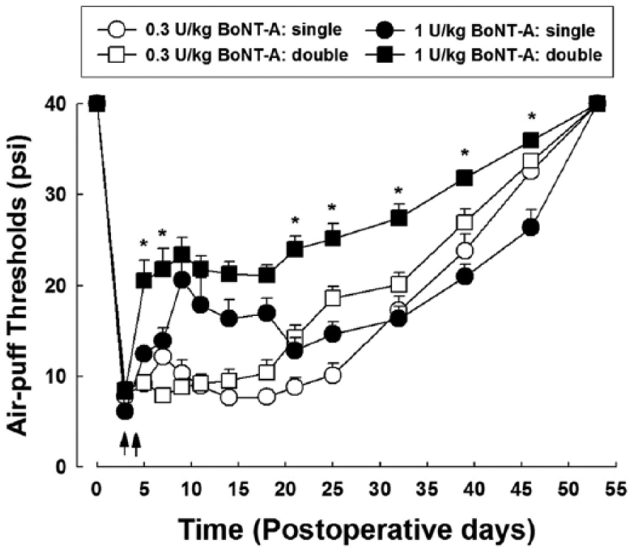
Comparison of the effects of single and double treatment with botulinum toxin type A (BoNT-A) on mechanical allodynia in rats with trigeminal neuropathic pain. Double treatments with 1 U/kg of BoNT-A produced significantly prolonged antiallodynic effects as compared with single treatments. n = 8 animals per group. Arrows, BoNT-A injections on postoperative days 3 and 4. *P < 0.05, single- vs. double-BoNT-A-treated groups. Results are mean ± SEM at each time point tested.
Our present findings also demonstrated antinociceptive effects following subcutaneous injection of BoNT-A (3 U/kg) on POD 7 or 12 (Fig. 3). Treatment with BoNT-A on PODs 7 and 12 significantly increased the air-puff threshold (F3,28 = 26.434, P < 0.001) when pain had already been established. The antiallodynic patterns arising from late treatments with BoNT-A on PODs 7 and 12 were similar to those found with early treatments on POD 3.
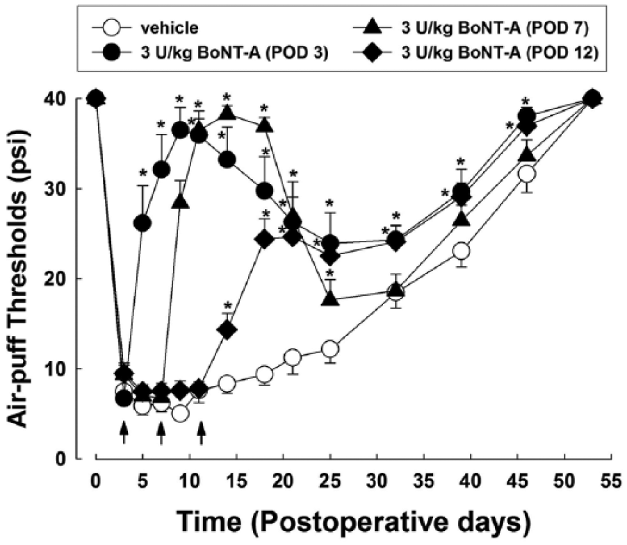
Long-term effects of treatment with botulinum toxin type A (BoNT-A) on mechanical allodynia induced by malpositioned dental implant in rats when pain had already been established. Treatment with BoNT-A (3 U/kg) on postoperative day (POD) 7 or 12 attenuated this mechanical allodynia. n = 8 animals per group. Arrows, BoNT-A injection on POD 3, 7, or 12. *P < 0.05, BoNT-A vs. vehicle-treated group. Results are mean ± SEM at each time point tested.
Effects of BoNT-A on the Expression of Navs in the Trigeminal Ganglion
Figure 4 illustrates changes in the expression of Nav1.3, Nav1.6, Nav1.7, and Nav1.8 in the trigeminal ganglion following the subcutaneous administration of BoNT-A (3 U/kg). The malpositioned dental implant produced significant increases in the expression of Nav1.6, Nav1.7, and Nav1.8, but nerve injury did not affect the expression of Nav1.3 in the trigeminal ganglion. Subcutaneous administration of 3 U/kg of BoNT-A did not affect the upregulation of Nav1.6 and Nav1.8 expression in the nerve-injured animals. However, subcutaneous administration of 3 U/kg of BoNT-A significantly inhibited the upregulation of Nav1.7 expression alone in the nerve-injured animals.
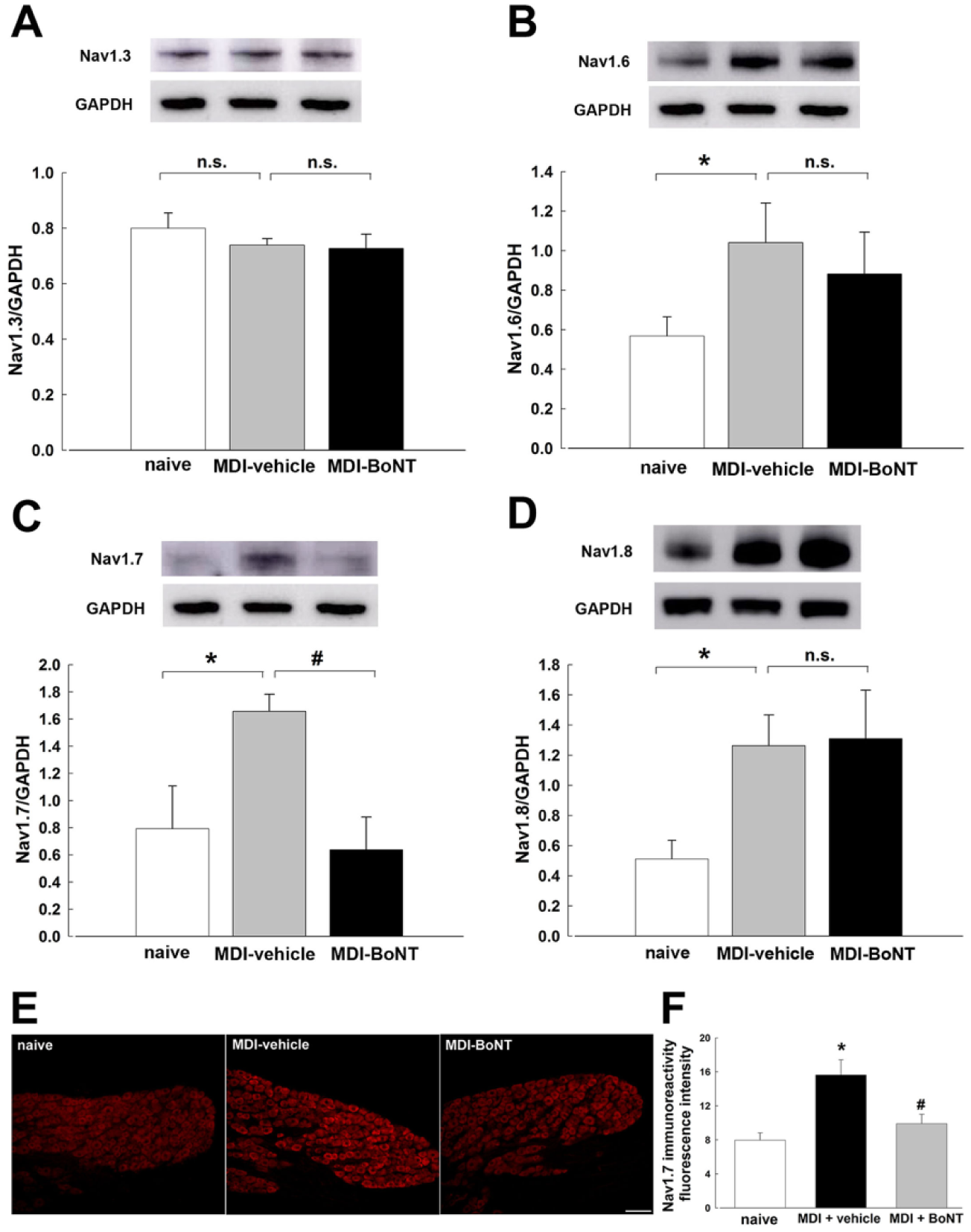
Effects of subcutaneous injection with botulinum toxin type A (BoNT-A; 3 U/kg) on expression of voltage-gated sodium channel isoforms in the ipsilateral trigeminal ganglion following the surgical placement of a malpositioned dental implant (MDI) in rats: (
Changes in the Nav1.7 level after BoNT injection were confirmed by immunohistochemical study. Nerve injury upregulated this isoform, which was blocked by subcutaneous injection of BoNT-A, in the mandibular (V3) division of the trigeminal ganglion in our rat model (F2,12 = 9.176, P < 0.05; Fig. 4E, F).
Effects of BoNT-A on Nav1.7 Expression in the Boundary Area and ATF3 Expression in the Trigeminal Ganglion
Figure 5A illustrates changes in Nav1.7 expression in the boundary area of the mandible (innervated by injured nerve) and the maxilla (innervated by uninjured nerve), which is a mixed innervation area. Representative immunofluorescence images revealed that nerve injury produced upregulation of Nav1.7 expression, which subcutaneous injection of BoNT-A blocked in the boundary area of the trigeminal ganglion.
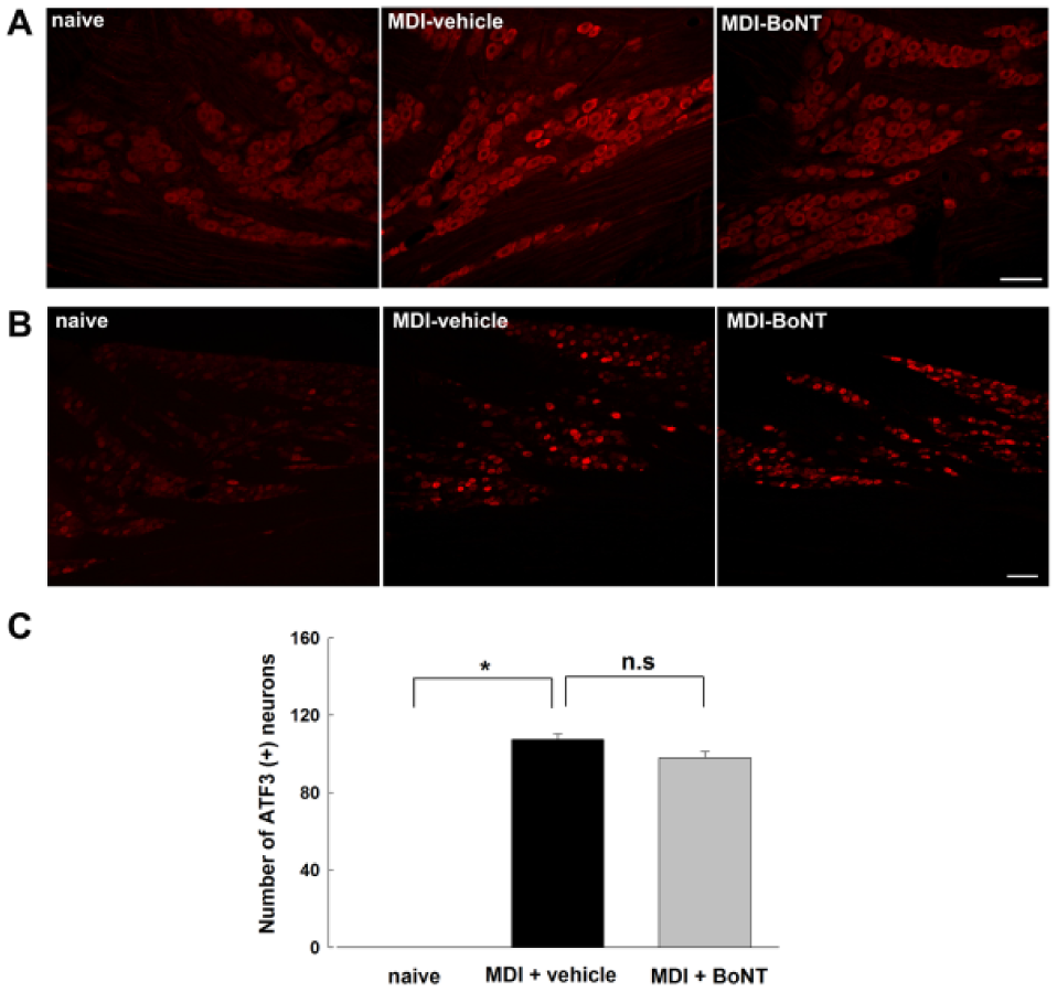
Effects of subcutaneous injection of botulinum toxin type A (BoNT-A; 3 U/kg) on expression of Nav isoform 1.7 (Nav1.7)– and ATF3-positive cells in the ipsilateral trigeminal ganglion following the surgical placement of a malpositioned dental implant (MDI) in rats. (
Figure 5B illustrates changes in the expression of ATF3, a marker of neuronal injury, in the trigeminal ganglion after the subcutaneous administration of 3 U/kg of BoNT-A. ATF3 immunoreactive cells were not observed in the trigeminal ganglion of the naïve rat. The malpositioned dental implant increased the level of ATF3-positive cells in the trigeminal ganglion on POD 9, but this was not affected by the subcutaneous injection of 3 U/kg of BoNT-A (Fig. 5C).
Discussion
The present study is the first to demonstrate that Nav1.7 participates in the antiallodynic effects of BoNT-A in rats with inferior alveolar nerve injury. A single treatment with BoNT-A produced prolonged antinociceptive effects in this rat model of trigeminal neuropathic pain. Moreover, late treatment with BoNT-A also produced significant antinociceptive effects when pain had already been established in these animals, blocking the increase in Nav1.7 expression in the trigeminal ganglion. These results suggest that BoNT-A has antinociceptive properties that are mediated by the regulation of peripheral Nav1.7 levels.
Our present findings demonstrate that a single subcutaneous administration of BoNT-A into the most sensitive area of the facial region produces prolonged antinociceptive effects in our rat model of trigeminal neuropathic pain induced by malpositioned dental implants. BoNT-A is therefore a potentially important new therapeutic approach for neuropathic pain. Importantly, anticonvulsants, tricyclic antidepressants, serotonin, norepinephrine reuptake inhibitor, pregabalin, and gabapentin have proven effective in the treatment of neuropathic pain in the clinic but do not induce prolonged antinociceptive effects after a single injection.
Moreover, the subcutaneous injection of BoNT-A (3 U/kg) into the hind leg of our rat model did not induce antiallodynic effects (data not shown). Thus, our current data suggest that the antinociceptive effects of BoNT-A are mediated by the primary afferent fibers of the trigeminal nerve, not via systemic action. We found that double treatment with 1 U/kg of BoNT-A has a higher efficacy than a single treatment. This finding suggests that double treatments with BoNT-A are more effective for neuropathic pain, with reduced side effects.
In previous clinical studies, BoNT-A induced analgesic effects in patients with chronic neuropathic pain (Ranoux et al. 2008; Yoon et al. 2010) and alleviated burning pain with a neuropathic quality (Jabbari et al. 2003) and trigeminal neurlagia (Borodic and Acquadro 2002). The antinociceptive effects of BoNT-A have also been reported in animal studies. A single peripheral injection of BoNT-A has been shown to be sufficient to reduce thermal and mechanical hyperalgesia in rats with neuropathic pain induced by partial sciatic nerve transaction (Bach-Rojecky et al. 2005) or by chronic constriction injury of the sciatic nerve (Luvisetto et al. 2007). These results with our present data suggest that BoNT-A has a potential therapeutic efficacy against trigeminal neuropathic pain.
A previous study reported that central hypersensitivity was established within POD 3 and may be independent of the peripheral input after pain has been established (Curatolo et al. 2001). These results suggested that pain should be treated early in its development to prevent it from becoming chronic (Hefferan et al. 2003; Verdi et al. 2013). Another previous study reported that early treatment with dexamethasone produces prolonged antinociceptive effects in rats with malpositioned dental implants, whereas delayed treatment has no effect (Han et al. 2010). These results indicated that once neuropathic pain is established, it tends to become more difficult to successfully manage, and more aggressive treatments may be required. These earlier results also suggest that neuropathic pain can be attributed to central hypersensitivity or neuronal plasticity and that late treatment would be ineffective after its establishment. Hence, early treatment is one of the most important considerations in preventing further development of neuropathic pain and achieving normal conditions.
Interestingly, our present findings demonstrated that treatment with BoNT-A produces antinociceptive effects on PODs 7 and 12, when nociceptive behavioral responses had been already established in our rats. These results provide a new strategy for neuropathic pain treatment. Although pain had already been established by central mechanisms, neuropathic pain could be controlled by an injection of BoNT-A. Furthermore, a single injection of BoNT-A was found to be sufficient to control neuropathic pain because its antinociceptive effects are maintained for a long period after injection, unlike anticonvulsants or tricyclic antidepressants.
It has been well established that Navs play a critical role in modulating the excitability of most neurons, including nociceptive sensory signaling. The distribution of individual subunits is specific to different tissues and cell types. Moreover, the differential expression of different α-subunits (Nav1.1 to Nav1.9) could lead to changes in the excitability of sensory neurons leading to some chronic pain conditions (Baker and Wood 2001; Catterall et al. 2005; Dib-Hajj et al. 2009). Hence, modulation of the α-subunits of Navs in sensory neurons may be a viable strategy for the treatment of neuropathic pain.
The findings of the present study indicate that malpositioned dental implants produce upregulation in expression of the Nav1.6, Nav1.7, and Nav1.8 in the trigeminal ganglion in our rat model of trigeminal neuropathic pain. The subcutaneous injection of BoNT-A, which produced significant antinociception, downregulated only Nav1.7 in the trigeminal ganglion. It is well known that Nav1.7 is expressed mainly in small-diameter nociceptive neurons, contributes to the amplification of generator potentials, and sets the gain on nociceptors (Djouhri et al. 2003; Waxman 2006). Previous studies demonstrated a key role of this isoform in inflammatory pain. Nav1.7 was also found to be upregulated in dorsal root ganglion neurons following injection of carrageenan or Freund’s complete adjuvant (Strickland et al. 2008), and its knockdown significantly prevented the development of hyperalgesia induced by injection of Freund’s complete adjuvant (Yeomans et al. 2005). Moreover, Nav1.7 was found to be upregulated in the dorsal root ganglion of rats with painful diabetic neuropathy (Huang et al. 2014) and with chronic constriction injury of the sciatic nerve (Liu et al. 2012), also suggesting an important role in the development of neuropathic pain. These results indicate that Nav1.7 plays an important role in the trigeminal ganglion during pain processing and suggest that a reduction in its activity might contribute to antinociception after BoNT-A injection. A recent study also showed that BoNT-A inhibited voltage-dependent Na+ currents of both TTX-sensitive and TTX-insensitive peripheral dorsal ganglion cells (Shin et al. 2012). These data, with the present data, suggest that BoNT-A produces antinociception in the orofacial area through the modulation of Nav1.7 expression in the trigeminal ganglion. However, the cellular mechanism of the inhibitory effects of BoNT-A on Nav1.7 is not clear, and further studies will be needed.
Interestingly, significant changes were observed in the boundary area of the mandible (innervated by injured nerve) and the maxilla (innervated by uninjured nerve). The changes in this area are one of the reasons why the maxillary innervated area showed mechanical allodynia after injury of the mandibular nerve branch. Moreover, previous studies already demonstrated that functional interactions between neurons and satellite glial cells or between neurons and neurons in the trigeminal ganglion are involved in orofacial extraterritorial pain associated with trigeminal nerve injury (Iwata et al. 2001; Iwata 2015). However, to fully elucidate the underlying mechanisms, future studies will be needed.
Our present study findings showed that the subcutaneous injection of 3 U/kg of BoNT-A did not affect the number of ATF3-positive cells in rats with malpositioned implants. These results suggest that BoNT-A did not alleviate injury of the inferior alveolar nerve produced by a malpositioned dental implant.
In summary, we have demonstrated here that a single treatment with BoNT-A in a rat model of trigeminal neuropathic pain produces prolonged antinociceptive effects both before and after pain has been established. These effects of BoNT-A are mediated by an inhibition of upregulated Nav1.7 expression in the trigeminal ganglion. Therefore, BoNT-A is a potential new therapeutic agent for chronic pain control, including neuropathic pain.
Author Contributions
K.Y. Yang, M.J. Kim, D.K. Ahn, contributed to conception, design, and data analysis, drafted and critically revised the manuscript; S.T. Kim, contributed to interpretation and critically revised manuscript; S.K Park, contributed to data analysis, drafted the manuscript; C.G. Lee, J.S. Ju, contributed to data analysis and interpretation. Y.C. Bae, contributed to conception and design, critically revised manuscript. All authors gave final approval and agree to be accountable for all aspects of the work.
Footnotes
Acknowledgements
The authors thank Megagen and Hugel, Inc., for providing miniature dental implants and Botulax, respectively.
This research was supported by the National Research Foundation of Korea (funded by the Ministry of Science, ICT and Future Planning; 2008-0062282 and 2012M3A9B6055414) and by Hugel, Inc.
The authors declare no potential conflicts of interest with respect to the authorship and/or publication of this article.