
Abstract
Streptococcus mutans in dental plaque biofilms play a role in caries development. The biofilm’s complex structure enhances the resistance to antimicrobial agents by limiting the transport of active agents inside the biofilm. The authors assessed the ability of high-velocity water microsprays to enhance delivery of antimicrobials into 3-d-old S. mutans biofilms. Biofilms were exposed to a 90° or 30° impact, first using a 1-µm tracer bead solution (109 beads/mL) and, second, a 0.2% chlorhexidine (CHX) or 0.085% cetylpyridinium chloride (CPC) solution. For comparison, a 30-s diffusive transport and simulated mouthwash were also performed. Confocal microscopy was used to determine number and relative bead penetration depth into the biofilm. Assessment of antimicrobial penetration was determined by calculating the killing depth detected by live/dead viability staining. The authors first demonstrated that the microspray was able to deliver significantly more microbeads deeper in the biofilm compared with diffusion and mouthwashing exposures. Next, these experiments revealed that the microspray yielded better antimicrobial penetration evidenced by deeper killing inside the biofilm and a wider killing zone around the zone of clearance than diffusion alone. Interestingly the 30° impact in the distal position delivered approximately 16 times more microbeads and yielded approximately 20% more bacteria killing (for both CHX and CPC) than the 90° impact. These data suggest that high-velocity water microsprays can be used as an effective mechanism to deliver microparticles and antimicrobials inside S. mutans biofilms. High shear stresses generated at the biofilm-burst interface might have enhanced bead and antimicrobial delivery inside the remaining biofilm by combining forced advection into the biofilm matrix and physical restructuring of the biofilm itself. Further, the impact angle has potential to be optimized both for biofilm removal and active agents’ delivery inside biofilm in those protected areas where some biofilm might remain.
Keywords
Introduction
Oral biofilms play an important role in the development and the persistence of caries, halitosis, gingivitis, and periodontitis (Marsh 2004). The addition of antiplaque agents or antimicrobials to toothpastes, mouthwashes, and varnishes to kill bacteria is one of the most common ways to control oral diseases (Marsh 2006). The challenge, however, is that dental plaque bacteria organize themselves into biofilms, which increases their tolerance to these active agents through diffusion limitation (Stoodley et al. 2008; von Ohle et al. 2010). The role of the hydrodynamics in the enhancement of the delivery of active agents inside the biofilm has become a topic of interest, since it might also be used to improve delivery of dentifrices to oral surfaces (teeth, gums, tongue) or to bacteria directly. Fluid-dynamic activity generated by powered toothbrushes can enhance antimicrobial/antiplaque agent delivery into remaining biofilm compared with simple diffusional transport (Stoodley et al. 2007; He et al. 2014; Jongsma et al. 2015). Microsprays are a useful strategy for removal since they require low liquid volume but also have an air-water interface moving over the solid surface, which facilitates biofilm removal. We previously investigated the ability of high-velocity water microsprays to mechanically remove interproximal biofilms (Rmaile et al. 2014, 2015) and discovered that the biofilm was physically “churned up” during the exposure (Fabbri et al. 2016). We hypothesized that this churning of the biofilm might improve the delivery of active agents into the biofilm extracellular polymeric substance (EPS) matrix. To test this hypothesis, we assessed the potential of high-velocity water microsprays for delivery into in vitro Streptococcus mutans biofilms, first using 1-µm-diameter fluorescent beads as tracer particles and, second, using chlorhexidine (CHX) and cetylpyridinium chloride (CPC), 2 antimicrobials commonly found as active ingredients in dentifrices. Delivery of these materials into the biofilm by microspray was compared with a static (diffusion-only) delivery and simulated swirling mouthwash.
Materials and Methods
Biofilm Growth Conditions
Biofilms were grown on glass microscope slides (75 × 25 mm; Corning, Sigma-Aldrich). The slides were first conditioned with 10 mL of 2% sucrose-supplemented brain-heart infusion (BHI+S) and 1% type II porcine gastric mucin (BHI+SM; Sigma-Aldrich) to simulate salivary proteins and establish a conditioning film. The slides were placed in a petri plate and inoculated with a S. mutans UA159 adjusted overnight culture (106 colony-forming units [CFU]/mL) and grown in 10 mL BHI+SM medium, then grown under static conditions for 72 h at 37 °C and 5% CO2 with medium replacement every 24 h.
Microbead Delivery
Carboxylate-modified polystyrene yellow/green fluorescent beads (λex/λem: 470/505, density = 2.5%, charge density ≥0.008 mEq; Sigma-Aldrich [L4655]) of a 1-µm-diameter stock solution was diluted down to 109 beads/mL. A Philips Sonicare AirFloss (AF), a commercially available oral hygiene device for interdental cleaning, was used to generate high-velocity water microsprays. The device delivers a volume of 130 ± 0.03 µL (n = 11) as a microspray in a single discrete shot. The device was filled with 3 mL of the beads working solution; therefore, approximately 1.3 × 10⁸ beads were delivered in each shot. First, S. mutans biofilm-covered slides were exposed to a single microspray shot at either a 90° or a 30° impact angle with the tip held a distance of 5 mm from the biofilm. For comparative purposes, the delivery of beads into the biofilm was compared by 1) diffusion alone in a static incubation by gently adding 130 µL of bead solution (1.3 × 10⁸ beads) over the biofilm and let to diffuse for 30 s or 2) by a simulated swirling mouthwash exposure to beads in an overlying liquid by placing the glass slide inside a petri plate filled with 3 mL of bead solution and then shaken for 30 s at 200 rpm. Three independent replicates were performed for each experiment. Immediately after each experiment (microspray, static, and shaking), the slide samples were washed once with 1% phosphate-buffered saline (PBS) solution to removing loose beads. One PBS tablet (Sigma-Aldrich) was dissolved in 200 mL distilled water to obtain 10 mM phosphate buffer, 2.7 mM potassium chloride, and 137 mM sodium chloride.
Quantification of Beads in the Biofilm
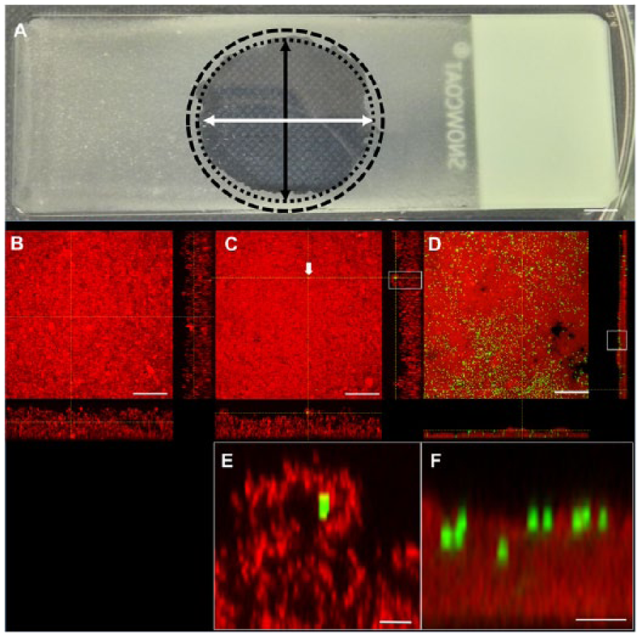
The penetration of beads into the biofilm was quantified by confocal microscopy and image analysis. After exposure, the biofilms were fixed with 4% paraformaldehyde (PFA) solution to preserve structure (von Ohle et al. 2010). The fixed biofilms were stained with the nucleic acid stain Syto 63 (Invitrogen) to visualize the total biomass and subsequently imaged under a confocal laser scanner microscope (CLSM; Leica TCS SP5). A nontreated biofilm was used as control. Confocal 3-dimensional stacks were collected for each of 3 independent replicates for the nontreated, static, shaking, and microspray experiments. For the biofilms exposed to the microsprays, we took confocal images from 3 locations 1 to 2 mm outside of the clearance zone caused by the microspray (Supplemental Material 1; Fig. 1A, Appendix Fig. 1A, B). For the 30° shot, we distinguished between the proximal area and distal areas with respect to distance from the device nozzle. For the nontreated, static, and shaking exposures, we collected data at 3 random positions on the slide (Supplemental Material 1; Appendix Fig. 1C). To quantify the penetration of the beads into the biofilm, we used a relative depth ratio (RDBEADS) to account for differences in biofilm thickness at each XY pixel location on the substratum, as explained previously (Miller et al. 2013; Supplemental Material 2).
(
Antimicrobial Delivery and Killing Depth in the Biofilm
To assess the ability of the microspray to enhance the delivery and killing of bacteria within the biofilm by antimicrobial agents, we used CHX and CPC (Sigma-Aldrich). The stock solutions were diluted in 1% PBS to 0.2% (wt/vol) CHX and 0.085% (wt/vol) CPC. The AF device was filled with 3 mL of the CHX or CPC solutions. PBS (1%) alone was used as a control. The biofilms were then exposed to a single 90° or a 30° impact angle microspray shot or a static incubation for each antimicrobial and the control. Simulated mouthwash (shaking experiments) were also performed using CHX. Static diffusion incubation was also performed with 1% PBS alone as a nontreated control. Three independent replicates were performed for each experiment. The influence of the various exposures on biofilm structure, depth of killing, and the zone of lateral killing (in the case of the microspray exposures) was assessed by confocal microscopy. Immediately after exposure, the biofilms were stained with live/dead stain (BacLight; Invitrogen) according to the manufacturer’s instructions. The stained samples were then rinsed with PBS to remove excess stain and immediately imaged under a CLSM. Live cells were stained green and dead and membrane-compromised cells were stained red (von Ohle et al. 2010). Confocal 3-dimensional stacks were taken on each of 3 independent replicates for the static, shaking, and microspray experiments in the same way as they were taken for the beads experiments (Supplemental Material 1, Appendix Fig. 1A–C). Since we could not directly visualize the antimicrobial agent within the biofilm, we used the depth of bacterial killing (measured from live/dead staining) as a relative indicator of antimicrobial penetration depth as described elsewhere (He et al. 2014; Supplemental Material 3). This will underestimate the absolute penetration depth since it more closely relates to the depth where the microbial biocidal concentration (MBC) was achieved, but for practical purposes, the depth of killing is arguably more relevant. In addition to measuring the depth of killing for those biofilms exposed to the microsprays, we also assessed the proportion of bacteria killed as a function of lateral distance from the edge of the zone of clearance by measuring the variation in the red/green (R/G) signal ratio (Supplemental Material 4). The lateral extent of the antimicrobial killing zone from the edge of the impact clearance zone for each antimicrobial was defined as the distance for which R/G was >1.5 (He et al. 2014).
Statistical Analysis
Statistical comparisons were performed using analysis of variance (ANOVA) with a Bonferroni post hoc test. Differences were considered significant for P < 0.05.
Results
Biofilm Structure, Thickness, and Bead Penetration
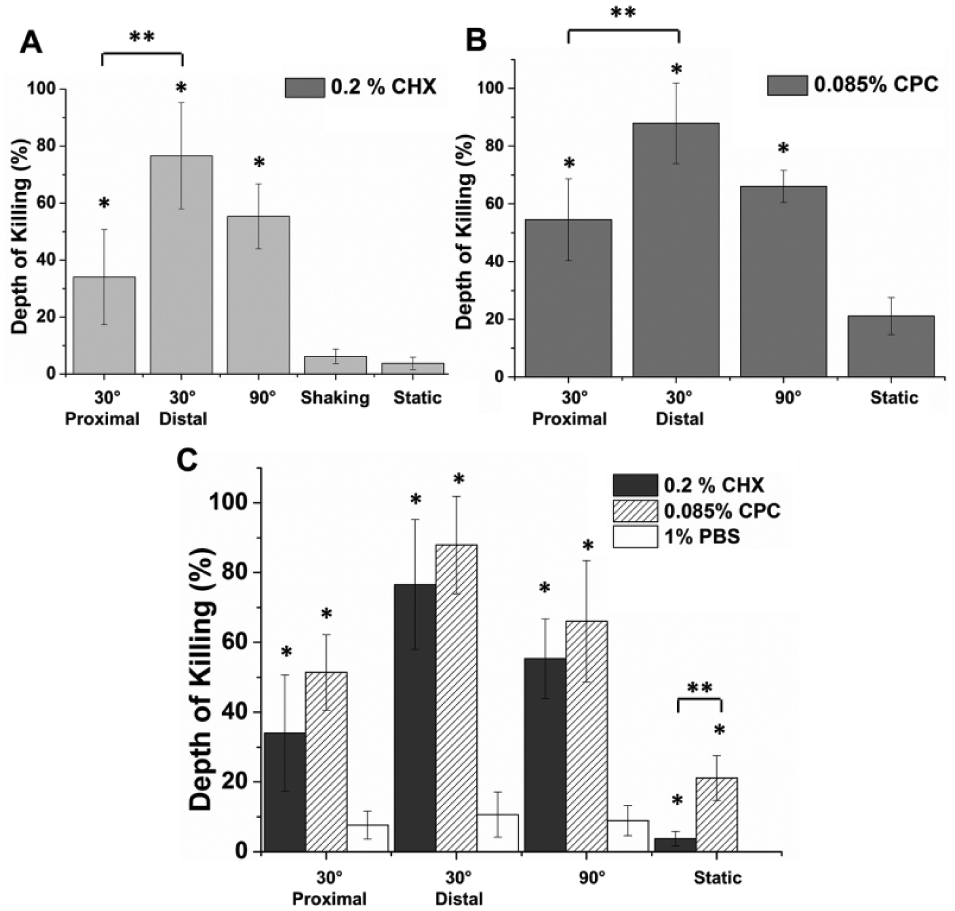
The untreated (control) biofilm was relatively uniform with a slightly undulating surface and an average thickness of 51.8 ± 4.9 µm (Table; Fig. 1B). The static incubation with the beads did not significantly change biofilm structure and thickness (Table; Fig. 1C). However, the shaking exposure to the beads resulted in some biofilm loss, probably due to shear stresses.
Average Biofilm Thickness (µm) as a Function of the Various Exposures and Treatment Solutions.
Data represented as mean and 1 SD from 3 independent replicates.
CHX, chlorhexidine; CPC, cetylpyridinium chloride; PBS, phosphate-buffered saline; ZOC, zone of clearance.
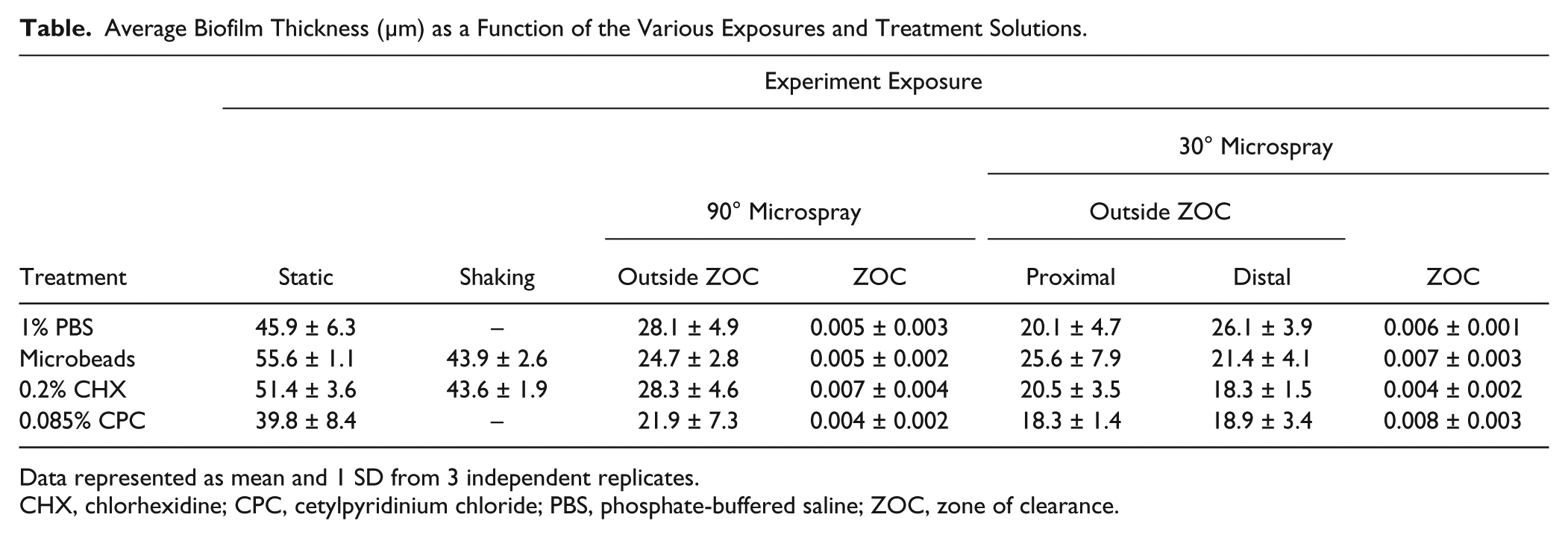
CLSM images of the biofilms exposed to microbeads showed more beads were present in the biofilm after exposure to the microspray (both the 30° and the 90° impact angles) than after their introduction by static or shaking means (Fig. 1C, D). Beads were observed in the confocal cross-section images, confirming their penetration inside the biofilm (Fig. 1E, F). Figure 2 shows the bead distribution inside the biofilm in each relative depth “slice” (0%–25%, 25%–50%, 50%–75%, and 75%–100%). In all cases, the 30° impact angle delivered significantly more beads than the 90° impact angle microspray, and both (90° and 30°) yielded a significantly higher bead penetration than the shaking or static incubations. In addition, the 30° impact angle microspray delivered significantly more beads in the distal zone than in the proximal zone.
Bar chart (logarithmic scale) showing bead distribution (N/cm2) inside Streptococcus mutans biofilm in terms of relative depth ratio for the static, shaking, and 90° and 30° experiments. Data represented as mean and 1 SE from 3 independent replicates. A relative depth value of 0% corresponded to a bead located near the biofilm surface, while a relative depth of 100% corresponded to a bead located in the biofilm substratum.
Biofilm Structure, Thickness, and Antimicrobial Killing Depth
Static treatment with CHX, CPC, and PBS had no significant effect on biofilm thickness compared with the nontreated biofilm (Table), but shaking exposure to CHX significantly (P < 0.05) reduced biofilm thickness to 43.6 ± 1.9 µm compared with the biofilm exposed to static diffusion using the same active agent (Table). The biofilm thickness after the microspray exposure (both 30° and 90° impact angles) to CHX, CPC, and PBS was significantly less than the thickness after the static exposure to the same antimicrobials.
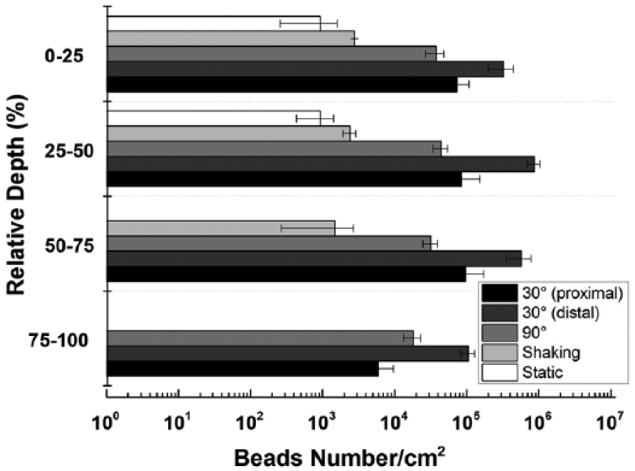
The 30° and 90° impact angle microspray caused a greater depth of killing with both antimicrobials than achieved with the simulated mouthwash shaking and static incubation (Fig. 3A, B). In addition, when the biofilm was exposed to the 30° impact angle microspray, both antimicrobials generated a significantly greater killing depth (KD) in the distal position than in the proximal position. Biofilm exposed to a static assay showed that CPC resulted in greater KD than CHX (Fig. 3C). No dead zones were observed in the biofilm samples after the static diffusion assay performed with PBS (Fig. 3C). Surprisingly, the microspray (both 90° and 30° impact angles) performed with PBS alone resulted in a zone of killing in the upper part of the biofilm, but this was significantly lower than that achieved with the antimicrobials (Fig. 3C).
Biofilm depth of killing caused by 0.2% chlorhexidine (CHX) (
Cross-sections of confocal images of biofilm samples exposed to microsprays and static assay using CHX, CPC, and PBS were in agreement with our results (Fig. 4). In particular, biofilm samples exposed to CHX and CPC revealed that both 30° and 90° impact angle microsprays caused an increase in the size of the red areas (dead biofilm) compared with static transport (Fig. 4A–H). In addition, no dead zones were observed in the PBS-treated samples, as shown by an increase of the green areas (live biofilm; Fig. 4I–L).
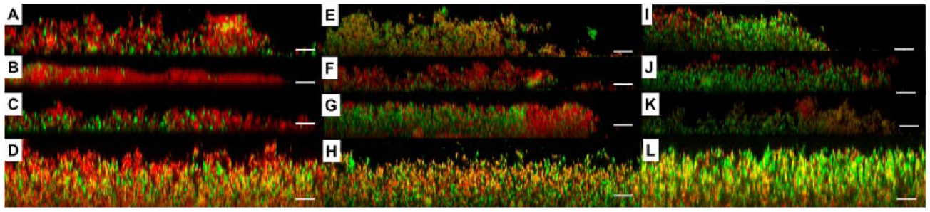
Confocal images in x–z cross-section view showing Streptococcus mutans biofilms after the 90° shooting, 30° shooting in the distal zone, 30° shooting in the proximal zone, and the static assay using 0.085% cetylpyridinium chloride (CPC) (
Finally, the 30° and 90° impact angle microsprays caused a higher lateral zone of killing (up to 200 µm from the ZOC edge) with both antimicrobials than the same experiments performed with PBS (~52 µm; Appendix Fig. 2A, B). No dead zones were observed in the PBS controls (Appendix Fig. 2C).
Discussion
The purpose of this study was to assess whether a high-velocity water microspray enhances the penetration of antimicrobials into biofilms using a laboratory-grown S. mutans biofilm. While human dental plaque is clearly more complex in terms of microbial diversity (Dewhirst et al. 2010) than the single-species S. mutans biofilm, the sticky glucans produced by glucosyltransferases released by S. mutans has a significant determination of the mechanical stability, ecological succession, and development of acidic and anaerobic gradients of cariogenic biofilm, even if present in relatively low numbers (Bowen and Koo 2011; Koo et al. 2013). As such, S. mutans biofilms have been widely used in dental and microbiology research as a biofilm model for the study of caries, biofilm mechanical properties, and detachment (Vinogradov et al. 2004; Hwang et al. 2014; Hashizume and Dariva 2015).
First, we demonstrated that the microspray was able to deliver significantly more microbeads deeper in the biofilm compared with a simple diffusion and a simulated mouthwash (Fig. 2). Next, we confirmed that the microspray yielded better penetration of CHX and CPC evidenced by a deeper bacteriostatic effect within the biofilm (Figs. 3, 4) and a wider killing zone around the zone of clearance (Appendix Fig. 2). The microspray used in our experiments was a turbulent complex 2-phase burst of water and air that lasted 60 ms (Fabbri et al. 2016). Once the spray impacts the surface, the flow can be divided in 2 regions: an impingement region in the center of the impact followed by a 2-phase wall-jet region where the flow spreads radially outward. Shear stresses generated at the jet-film interface rise as a function of radial distance, reaching a maximum value before decreasing again afterward (Deshpande and Vaishnav 1983; Phares et al. 2000). Our data suggest that the stress generated is responsible not only for the biofilm detachment but also for structural deformations (what we define as biofilm “churning up”) of the unremoved biofilm at edges of the clearance zone. In addition, studies on rinsing flows, where a water jet impinges on a flat surface coated with a second fluid at higher viscosity, demonstrated the formation of recirculation zones downstream at the interface between the 2 liquids (Hsu et al. 2011; Walker et al. 2012). The formation of eddies at the edges of the clearance zone could have contributed to the increased mixing between biofilm and the beads or antimicrobial solutions. Interestingly, a 30° nozzle inclination was more powerful (in terms of bead delivery and bacterial killing caused by the antimicrobials) in the distal part of the exposed area compared with the proximal part and to a 90° microspray. In the 30° microspray, there would be an expected increase in the velocity in the direction of shooting (i.e., distal position) and a reduction in the opposite direction (i.e., proximal position). Such an asymmetrical velocity distribution may “focus” the impact and create cohesive failure at the biofilm-substratum interface (i.e., the low-impact angle allows the spray to go between the biofilm-substratum interface in the distal position, which could enhance bead and dentifrice penetration).
After the static experiment, only 925 beads/cm2 penetrated 30% of the biofilm depth. Previous studies in biofilm permeability using fluorescent beads are consistent with our data showing that the bead penetration was limited to the outer biofilm layers (20%–30% of the biofilm thickness; Drury, Characklis, et al. 1993; Drury, Stewart, et al. 1993) or needed timescales up to hours to reach up 90% to 95% biofilm substratum by diffusion alone (Miller et al. 2013), in which case some of the effect could be “overgrowth” of the biofilm as seen by Chew et al. (2014). The same trend was observed for the antimicrobial experiments where the bacterial KDs yielded by CPC and CHX after a simple diffusion transport were approximately 5% and 20%, respectively. These data are consistent with previous studies on oral biofilms showing that CHX and CPC antimicrobial efficacy decreased with increasing biofilm depth (Hope and Wilson 2004; von Ohle et al. 2010) with penetration times up to 20 min to reach half of the biofilm thickness (Corbin et al. 2011; Nance et al. 2013). It is well known that the biofilm matrix confers antimicrobial tolerance (Costerton et al. 1999). In particular, S. mutans biofilm matrix’s sticky glucans are known to limit diffusion of CPC and CHX (Bowen and Koo 2011). CPC and CHX biological activity for S. mutans biofilms is mainly limited to bactericidal effects rather than to the degradation of glucans or the inhibition of EPS production (Pandit et al. 2015; Tawakoli et al. 2015; Gao et al. 2016). Therefore, the microspray impact might have changed the diffusion-limiting properties of the glucan matrix, causing increased delivery. Future work on staining the glucan matrix to assess the effects of microsprays on its structure could be an interesting complement to the bacterial killing and fluorescent bead visualization.
Another important physiological property of biofilms is the viscoelastic mechanical behavior (He et al. 2013). Viscoelasticity means the material behaves either as an elastic solid or a viscous fluid depending how fast it is deformed and how quickly it recovers (Banks et al. 2010). The EPS polymers that are kept together by physicochemical interactions (electrostatic interactions, van der Waals forces, and hydrogen bonding) appear to be the main contributors to biofilm viscoelasticity (Korstgens 2001). Recent studies on biofilms exposed to a noncontact brushing routine showed a change in viscoelastic properties linked to an increase of antimicrobial penetration (He et al. 2013, 2014; Jongsma et al. 2015). We hypothesize that a combined effect of shear, which left the unremoved biofilm structure in a more vulnerable state enabling better chemical penetration, and antimicrobial delivery yielded an increase in bacteria killing.
Our data showed that the microspray alone resulted in killing of bacteria in the remaining biofilm outside the zone of clearance (Fig. 3). It is not clear why this is. Powered and mechanical toothbrushes caused damage to cell surface structures but did not affect the cell viability (McInnes et al. 1993). However, no studies examined viability of biofilm-associated bacteria when exposed to hydrodynamic phenomena. It is possible that the remaining cells could be not completely dead but metabolically compromised and not have the necessary energy reserves to repair essential channels, walls, membranes, and receptors. Another possibility is that the microspray could have introduced oxygen into the biofilm, causing oxidative stresses. It has been recently demonstrated that the presence of oxygen can alter cell surface composition in S. mutans biofilms (Ahn et al. 2007).
In conclusion, low-volume, high-velocity water microsprays are effective at removing S. mutans biofilms from areas relevant to that of a tooth surface and have the additional benefit as a potential delivery method for antimicrobials inside dental biofilm that might remain in or adjacent to the zone of clearance. There are other potential clinical benefits that we did not test but might explain the clinical efficacy of powered interdental devices using high-velocity microsprays in improving gum health (Ward et al. 2015). It is reasonable to assume that if the microspray is effectively mixing the biofilm up to drive fluids into the biofilm, then potentially proinflammatory mediators such as bacterial toxins would get driven out. Furthermore, dental biofilm is a complex ecosystem made of oxygen-intolerant organisms, and it is possible that the turbulent mixing of the biofilm also disrupts the oxygen gradient, thereby inhibiting anaerobic growth of periopathogens. It is not still clear what force threshold is required by interdental cleaning devices to produce advective mixing or what force is required to eliminate biofilms. The further improvement of the synergy between microsprays and antibacterial activity, either by changing the design of the dental device or the use of different drug delivery methods such as antimicrobial-coated nanoparticles or micro/nanoemulsions, is of interest in future research.
Author Contributions
S. Fabbri, contributed to conception, design, data acquisition, analysis, and interpretation, drafted and critically revised the manuscript; D.A. Johnston, contributed to data acquisition, critically revised the manuscript; A. Rmaile and B. Gottenbos contributed to conception, data interpretation, critically revised the manuscript; M. De Jager, contributed to data interpretation, critically revised the manuscript; M. Aspiras, E.M. Starke, and M.T. Ward, contributed to conception and data interpretation, critically revised the manuscript; P. Stoodley, contributed to conception, design, data analysis, and interpretation, drafted and critically revised the manuscript. All authors gave final approval and agree to be accountable for all aspects of the work.
Footnotes
Acknowledgements
This work was financially funded in part by EPSRC DTP EP/K503130/1 award and in part by Philips Oral Healthcare, Bothell, WA, USA. The authors thank the Southampton Biomedical Imaging Unit for their support in imaging the biofilms.
All data supporting this study are openly available from the university of Southampton repository at ![]() . A.R., M.J. and B.G. are employed by Philips Research Eindhoven, Netherlands. M.S. and M.W. are employed by Philips Oral Healthcare, Bothell, WA, USA. M.A. was a former employee of Philips Oral Healthcare, Bothell, WA, USA. P.S. has served as a private consultant for POH.
. A.R., M.J. and B.G. are employed by Philips Research Eindhoven, Netherlands. M.S. and M.W. are employed by Philips Oral Healthcare, Bothell, WA, USA. M.A. was a former employee of Philips Oral Healthcare, Bothell, WA, USA. P.S. has served as a private consultant for POH.
The other authors declare no potential conflicts of interest with respect to the authorship and/or publication of this article.
References
Supplementary Material
Please find the following supplemental material available below.
For Open Access articles published under a Creative Commons License, all supplemental material carries the same license as the article it is associated with.
For non-Open Access articles published, all supplemental material carries a non-exclusive license, and permission requests for re-use of supplemental material or any part of supplemental material shall be sent directly to the copyright owner as specified in the copyright notice associated with the article.