






Abstract
The pulp is a highly vascularized tissue situated in an inextensible environment surrounded by rigid dentin walls, with the apical foramina being the only access. The pulp vascular system is not only responsible for nutrient supply and waste removal but also contributes actively to the pulp inflammatory response and subsequent regeneration. This review discusses the underlying mechanisms of pulp vascularization during tooth development, regeneration, and therapeutic procedures, such as tissue engineering and tooth transplantation. Whereas the pulp vascular system is established by vasculogenesis during embryonic development, sprouting angiogenesis is the predominant process during regeneration and therapeutic processes. Hypoxia can be considered a common driving force. Dental pulp cells under hypoxic stress release proangiogenic factors, with vascular endothelial growth factor being one of the most potent. The benefit of exogenous vascular endothelial growth factor application in tissue engineering has been well demonstrated. Interestingly, dental pulp stem cells have an important role in pulp revascularization. Indeed, recent studies show that dental pulp stem cell secretome possesses angiogenic potential that actively contributes to the angiogenic process by guiding endothelial cells and even by differentiating themselves into the endothelial lineage. Although considerable insight has been obtained in the processes underlying pulp vascularization, many questions remain relating to the signaling pathways, timing, and influence of various stress conditions.
Keywords
Introduction
The vasculature is the earliest functional organ system of the human body (Udan et al. 2012) and is formed by vasculogenesis during embryonic development (Fig. 1A–D). Endothelial cells derived from the mesoderm aggregate and form vascular structures that are completed with a basement membrane and supporting cells, such as pericytes and vascular smooth muscle cells. At later developmental stages and in adult life, new blood vessels are mainly formed by angiogenesis, which is the sprouting of new vessels from existing ones (Fig. 1E–G). Both processes are controlled by angiogenic growth factors (Grant and Janigro 2006).

Vasculogenesis and angiogenesis. Vasculogenesis is the formation of new blood vessels during embryogenesis. (
A well-functioning vascular system is vital, as it ensures gas exchange, nutrient supply, and waste removal for all organs, including the tooth. The tooth pulp tissue is highly vascularized and situated in an inextensible environment surrounded by rigid dentin walls with the apical foramina being the only access (Vongsavan and Matthews 1992). This renders unique properties to the pulp vasculature, which have been described previously (About 2014).
Pulp Vascularization: Parallels between Tooth Development and Engineering
During tooth development, vascularization of the tooth germ occurs by vasculogenesis, which has been demonstrated through the slice culture method. Indeed, it was observed that mesoderm-derived endothelial cell precursors invaded the developing papilla during the early bell stage, where they aggregate to form vascular structures. This invasion coincided with dental papilla size exceeding 200 µm (Rothova et al. 2011). It is known that oxygen diffusion through tissues is limited to 100 to 200 µm and that a vascular network is required to ensure all cells to be within this distance (Hoeben et al. 2004). Interestingly, the vascularization during entire tooth engineering procedures resembles what happens in vivo during tooth development (Fig. 2). Mesenchymal- and epithelial-derived stem cells cultured in vitro in a collagen gel allowed the formation of a tooth germ–like construct, similar to the bud and cap stage (Oshima et al. 2011). When this construct grows and exceeds 200 µm in size, vascularization becomes essential for its survival. To this end, the construct has to be implanted in vivo given that the in vitro culture lacks the necessary vascular supply. This step recalls the early bell stage—with the difference that vascularization of the construct occurs by invasion of the host vasculature, most likely by angiogenesis.
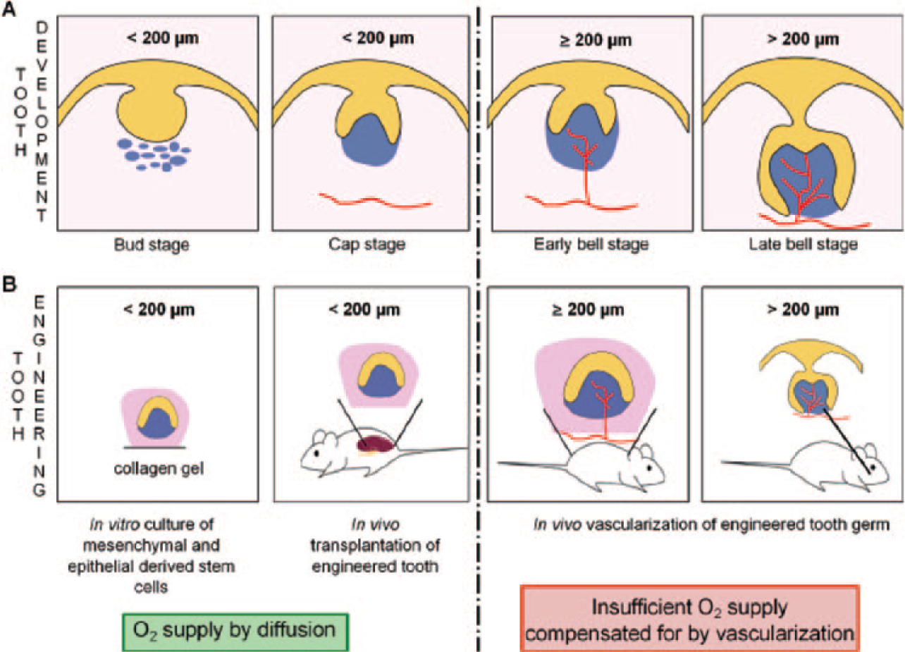
Parallels between tooth development and tooth engineering. (
The underlying pathways responsible for vascularization of developing and engineered teeth are not fully elucidated yet. Nevertheless, hypoxia can be considered a common driving force given that in both cases the growing tissue experiences a moment of oxygen (and nutrient) deprivation as a result of inadequate diffusion. It is thus hypothesized that hypoxic cells secrete proangiogenic factors that either act on neighboring endothelial cells to induce angiogenesis of engineered teeth or stimulate the invasion of endothelial cell precursors in the dental papilla during tooth development. Whereas more research is needed regarding these hypoxic processes in both tooth development and engineering, hypoxia as the driving force of angiogenesis in injured dental pulp tissue has been well established (Aranha et al. 2010).
Hypoxia Is the Driving Force of Angiogenesis in Injured Dental Pulp Tissue
The prevalent cause of dentin-pulp tissue injury is bacterial infection or caries, leading to inflammatory reaction. A hypoxic tissue with a lot of debris can be found at the inflammation site and in mechanically injured pulp tissue due to restoration procedures (Fig. 3A).
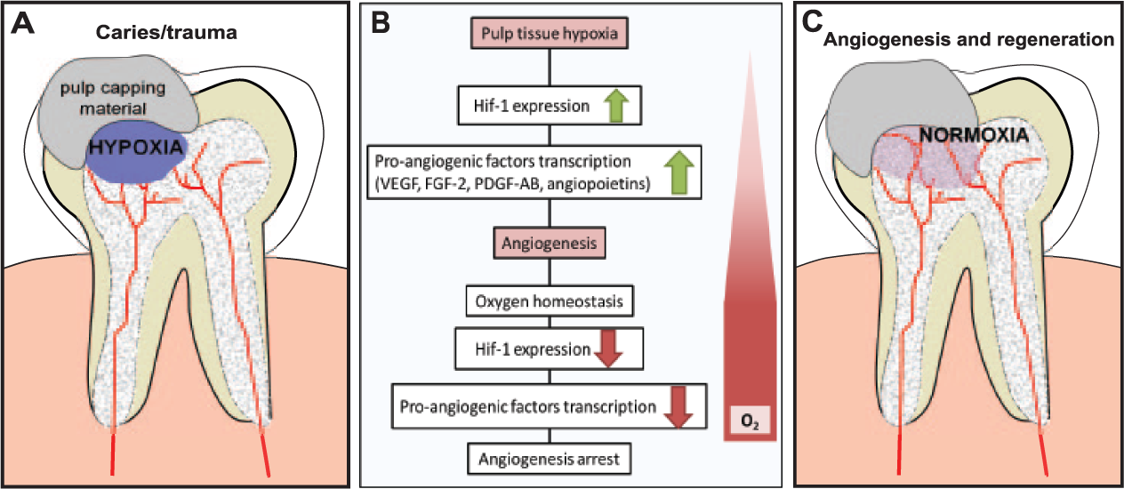
Hypoxia is the driving force for angiogenesis. (
Under hypoxic conditions, dental pulp cells rapidly increase their expression of hypoxia-inducible transcription factor 1 (Hif-1; Aranha et al. 2010). Hif-1 mediates increased transcription of various angiogenic genes, such as vascular endothelial growth factor (VEGF), platelet-derived growth factor AB, placental growth factor, and angiopoietins. It also regulates the expression of angiogenic chemokines, such as stromal cell–derived factor 1, sphingosine-1-phosphate, and their receptors (Semenza 2002; Zimna and Kurpisz 2015). When new blood vessels are formed and oxygen homeostasis is restored, Hif-1 expression decreases, followed by an arrest of angiogenesis (Fig. 3B, C). Hypoxia is thus a major regulator of angiogenesis, particularly by stimulating the paracrine angiogenic activity of dental pulp cells.
Paracrine Angiogenic Activity of Pulp Cells during Regeneration
Under severe carious injury affecting the pulp, a local collapse of blood vessels leads to a situation of hypoxia and nutrient deprivation in the pulp. Consequently, paracrine angiogenic activity of dental pulp cells can be observed. These stress situations can be simulated in vitro by culturing dental pulp cells in hypoxic (2% O2), low-serum, or low-glucose conditions (Bakopoulou et al. 2015). Under these conditions, increased VEGF secretion was observed in stem cells obtained from apical papilla (SCAP; Vanacker et al. 2014; Bakopoulou et al. 2015), pulp fibroblasts, and dental pulp stem cells (DPSCs; Aranha et al. 2010; Bronckaers et al. 2013). Similarly, VEGF secretion was increased by injured dental pulp fibroblasts (Tran-Hung et al. 2008). Vascular network formation in cocultures of DPSCs and human umbilical vein endothelial cells confirmed the paracrine angiogenic activity of DPSCs (Janebodin et al. 2013; Yuan et al. 2015), which could be attributed to increased VEGF secretion and associated VEGF receptor 2 (VEGFR-2) signaling in the endothelial cells.
Besides VEGF, other proangiogenic factors were secreted in stress-simulated culture conditions—including angiogenin, insulin-like growth factor binding protein 3, monocyte chemoattractant protein 1, platelet-derived growth factor AB, angiopoietin 1, chemokine (C-X-C motif) ligand 16, insulin-like growth factor binding protein 2, hepatocyte growth factor, PIGF, fibroblast growth factor 7, and tissue inhibitor of metalloproteinase 4 (Aranha et al. 2010; Bronckaers et al. 2013; Bakopoulou et al. 2015). Also, fibroblast growth factor 2 (FGF-2) was shown to be secreted by pulp fibroblasts in response to mechanical injury (Tran-Hung et al. 2008). In hypoxic and serum-deprived conditions, however, FGF-2 was not secreted by SCAP (Bakopoulou et al. 2015), pulp fibroblasts (Aranha et al. 2010), and DPSCs (Bronckaers et al. 2013). However, it was abundantly present in cell lysates suggesting that this growth factor is restricted to cellular expression and is not secreted (Bronckaers et al. 2013). Antiangiogenic factors were detected as well, including endostatin, plasminogen activator inhibitor 1 (Bronckaers et al. 2013), tissue inhibitor of metalloproteinase 1, thrombospondin 1, pentraxin 3, serpin E1, and serpin F1 (Bakopoulou et al. 2015). The presence of these antiangiogenic molecules can explain why, in several studies, conditioned media of DPSCs did not induce endothelial cell proliferation and sprouting (Aranha et al. 2010; Bronckaers et al. 2013).
The composition and concentration of “secretome” from dental pulp cells varies under different stress conditions. For instance, a combination of oxygen, serum, and glucose-deprived medium, which mimics most closely the ischemic conditions in vivo, led to the highest increase in proangiogenic VEGF and angiogenin secretion by SCAP (Bakopoulou et al. 2015), whereas lower secretion was seen for antiangiogenic serpin E1, tissue inhibitor of metalloproteinase 1, and thrombospondin 1, compared with the other stress conditions. The secretion of such modulation factors underlines the significant role of the pulp angiogenic capacity during its own regeneration.
DPSCs and Angiogenesis
Investigating the angiogenic potential of pulp stem cells was initiated several years after postnatal human DPSCs were isolated as a source of adult mesenchymal stem cells (MSCs; Gronthos et al. 2000). In accordance with the International Society for Cellular Therapy, these MSCs express (≥95%) CD105, CD73, and CD90 and do not express (≤2%) CD45, CD34, CD14 or CD11b, CD79α or CD19, and HLA-DR. Furthermore, they adhere to plastic and give rise to 3 lineages: osteoblastic, adipogenic, and chondroblastic (Dominici et al. 2006). On top of these minimal properties, DPSCs have also demonstrated neuronal and endothelial differentiation potential (Nakashima et al. 2009).
Endothelial Differentiation Potential of DPSCs
Endothelial differentiation of DPSCs depends on their microenvironment. It has been shown that short exposure to stress conditions was more efficient at inducing transformation of SCAP toward an endothelial-like cell type as compared with 28 d of exposure to angiogenic medium (Bakopoulou et al. 2015). VEGF seems to play a pivotal role in endothelial differentiation of DPSCs. Indeed, in vitro endothelial differentiation of DPSCs (Marchionni et al. 2009) and stem cells from human exfoliated deciduous teeth (SHED; Bento et al. 2013) was obtained by supplementing culture medium with VEGF. Furthermore, DPSCs showing the ability to differentiate into endothelial-like cells, in vivo and in vitro, expressed the VEGFR-2 receptor (Iohara et al. 2008; Sakai et al. 2010). It should be noted that some studies (Marchionni et al. 2009) claiming to use DPSCs were utilizing a heterogeneous dental pulp cell population in which the presence of endothelial cells cannot be excluded.
Various in vivo studies confirmed this endothelial differentiation of DPSCs. For instance, it has been demonstrated that DPSCs synergistically differentiate into osteoblast-like (VEGFR-2+/STRO-1–/CD44+/RUNX-2+) and endothelial-like cells (VEGFR-2+/STRO-1+/CD44+/CD54+), as indicated by different surface markers (D’Aquino et al. 2007). Implantation of these differentiated cells into immunocompromised rats resulted in the formation of bone-like tissue with newly formed blood vessels, which were efficiently integrated with the host vasculature. Implantation of a DPSC subfraction (CD31–/CD146–/CD34+/VEGFR-2+) into a mouse model of hind limb ischemia led to an increase of capillary density with reestablishment of the blood flow after 14 d (Iohara et al. 2008). Furthermore, the secretome of this subfraction containing proangiogenic factors such as VEGF demonstrated angiogenic-inducing capacities when applied on human umbilical vein endothelial cell cultures. In other studies, human tooth slices containing biodegradable scaffolds with LacZ-transduced SHED were implanted in immunocompromised mice. These SHED showed endothelial differentiation and lined the blood vessels in the newly formed pulp-like tissue (Cordeiro et al. 2008; Sakai et al. 2010). Recently, evidence has been given that DPSCs are capable of de novo blood vessel formation, similar to embryonic vasculogenesis, by differentiating into vascular endothelial cells (Zhang et al. 2016). Furthermore, this study demonstrated that the canonical Wnt-β-catenin signaling is decisive for DPSC endothelial differentiation, which implies that blocking this pathway will leave room for other differentiation fates of DPSCs.
Overall, some DPSC subsets seem to have a higher endothelial differentiation potential, including granulocyte colony-stimulating factor–mobilized DPSCs (Murakami et al. 2013) and CD31–/CD146–/CD34+/VEGFR-2+ DPSCs (Iohara et al. 2008). Many studies used SHED or SCAP, which may predispose to higher endothelial differentiation potential. However, this claim needs to be confirmed in future research. To this end, it is important to use homogenous stem cell populations that can be obtained through specific antibody-based sorting protocols.
Roles of DPSCs in Angiogenesis
There is no doubt regarding the necessity of sprouting angiogenesis by endothelial cells in dentin pulp regeneration (Fig. 4A). Recent data indicate that DPSCs are not only recruited for lost tissue regeneration but may also be involved in this angiogenic process. They can support angiogenesis by guiding endothelial cells in a paracrine fashion and by stabilizing the newly formed blood vessels by adopting a pericyte-like location (Fig. 4B), which was reported in various in vitro studies (Dissanayaka et al. 2012; Janebodin et al. 2013; Yuan et al. 2015). It should be noted though that the Matrigel angiogenesis assays used in vitro do not provide solid proof for the pericyte-like function of DPSCs. However, DPSC perivascular location and expression of pericyte marker 3G5 suggest that pericytes may represent a DPSC subpopulation (Shi and Gronthos 2003).
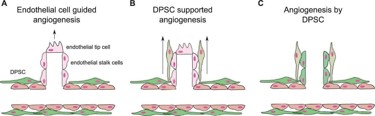
Dental pulp stem cell (DPSC) involvement in sprouting-angiogenesis hypotheses. Three hypotheses can be put forward for the formation of new blood vessels in regenerating dental pulp tissue: (
Recent investigations suggest that they can even actively contribute by differentiating themselves into endothelial-like cells (Cordeiro et al. 2008; Iohara et al. 2008; Sakai et al. 2010; Zhang et al. 2016). It can therefore be hypothesized that DPSCs residing in perivascular regions differentiate into endothelial-like cells and initiate the sprouting of new blood vessels from the adjacent ones (Fig. 4C). The last hypothesis should, however, be considered with caution since the major direct evidences of endothelial cell differentiation of MSCs have been obtained in vitro and are based on phenotypic identification (expression of CD31, CD34, VEGFR-2, and von Willebrand factor) and functional tests in vitro (capillary-like structure formation in Matrigel and uptake of acetylated low-density lipoprotein; Pacini and Petrini 2014).
DPSCs can clearly be considered an essential part of the angiogenic process during dentin-pulp tissue regeneration. More research is, however, required to fully establish their role in this process and to confirm their endothelial differentiation potential/pericyte-like function.
Dental Pulp Tissue Engineering: Vascularization Is Key
Pulp vitality preservation is a major objective in endodontics, as devitalized teeth are more vulnerable and prone to tooth loss later in life (Ajay Sharma et al. 2013). An adequate revascularization is a determining element of success for dental pulp tissue engineering, and the advancements made in this field are discussed below.
Cell-Free Approaches for Dental Pulp Tissue Regeneration
Dental pulp tissue regeneration takes place in a confined space with a sole access for nerve and vasculature supply via the foramen. A well-developed biodegradable scaffold that will promote timely vascularization is thus essential for successful regeneration (Demarco et al. 2011). Various in vivo studies have demonstrated the efficacy of scaffolds with embedded growth factors. For instance, human root canals treated with collagen scaffold containing FGF-2 showed cellular colonization and well-vascularized pulp tissue-like formation (Suzuki et al. 2011). Similar results were obtained when amputated rat pulp were treated with gelatin hydrogels containing FGF-2 (Ishimatsu et al. 2009). Enhanced neovascularization was observed in human tooth slices treated with VEGF before subcutaneous implantation in immunocompromised mice, suggesting a beneficial effect of topical VEGF administration (Mullane et al. 2008). Combinations of VEGF or FGF-2, bone morphogenetic protein 7, and nerve growth factor in collagen scaffolds were efficient in regenerating pulp-like tissues in endodontically treated human root canals subcutaneously implanted in mice (Kim et al. 2010). This study is of particular interest as it showed complete regeneration of dentin-pulp-like vascularized tissue in a relatively large space (8 to 10 mm in length). Although the presence of 2 access points would have facilitated the revascularization process, the use of control root canals prepared under similar conditions confirmed the benefit of added growth factors.
Currently used dental materials can also affect cell homing either indirectly by stimulating the secretion of bioactive molecules by residual pulp cells or directly by their chemical composition. For instance, conditioned medium of DPSCs exposed to mineral trioxide aggregate and growth hormone induced endothelial tube–like formation and migration (Yun et al. 2016). Furthermore, mineral trioxide aggregate was shown to augment angiopoietin 1 and von Willebrand factor expression in DPSCs (Huang et al. 2015).
Cell-Based Approaches for Dental Pulp Tissue Regeneration
When the lost pulp tissue exceeds a critical size, cell homing may not suffice to regenerate the whole dentin-pulp tissue, and cell-based treatments may represent an alternative approach. Considering ease of isolating DPSCs and their multilineage ability, the majority of studies applied these cells for dental pulp regeneration purposes.
Successful DPSC transplantations have been obtained despite the fact that the number of cells surviving after transplantation was low. It is thus believed that the interaction between DPSCs and their environment is a critical factor of the engraftment success (Bronckaers et al. 2014; Tran and Damaser 2015). In particular, their impact on proangiogenic factors expression seems to play an important role in vasculature formation in the newly generated tissues. Indeed, increased expression of proangiogenic genes has been observed following transplantation of DPSCs (Iohara et al. 2009). Furthermore, a higher blood vessel concentration was seen close to the dentin wall (Rosa et al. 2013), which could be explained by the presence of proangiogenic factors sequestered in the dentin matrix (Roberts-Clark and Smith 2000).
Often, DPSCs are transplanted with proangiogenic factors to enhance the vascularization of the newly formed dentin-pulp tissue. For instance, pulp tissue with nerves and vasculature was formed in dog root canals engrafted with dental pulp CD105+ cells with stromal cell–derived factor 1. The CD105+ cells were localized close to the newly formed capillaries and expressed additional angiogenic factors (Iohara et al. 2011). Implantation of DPSCs and platelet-rich fibrin constructs in root fragments in nude mice and canine endodontically treated root canals led to more pulp-like tissue generation with better vascularization as compared with DPSCs or platelet-rich fibrin alone. This may be attributed to the slow release of growth factors from platelet-rich fibrin acting on the DPSCs (Chen et al. 2015). A recent study also demonstrated the benefit of combining DPSCs with a VEGF-loaded microspheres-based scaffolding system for full-length human tooth root generation (Li et al. 2016). The addition of DPSCs was necessary to regenerate blood vessels throughout the whole root canal, which could be explained by DPSC-induced endothelial cell migration and increased growth factor expression.
Clinical Translation of Dental Pulp Tissue Regeneration Procedures
While the aforementioned studies have reported successful dental pulp–like tissue regeneration following the implantation of cell-free scaffolds or DPSC transplantation, translation to the clinic is more complicated. Indeed, the majority of these studies are based on mice subcutaneous implantation models with human healthy teeth without any signs of inflammation. However, in the clinic, dental pulp regeneration may be required in pathologic conditions with pulp inflammation, necrosis, and apical periodontitis. Before proceeding with regenerative procedures, the clinician should remove necrotic tissue and disinfect the transplantation site while maintaining a biologically favorable environment for regeneration (Galler 2015). Studies mimicking these clinical situations are thus needed to ascertain successful dentin-pulp regeneration with these procedures.
Vascularization of Engrafted Engineered Pulp Tissue Constructs
Besides DPSCs containing scaffolds, in vitro prepared tissue constructs have been studied for regeneration purposes. Tissue constructs with sizes larger than 2 to 3 mm3 are doomed to fail upon implantation when they lack a functional vasculature network. Thus, ideally, a vascular network should be present already before implantation and should connect with the host vasculature. Alternatively, timely formation of a vascular network should be stimulated before the tissue construct starts to deteriorate due to a lack of oxygen and nutrients (Shieh and Vacanti 2005). A scaffold-free approach has been studied where DPSCs and endothelial cells were cultured together, leading to the self-assembly of microtissue spheroids with an extracellular matrix–like environment (Dissanayaka et al. 2015). Interactions between the 2 cell types led to the survival and angiogenic stimulation of endothelial cells, which formed vessel-like structures, and the odontogenic differentiation of DPSCs. When these microtissues were inserted into human tooth slices and transplanted in vivo, a well-vascularized pulp-like tissue was formed with anastomoses between the in vitro preformed vessel-like structures and the in vivo vasculature (Dissanayaka et al. 2014). A more recent study used DPSCs, predifferentiated toward odontoblastic and osteoblastic lineages, and gingival epithelial cells seeded onto gelatin-chondroitin-hyaluronan cylindrical scaffolds, which were kept in stirred cultured conditions during 7 d (Yang et al. 2016). The osteoblast-like cells were seeded on the bottom to allow connection with the alveolar bone, followed by odontoblast-like cells for regeneration of dentin-pulp tissue. Epithelial cells were seeded on the top of the cylinder. Implantation of this cell/scaffold construction for 13.5 mo in the alveolar sockets of pigs showed regenerated teeth with enamel-like tissue, dentin, pulp, cementum, and periodontal tissues. The pulp tissue was well vascularized, and VEGF expression was detected on the endothelial cells.
Whole Tooth Engineering
A final aspect of dentin-pulp tissue engineering is the bioengineering of a whole tooth where significant advancements have been made. A molar tooth germ, equivalent to the bell stage of a natural tooth germ, was cultured in vitro with MSCs and epithelial-derived stem cells (Oshima et al. 2011). Implantation of this in vitro cultured tooth germ in a subrenal capsule in immunocompromised mice allowed further development of a whole tooth. Through a special ring-shaped device, the size and form of the growing bioengineered tooth could be controlled. This was followed by successful implantation of these bioengineered teeth in artificially created alveolar bone defects in mice, which was confirmed by restored masticatory potential and functionality. Although not discussed in the study, their success could not be obtained without a proper vascularization of the bioengineered tooth and connection with the host vasculature. Indeed, when these in vitro cultured tooth germs were implanted subcutaneously, they became vascularized with completed growth and root formation (Nait Lechguer et al. 2008). This can be explained at least in part by the secretion of angiogenic factors from the tooth germ in its surroundings. These factors may attract endothelial cells from the surrounding tissues and induce their proliferation and organization into vessels that invade the tooth germ, leading to its vascularization. It should be noted, however, that this is hypothesized but not clearly demonstrated.
Three-Dimensional Bioprinting
Bioprinting can be defined as the 3-dimensional printing of all components (cells and matrix) required for a functional organ (Obregon et al. 2015). Bioprinting of dentin-pulp constructs have not been carried out yet, but the technical know-how is available. Sufficient oxygen and nutrient supply for tissue survival remains a big challenge for the printing of large living constructs and requires a functional vascular system. Recent studies have shown the possibility to print a biocompatible vascular template, which is removed afterward, into tissue constructs. Printing of such a template has been performed with, among others, carbohydrate glass coated with a thin layer of poly(D-lactide-co-glycolide) (Miller et al. 2012) and agarose (Bertassoni et al. 2014). Next, cell-laden extracellular matrix–like hydrogels have been casted over these 3-dimensional printed vascular templates, followed by removal of the templates. This was done by dissolution of the carbohydrate lattice, whereas the agarose lattice was removed mechanically. Both studies obtained a successful perfusion of the “vascularized” hydrogels that maintained cellular viability. These approaches could be applied in the future to obtain bioprinted vascularized dental-pulp tissue constructs.
Vascularization Is Necessary for Transplanted Tooth Survival
Tooth transplantations have a long clinical history and are mainly used for the replantation of avulsed teeth and during orthodontic treatments. In a nutshell, 3 approaches can be discerned: transplantation of 1) an immature tooth with pulp tissue, 2) an apicoectomized mature tooth, and 3) a tooth with removed pulp tissue (Fig. 5). Ideally, pulp tissue functionality should be conserved after transplantation, which implies the presence of a functional vascular network.
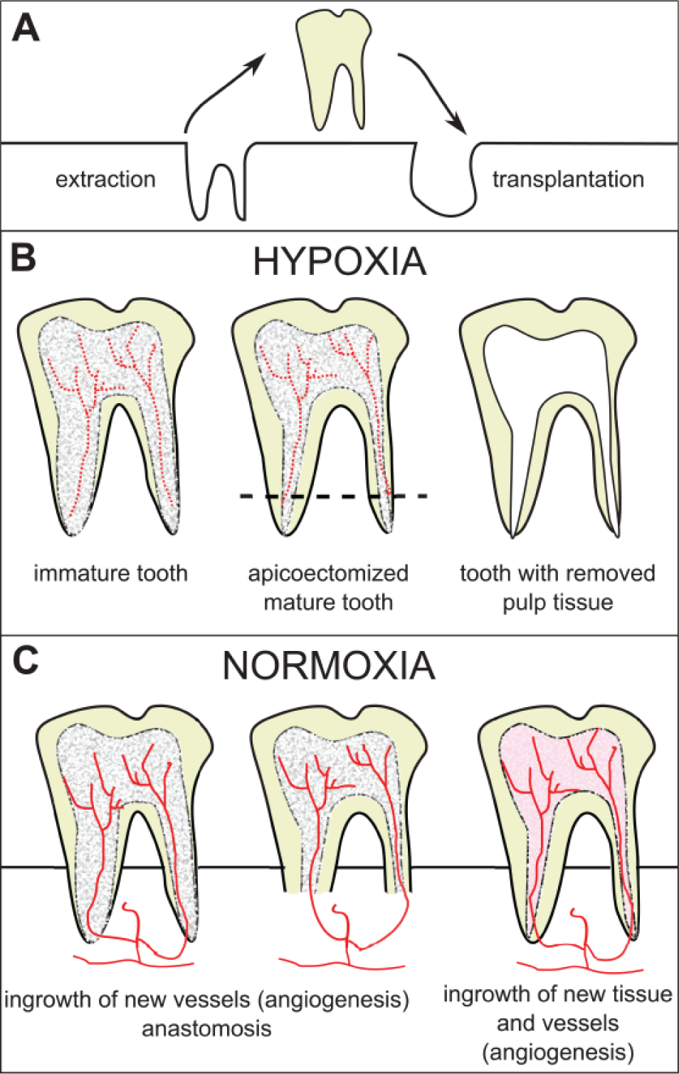
Tooth transplantation. (
In the first 2 approaches, it is believed that the vasculature of the transplanted tooth pulp tissue needs to be connected with the general blood supply for tissue survival. However, in an in vivo study in dogs, anastomosis between preexisting vessels of transplanted pulp tissue and ingrowing blood vessels was seen in only a few teeth. Revascularization of the pulp tissue was primarily the result of ingrowth of newly formed vessels (Skoglund et al. 1978). On the long term, it was observed that the number of blood vessels decreased, which was associated with pulp necrosis and formation of bone or cementum-like tissue in the pulp cavity leading to obliteration. Therefore, it was suggested to perform transplantations with teeth from which the pulp tissue was removed (Claus et al. 2004). Transplantation of these teeth in dogs revealed ingrowth of vascularized cell-rich connective tissue with vascularization being equal to the control teeth (nontransplanted; Laureys et al. 2001; Claus et al. 2004). Although these results are very promising, long-term follow-up is needed in future studies to rule out obliteration.
Conclusion
The driving force of angiogenesis is hypoxia, which can be met in situations where the pulp tissue is subjected to oxygen supply disrupt: tooth germ growth during the embryonal development process; engineered tooth germ growth after engraftment in vivo; and pathologic situations met during carious/traumatic pulp injuries and during therapeutic procedures, including tissue regeneration and tooth transplantation. Under all these situations, hypoxic cells secrete proangiogenic factors that induce sprouting angiogenesis or vasculogenesis, leading to vascular network formation in developing, engineered, and regenerating tissues.
Recent investigations indicate that DPSCs can support angiogenesis by guiding endothelial cells in a paracrine fashion and by stabilizing the newly formed blood vessels by adopting a pericyte-like location. Moreover, it is suggested that they can actively contribute angiogenesis by differentiating themselves into endothelial-like cells.
Although there is considerable insight into the complex orchestrated angiogenic process in the dental pulp, many questions remain relating to the signaling pathways, timing, and influence of various stress conditions, which are worthy of further research.
Author Contributions
C. Rombouts, T. Giraud, C. Jeanneau, and I. About, contributed to conception and design, drafted and critically revised the manuscript. All authors gave final approval and agree to be accountable for all aspects of the work.
Footnotes
This work was supported by Aix-Marseille Université and CNRS.
The authors declare no potential conflicts of interest with respect to the authorship and/or publication of this article.