
Abstract
Previous studies revealed that cementum formation is tightly regulated by inorganic pyrophosphate (PPi), a mineralization inhibitor. Local PPi concentrations are determined by regulators, including ectonucleotide pyrophosphatase/phosphodiesterase 1 (ENPP1), which increases PPi concentrations by adenosine triphosphate hydrolysis. Orthodontic forces stimulate alveolar bone remodelling, leading to orthodontic tooth movement (OTM). To better understand how disturbed mineral metabolism and the resulting altered periodontal structures affect OTM, we employed Enpp1 mutant mice that feature reduced PPi and increased cervical cementum in a model of OTM induced by a stretched closed-coil spring ligated between the maxillary left first molar and maxillary incisors. We analyzed tooth movement, osteoclast/odontoclast response, and tooth root resorption by micro–computed tomography, histology, histomorphometry, and immunohistochemistry. Preoperatively, we noted an altered periodontium in Enpp1 mutant mice, with significantly increased periodontal ligament (PDL) volume and thickness, as well as increased PDL-bone/tooth root surface area, compared to wild-type (WT) controls. After 11 d of orthodontic treatment, Enpp1 mutant mice displayed 38% reduced tooth movement versus WT mice. Molar roots in Enpp1 mutant mice exhibited less change in PDL width in compression and tension zones compared to WT mice. Root resorption was noted in both groups with no difference in average depths, but resorption lacunae in Enpp1 mutant mice were almost entirely limited to cementum, with 150% increased cementum resorption and 92% decreased dentin resorption. Osteoclast/odontoclast cells were reduced by 64% in Enpp1 mutant mice, with a predominance of tartrate-resistant acid phosphatase (TRAP)–positive cells on root surfaces, compared to WT mice. Increased numbers of TRAP-positive cells on root surfaces were associated with robust immunolocalization of osteopontin (OPN) and receptor-activator of NF-κB ligand (RANKL). Collectively, reduced response to orthodontic forces, decreased tooth movement, and altered osteoclast/odontoclast distribution suggests Enpp1 loss of function has direct effects on clastic function/recruitment and/or indirect effects on periodontal remodeling via altered periodontal structure or tissue mineralization.
Introduction
During orthodontic tooth movement (OTM), mechanical forces trigger stress/strain distribution in the periodontal ligament (PDL), initiating a signaling cascade resulting in alveolar bone remodeling allowing for tooth movement. Localized vascular disturbances and cell stress evoke cell necrosis, initiating a host immune response, including chemoattraction of immune-competent cells and their differentiation along the monocyte/macrophage lineage, to clear cellular debris and facilitate structural reorganization of the periodontium in the first stage and tooth movement in the later stage (Jäger et al. 1993; Kim et al. 2010; Wolf et al. 2016). However, excessive orthodontic forces or immune responses sometimes lead to adverse effects, including loss of alveolar bone height and external tooth root resorption of cementum and dentin by osteoclast/odontoclast cells (Yamaguchi et al. 2006; Koide et al. 2010).
Predictable repair and regeneration of periodontal tissues are major unrealized goals of periodontal therapy (Bosshardt and Sculean 2009), including cases of OTM-associated root resorption. There is continued interest in better understanding molecular regulators of cementogenesis and how these might restrict the magnitude of root resorption and/or promote reparative cementum formation. Studies in mice revealed cementum to be exceptionally sensitive to regulation by inorganic pyrophosphate (PPi), a mineralization inhibitor. Local PPi concentrations are determined by factors including tissue-nonspecific alkaline phosphatase (gene: Alpl; protein: TNAP), which hydrolyzes PPi to promote mineralization; progressive ankylosis protein (Ank; ANK), which regulates PPi transport to the extracellular space; and ectonucleotide pyrophosphatase/phosphodiesterase 1 (Enpp1; ENPP1), which hydrolyzes adenosine triphosphate (ATP) to PPi (Millán 2013). Reduction of PPi by loss of function of Ank or Enpp1 in mice results in hypermineralization, including ectopic calcification of joints and soft tissues (Harmey et al. 2004; Millán 2013). In dentoalveolar tissues, Ank or Enpp1 loss of function increases acellular cementum (Nociti et al. 2002; Foster et al. 2012; Zweifler et al. 2015; Thumbigere-Math et al. 2018). Studies employing surgical fenestration defects in Ank knockout mice revealed that genetically reducing PPi enhanced cementum repair (Rodrigues et al. 2011). However, how reduced PPi would alter periodontal remodeling in response to OTM has remained unexplored.
We hypothesized that altered mineral metabolism in Enpp1 mutant mice with decreased PPi concentrations and hypercementosis would affect periodontal remodeling, reducing cementum resorption that arises as an unwanted consequence of OTM. We used a novel orthodontic model in Enpp1 mutant mice to analyze OTM, periodontal remodeling, osteoclast response, and tooth root resorption.
Materials and Methods
Mice
Animal experiments were approved by the National Institute of Arthritis and Musculoskeletal and Skin Diseases (NIAMS)Animal Care and Use Committee. Two lines of Enpp1 mutant mice were used in the current study (Jackson Labs). Phenotyping and genotyping have been previously described for Enpp1asj (C57BL/6J-Enpp1asj/GrsrJ) mice (Li et al. 2013) and for Enpp1asj-2J (BALB/cJ-Enpp1asj-2J/GrsrJ) mice (Li et al. 2014). Both lines harbor Enpp1 loss-of-function mutations causing 80% reduced plasma PPi and hypermineralization (Li et al. 2013; McKee et al. 2013; Li et al. 2014). Enpp1 mutant mice were compared to littermate wild-type (WT) mice. Mice were fed standard rodent chow and provided access to water ad libitum. One cohort (3 WT and 4 Enpp1asj mice, including males and females) was analyzed without treatment at 60 d postnatal (dpn), while another cohort of Enpp1asj-2J mice underwent OTM.
Orthodontic Tooth Movement
At 60 dpn, isoflurane-anesthetized WT and Enpp1asj-2J mice (5/group, including males and females) were fixed with an orthodontic appliance according to a modified method described previously for rats (Ong et al. 2000; Zhang et al. 2003; Jäger et al. 2005). The appliance consisted of a stretched closed-coil spring (0.012-inch nickel-titanium wire; Dentaline GmbH) ligated between the maxillary left first molar and maxillary incisors using dental composite restoration (Fig. 1). A split-mouth design employed right maxillary first molars as contralateral controls (CCs). The appliance delivered 0.5 N of force in the mesial/anterior direction, causing mesial tipping of the first molar. After the 11-d experimental period, animals were euthanized by CO2. Crania were harvested and fixed in 4% paraformaldehyde (Lux et al. 2009).
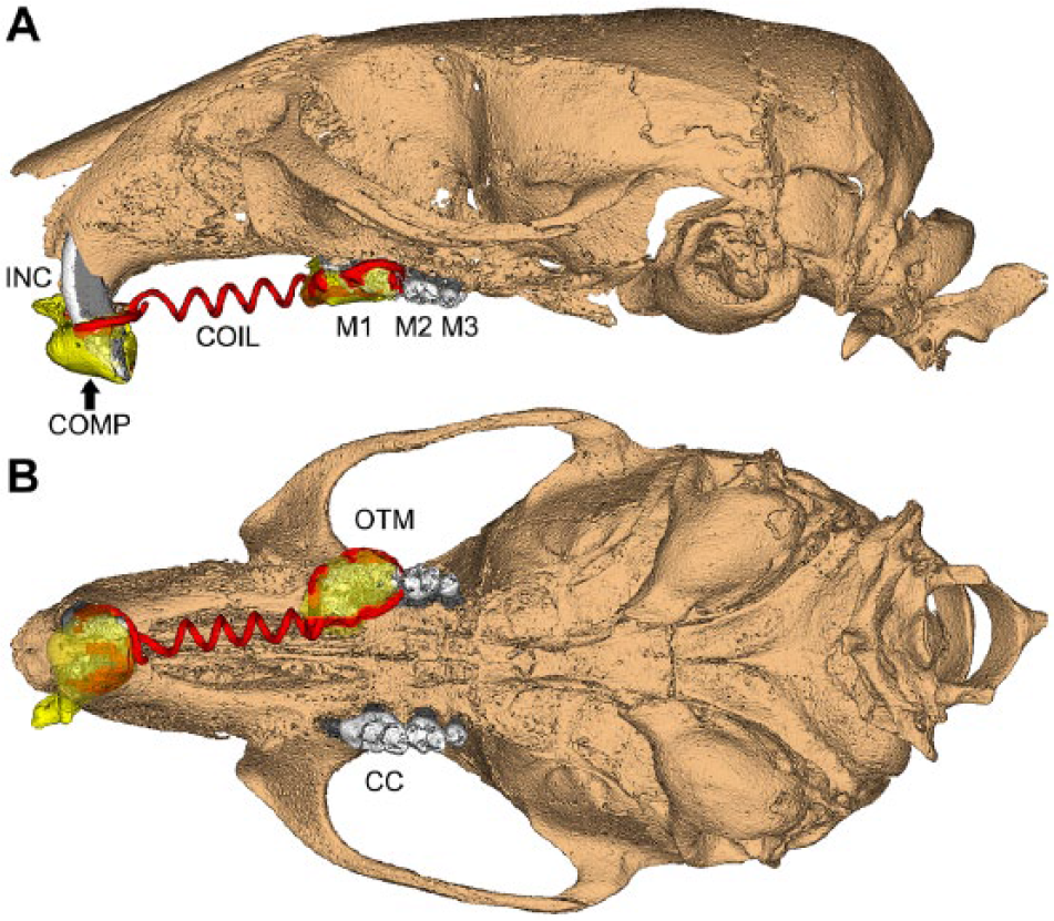
Orthodontic apparatus employed in mouse maxillae. Schematic based on micro–computed tomography scan showing 3-dimensional (
Micro–Computed Tomography
Samples were scanned in a µCT 50 (Scanco Medical) at 70 kVp, 76 µA, 0.5 Al filter, 300- to 900-ms integration time, and 6- to 17-µm voxel dimension. DICOM files were exported, and reconstructed images were analyzed using Amira (version 6.1.2; FEI, Berlin) or AnalyzePro (version 1.0; AnalyzeDirect). Detailed methods for micro–computed tomography (CT) analyses and description of terms are included in the Appendix, and regions of interest (ROI) for analysis are shown in Appendix Figure 1.
Histology
Crania were fixed, hemisected, decalcified, and processed for paraffin histology, and 2- to 5-µm serial sagittal sections were prepared for hematoxylin and eosin (H&E) staining (Götz et al. 2006). Tartrate-resistant acid phosphatase (TRAP) staining was performed to identify osteoclast/odontoclast-like cells (Foster et al. 2017). Two micrographs of molar roots were randomly selected from each animal for resorption measurements. Resorption depths (µm) into cementum and dentin in compression zones were quantified using a modification of a previously described method (Lu et al. 1999). Compression zones were located mesio-coronally to the molar root as previously described (Kawarizadeh et al. 2004; Kawarizadeh et al. 2005).
In Situ Hybridization and Immunohistochemistry
In situ hybridization (ISH) with probes for Spp1, Tnfsf11, Mcsf1, and Vegf was performed on deparaffinized sections and visualized with fast red dye (Advanced Cell Diagnostics), as previously described (Zweifler et al. 2016). Immunohistochemistry (IHC) was performed on deparaffinized sections using an avidin-biotinylated peroxidase (ABC)–based kit with a
Statistical Analysis
Mean ± standard deviation (SD) are shown in graphs. Data were analyzed using an independent samples t test or 1-way analysis of variance (ANOVA) with post hoc Tukey test (Prism version 6.01; GraphPad Software), where P < 0.05 was considered statistically significant.
Results
Altered Periodontium in Enpp1 Mutant Mice
Enpp1asj mice carry loss-of-function mutations causing 80% reduced plasma PPi and hypermineralization (Li et al. 2013). They exhibit an identical hypercementosis phenotype previously reported in Enpp1–/– and Enpp1ttw/ttw mutant mice (Fig. 2A, B and Appendix Fig. 2) (Nociti et al. 2002; Foster et al. 2012; Zweifler et al. 2015). In a recent study (Thumbigere-Math et al. 2018), micro-CT revealed a 4-fold increased acellular cementum thickness, a 5-fold increased acellular cementum volume, and 50% increased cellular cementum volume in 12-wk Enpp1asj versus WT mice. However, the status of other periodontal tissues (PDL and alveolar bone) was not previously analyzed in detail for Enpp1asj or other Enpp1 loss-of-function mice. Because of the significance of periodontal structures and their alterations to the planned orthodontic experiments, we performed high-resolution micro-CT analysis of mandibular bone and PDL space in untreated Enpp1asj versus WT mice at 60 dpn (Fig. 2C–E). While alveolar bone volume showed no significant differences between genotypes, mandibular basal bone volume was increased 20% (*P = 0.01) in Enpp1asj versus WT mice (Fig. 2F, G). No significant differences (P = 0.66) were found in bone density between groups (Appendix Fig. 3A). Despite featuring expanded cementum, Enpp1asj first molars displayed 20% increased (*P = 0.01) PDL volume (Fig. 2H), with no differences in PDL density (Appendix Fig. 3B). We reasoned that increased PDL space reflected an enlarged tooth socket and overall larger PDL interface with both bone and tooth root surfaces. Measurement of this entire PDL surface area (SA; including inner and outer PDL surfaces) revealed a significant 15% increase (**P = 0.002) in Enpp1asj versus WT mice (Fig. 2I). To explore these differences in a more defined anatomical region, we identified a 300-µm ROI midway from the cementum-enamel junction to apex around the mesial root (Fig. 2J–M). Compared to WT, Enpp1asj mice displayed significantly increased PDL thickness (10%; **P = 0.009) and PDL-bone/tooth SA (28%; ***P = 0.0001) (Fig. 2J, K) within this ROI. Separating bone and tooth sides in the ROI confirmed that SA for both bone-PDL and cementum-PDL was significantly increased in Enpp1asj versus WT mice (15% and 21%, respectively; **P = 0.004 and ***P = 0.0004, respectively) (Fig. 2L, M).
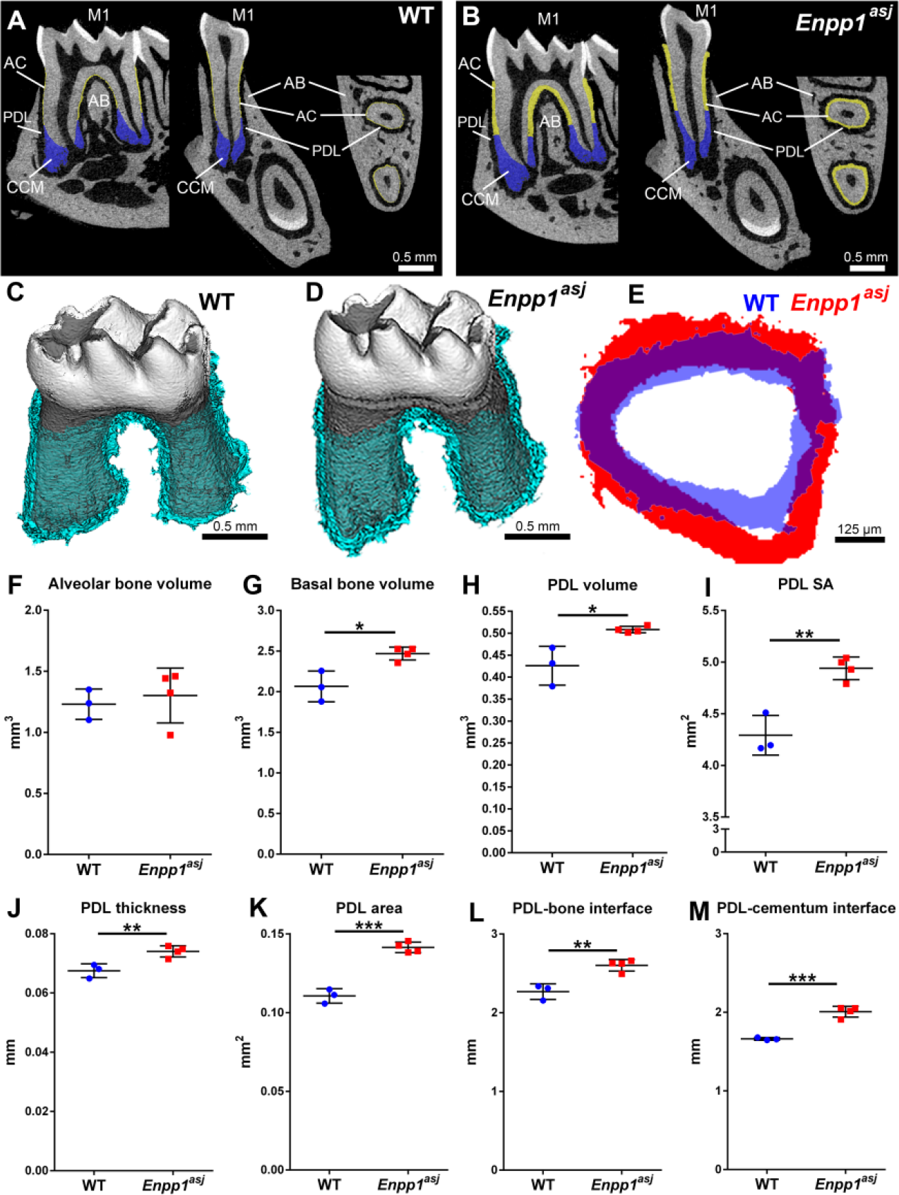
Altered periodontium in Enpp1 mutant mice. (
Reduced OTM in Enpp1 Mutant Mice
Enpp1asj-2J mice are allelic with Enpp1asj mice, with both featuring ENPP1 loss of function, 80% reduced plasma PPi, and hypermineralization (Li et al. 2014). Orthodontic appliances were placed in 60 dpn WT and Enpp1asj-2J mice. All animals (n = 5/genotype) completed 11-d experiments in good health with orthodontic appliances intact. In all mice, orthodontic force application resulted in observable mesial movement of maxillary first molars on OTM sides. Compared to WT, Enpp1asj-2J mice displayed reduced tooth movement (Fig. 3A–F) and increased vertical alveolar bone loss (Fig. 3G, H). Quantitative analysis confirmed a 38% reduction (*P = 0.02) in mesial tooth movement (Fig. 3I) and 50% greater bone loss on the OTM side of Enpp1asj-2J versus WT mice, a trend that did not reach statistical significance (P = 0.12) (Fig. 3J). Untreated CC sides showed no differences between groups (Appendix Fig. 4).
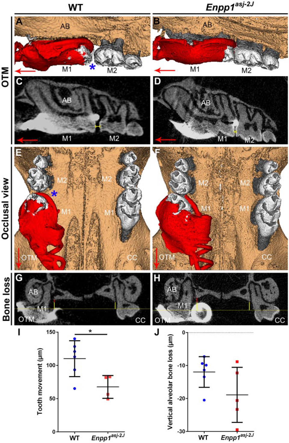
Reduced orthodontic tooth movement in Enpp1 mutant mice. Three-dimensional and 2-dimensional micro–computed tomography (CT) reconstructions of mouse maxillary molars under orthodontic tooth movement (OTM) or on contralateral control (CC) sides. (
To provide further insights into altered tooth movement, PDL and bone in compression (mesial) and tension (distal) zones around molars on OTM sides were assessed and compared to the same anatomic regions on CC sides. One Enpp1asj-2J maxillary CC side was removed from analysis due to suspected periodontitis secondary to food impaction. In OTM sites, Enpp1asj-2J mice showed no differences in alveolar bone density in either compression (P = 0.24) or tension zones (P = 0.67) compared to WT mice (Fig. 4A). However, mean PDL width on mesial aspects of OTM sites was greater (*P = 0.02) in Enpp1asj-2J than WT molars, while on the distal (tension) side, Enpp1asj-2J molars exhibited a nonsignificant (P = 0.12) trend of decreased PDL width (Fig. 4B). When changes in PDL width were calculated as differences between OTM and CC sides (i.e., Δ [PDLOTM – PDLCC]), Enpp1asj-2J molars displayed trends indicating less change on both mesial and distal aspects (Fig. 4C), suggesting they were less responsive to orthodontic forces than WT mice.
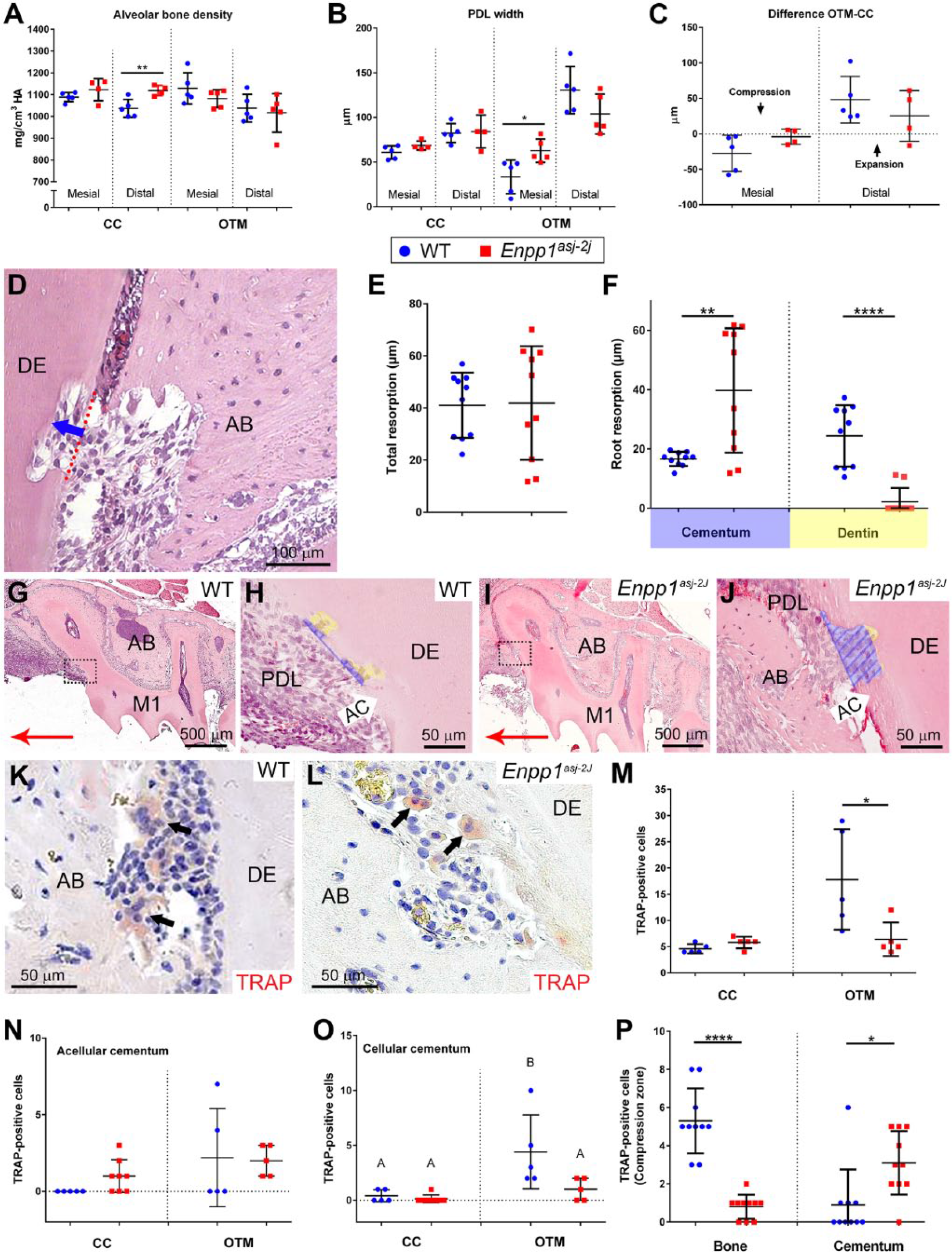
Altered periodontal parameters, resorption, and osteoclast distribution in Enpp1 mutant mice under orthodontic loading. (
Reduced Dentin Resorption in Orthodontically Treated Enpp1 Mutant Mice
Reduced tooth movement in Enpp1asj-2J versus WT mice suggested altered osteoclast response, prompting examination of tooth and bone resorption. Loading forces induced root resorption in both WT and Enpp1asj-2J mice, evident as resorption lacunae in compression zones of OTM sites (Fig. 4D). When average resorption depths were compared, no difference was found between WT and Enpp1asj-2J mice (P = 0.92) (Fig. 4E). However, differences were noted when resorption depths were separated for cementum and dentin. While resorption in WT was observed in both cementum and dentin, resorption in Enpp1asj-2J mice was almost entirely limited to cementum (Fig. 4F–J). Enpp1asj-2J mice featured 150% increased cementum resorption (P = 0.003) and 92% decreased dentin resorption (P < 0.0001) (Fig. 4F).
Altered Osteoclast Distribution in Orthodontically Treated Enpp1 Mutant Mice
Based on reduced tooth movement and dentin resorption in Enpp1asj-2J mice versus WT, we analyzed number and distribution of TRAP-positive osteoclast/odontoclast-like cells in periodontal tissues (Fig. 4K, L). On the CC side, Enpp1asj-2J mice featured a nonsignificant 26% increase in the number of TRAP-positive cells (P = 0.09) (Fig. 4M). On the OTM side, Enpp1asj-2J mice exhibited a significant 64% reduction (*P = 0.04) in TRAP-positive cells versus WT. TRAP-positive cell counts separated by cementum type revealed no significant differences in numbers on the acellular cementum, but OTM sites showed an increased mean number of osteoclasts/odontoclasts in both WT and Enpp1asj-2J mice (Fig. 4N). There was no difference in TRAP-positive cells on the cellular cementum of Enpp1asj-2J versus WT mice on the CC side, but OTM sites showed increased osteoclast-like cells in WT but not Enpp1asj-2J mouse cellular cementum (Fig. 4O).
Focusing only on OTM compression zones, we noted an 85% reduction in TRAP-positive cells on alveolar bone surfaces (***P < 0.0001) and more than a 200% increase in TRAP-positive cells on root surfaces (*P = 0.01) of Enpp1asj-2J versus WT mice (Fig. 4P). Whereas WT mice featured greater numbers of osteoclast-like cells on bone versus tooth root surfaces (5.3 vs. 0.9 cells, respectively), this trend was inverted in Enpp1asj-2J mice, where alveolar bone harbored fewer TRAP-positive cells than root surfaces (0.8 vs. 3.1 cells, respectively).
Regulators of osteoclast/odontoclast migration/attachment (Spp1/OPN, Vegf) and differentiation (Tnfsf11/RANKL, Mcsf1) were assessed. ISH in preoperative 60 dpn mandibular molars showed increased Spp1 messenger RNA (mRNA) in Enpp1asj-2J cementoblasts compared to WT (Appendix Fig. 5A–C). IHC identified intense OPN immunolocalization in Enpp1asj-2J acellular cementum and associated cementocyte-like cells and lining cementoblasts in mesial regions of maxillary molars on both CC and OTM (compression) sides compared to WT, while alveolar bone OPN labeling was similar between genotypes and locations (Fig. 5A–D). Tnfsf11 mRNA was not found in association with preoperative dental or periodontal cells in Enpp1asj-2J or WT mice (Appendix Fig. 5D–F). However, RANKL immunolocalization was associated with sites of bone/tooth resorption on OTM sides of Enpp1asj-2J and WT mice (Fig. 5E–H). Positive RANKL staining was observed in some cementocyte-like cells in Enpp1asj-2J mice, as well as in osteocytes of both genotypes. Mcsf1 and Vegf mRNAs were not found in association with preoperative Enpp1asj-2J or WT molars (Appendix Fig. G–L).
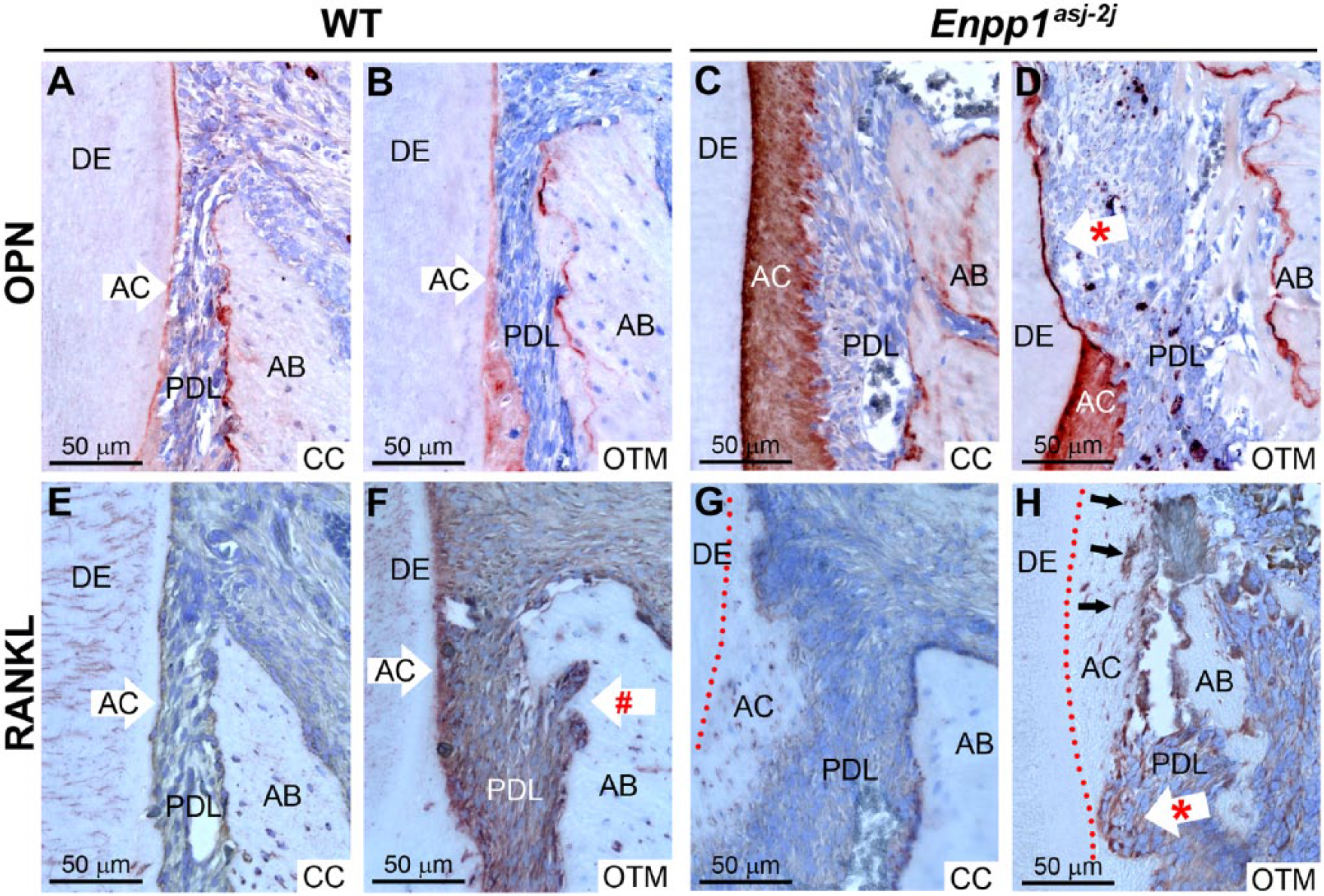
Altered immunostaining of osteopontin and receptor-activator of NF-κB ligand (RANKL) in Enpp1 mutant mice. Immunohistochemistry (IHC) identified osteopontin (OPN) in wild-type (WT) acellular cementum (AC) and alveolar bone (AB) on (
Discussion
To our knowledge, this is the first study focused on effects of orthodontic force on periodontal tissues in genetically altered mice harboring substantially altered cementum and periodontal structures. Enpp1 mutant mice feature decreased PPi, leading to significantly increased acellular cementum and expanded PDL volume with larger PDL-bone and PDL-tooth surface areas. Enpp1 mutant mice undergoing OTM exhibited reduced tooth movement, less evidence of PDL compression, reduced dentin but increased cementum resorption, and altered distribution of osteoclasts/odontoclasts on root and bone surfaces compared to WT mice. While the hypothesis of reduced cementum resorption in Enpp1 mutant mice undergoing OTM was not validated, observations of decreased tooth movement and altered osteoclast/odontoclast distribution provide novel insights into the intersection of mineral metabolism and periodontal remodeling.
ENPP1 is a regulator of extracellular levels of PPi, an inhibitor of physiological and pathological mineralization (Millán 2013). ENPP1 is selectively highly expressed by cementoblasts during acellular cementum formation, and Enpp1 loss of function in mice causes increased acellular cementum thickness (Nociti et al. 2002; Foster et al. 2012; Zweifler et al. 2015). In mice, Ank loss of function also reduces PPi levels, resulting in hypercementosis (Nociti et al. 2002; Foster et al. 2012). We have identified targeted PPi depletion as a novel approach to promote cementum regeneration using a surgical defect model (Rodrigues et al. 2011). Because cementum is a target of undesirable root resorption resulting from OTM, we aimed to understand whether altered PPi metabolism and/or periodontal and cementum properties would prove beneficial in this context. The OTM model employed has been characterized as a reliable approach for studying periodontal remodeling induced by compression and tension, producing well-described changes in mesial tooth movement, alveolar bone height, osteoclast regulation, and root resorption (Ong et al. 2000; Pavlin et al. 2000a, 2000b; Jäger et al. 2005; Nakamura et al. 2008; Gonzales et al. 2009; Kitaura et al. 2009; Wolf et al. 2013; Wolf et al. 2014).
We discovered significantly reduced tooth movement in Enpp1 mutant versus WT mice. Several hypotheses present themselves based on previous findings and data reported here. Because of the critical role of osteoclast-driven bone remodeling in OTM, potential osteoclast defects should be considered. However, normal osteoclast formation and function was reported in Enpp1 knockout mice versus controls (Hajjawi et al. 2014). Our previous study indicated increased osteoclast numbers on alveolar bone surfaces of Enpp1 knockout versus WT mice, allowing for bone remodeling and maintenance of PDL space despite cementum expansion (Foster et al. 2012). Three-dimensional micro-CT analyses of PDL volume and thickness reported here confirm maintenance and even expansion of PDL space. However, while Enpp1 mutant mice showed similar or slightly greater osteoclast numbers in CC (unloaded) periodontia, total TRAP-positive cells were decreased on OTM sides. Furthermore, preference of clastic cells appeared to switch from bone to root surfaces in Enpp1 mutant mice. While WT molars undergoing OTM presented the expected combination of dentin and cementum resorption, Enpp1 mutant mouse molars exhibited resorption lacunae limited almost exclusively to the cementum. While one reason for this is the relative cervical cementum thickness in Enpp1 mutant mice, it is intriguing to consider that cementum maintains a high regenerative potential, and resorptive defects in cementum are classified as less severe and more likely to spontaneously regenerate (Lossdörfer et al. 2010; Rodrigues et al. 2011).
We recently compiled the first report of cervical hypercementosis resulting from generalized arterial calcification of infancy (GACI; OMIM# 208000) in human subjects caused by ENPP1 mutations (Thumbigere-Math et al. 2018). Intriguingly, several GACI patients exhibited dental infraocclusion (lack of normal eruption/maintenance of occlusal position), failure of primary tooth exfoliation, ankylosis, and/or unusual reparative cementum on primary tooth roots. One GACI subject’s dental history highlighted difficulty in achieving OTM. In all of these manifestations, altered regulation of mineralization (e.g., ankylosis) and/or resorption may be suspected. While defective osteoclast/odontoclast resorption has not been previously documented in Enpp1 loss-of-function mice or GACI subjects, loss of function of PPi regulator ANK/ANKH contributes to osteoclast dysfunction in craniometaphyseal dysplasia (Chen et al. 2014). These findings implicating altered osteoclast/odontoclast activities in mice and humans with ENPP1 loss of function warrant further investigation.
Additional hypotheses for altered OTM in Enpp1 mutant mice may involve the unusual inclusion of cementocyte-like cells in the thick cervical cementum that under normal conditions remains acellular. Some of these unusually located cementocytes express RANKL, possibly promoting migration and activation of osteoclast precursors to root rather than bone surfaces. We show increased expression of Spp1/OPN in cementoblasts and cervical cementocytes of Enpp1asj versus WT mice. OPN is an extracellular matrix protein that promotes cell migration and attachment, and absence of OPN has caused defective osteoclast recruitment and reduced OTM in mice (Chung et al. 2008; Walker et al. 2010). Conversely, increased OPN expression by cementum-associated cells in Enpp1 loss-of-function mice could influence osteoclast recruitment toward the root surface and away from bone, affecting resorptive activity at these locations. In a report on hypophosphatemic Hyp mutant mice, Coyac and colleagues found that increased OPN played a role in mineralization defects in dentin and alveolar bone, and sustained osteoclastic activity following ligature-induced periodontitis was associated with OPN-rich osteoid (Coyac et al. 2017). However, OPN was not localized to the hypomineralized cellular cementum. The potential for changes in inorganic phosphate and PPi concentrations to affect osteoclasts and bone remodeling should be explored in more detail in future studies.
We propose an additional hypothesis to potentially explain reduced OTM in Enpp1 mutant mice. Micro-CT analysis revealed increased PDL volume and thickness, as well as nearly 30% increased surface area between PDL and tooth root/alveolar bone of Enpp1 mutant versus WT mice. These interfaces incorporate high-density Sharpey’s fiber insertions into both cementum and bone surfaces. Assuming the density of Sharpey’s fibers remains similar, this translates to an increase of almost 30% more inserted fibers in Enpp1 mutant mice. This altered periodontal attachment would be anticipated to increase the strength of PDL-bone/tooth attachment, affecting both mechanical deformation of PDL and periodontal remodeling, which may be observed as a reduced response to orthodontic loading. Enpp1 mutant mouse molars exhibited less compression and expansion than similarly loaded WT molars. Less compression mesial to the root would be expected to produce less vascular disturbance and attract fewer osteoclast precursors, and Enpp1 mutant mice exhibited fewer osteoclasts on OTM sides. While speculative, this observation of altered periodontal structure in Enpp1 mutant mice may explain reduced deformation under mechanical stress, reduced tooth movement, and fewer osteoclasts, as well as altered tooth movement and delayed primary tooth exfoliation in GACI subjects (Thumbigere-Math et al. 2018). Mechanical testing of Enpp1 mutant periodontia may provide further insights.
Author Contributions
M. Wolf, V. Thumbigere-Math, B.L. Foster, contributed to conception, design, data acquisition, analysis, and interpretation, drafted and critically revised the manuscript; M. Ao, contributed to data acquisition, critically revised the manuscript; M.B. Chavez, T.N. Kolli, contributed to data acquisition, analysis, and interpretation, drafted and critically revised the manuscript; K. Becker, contributed to conception and data interpretation, critically revised the manuscript; E.Y. Chu, contributed to data acquisition and analysis, critically revised the manuscript; A. Jäger, contributed to data acquisition, analysis, and interpretation, critically revised the manuscript; M.J. Somerman, contributed to conception, design, data analysis and interpretation, critically revised the manuscript. All authors gave final approval and agree to be accountable for all aspects of the work.
Supplemental Material
DS_10.1177_0022034518759295 – Supplemental material for Reduced Orthodontic Tooth Movement in Enpp1 Mutant Mice with Hypercementosis
Supplemental material, DS_10.1177_0022034518759295 for Reduced Orthodontic Tooth Movement in Enpp1 Mutant Mice with Hypercementosis by M. Wolf, M. Ao, M.B. Chavez, T.N. Kolli, V. Thumbigere-Math, K. Becker, E.Y. Chu, A. Jäger, M.J. Somerman, and B.L. Foster in Journal of Dental Research
Footnotes
Acknowledgements
We thank Dr. Kristina Zaal (Light Imaging Section, NIAMS/NIH) for assistance in slide scanning and Alyssa Coulter (NIAMS/NIH) for assistance with histological sectioning. We thank the editor and anonymous reviewers for their critiques and suggestions that improved this manuscript.
A supplemental appendix to this article is available online.
This research was supported by the German Orthodontic Association (DGKFO) and Medical Faculty University of Bonn, Germany (to M.W.); grants AR 066110 to B.L.F. and AR 069643 to V.T.-M. from the National Institute of Arthritis and Musculoskeletal and Skin Diseases (NIAMS), National Institutes of Health (NIH, Bethesda, MD); and intramural funding from NIAMS/NIH (to M.J.S).
The authors declare no potential conflicts of interest with respect to the authorship and/or publication of this article.
References
Supplementary Material
Please find the following supplemental material available below.
For Open Access articles published under a Creative Commons License, all supplemental material carries the same license as the article it is associated with.
For non-Open Access articles published, all supplemental material carries a non-exclusive license, and permission requests for re-use of supplemental material or any part of supplemental material shall be sent directly to the copyright owner as specified in the copyright notice associated with the article.