
Abstract
Periodontal ligament derived stem cells (PDLSCs) are capable of differentiating into multiple cell types and inducing a promising immunomodulation for tissue regeneration and disease treatment. However, it is still challenging to develop a practical approach to activate endogenous stem cells for tissue self-healing and regeneration. In this study, transcriptome analysis reveals that resveratrol promotes PDLSC stemness through activation of stem cell, osteoprogenitor, and chondroprogenitor markers. Self-renewal and multipotent differentiation abilities are also improved in resveratrol-treated PDLSCs. In addition, immunomodulation of PDLSCs is dramatically increased after resveratrol treatment. Mechanistically, we show that resveratrol activates ERK/WNT crosstalk through elevation of olfactory and growth factor signaling pathways to upregulate the expression levels of RUNX2 and FASL for osteogenesis and immunomodulation, respectively. By using a periodontitis animal model, administration of resveratrol partially rescues bone loss through activation of endogenous somatic stem cells and inhibition of inflammatory T-cell infiltration. Taken together, our findings identify a novel pharmacological approach to achieve autotherapies for endogenous tissue regeneration.
Keywords
Introduction
Tissue regeneration is a dynamic remodeling process through the external stimulations and the internal processes. To maintain body homeostasis, continual tissue renewal and regeneration are necessary, which are mainly attributed to adult stem cells (Zhang et al. 2016). Mesenchymal stem cells (MSCs), originating from both the mesoderm and the neural crest, constitute a population of self-renewal stem cells that can give rise to multiple specialized cell types (Pittenger et al. 1999). Along with their extensive distribution in many adult tissues, MSCs are required for continuous tissue homeostasis maintenance within diverse organs, which have made them an attractive target for tissue engineering (Prockop 1997). MSCs derived from orofacial sources, such as periodontal ligament stem cells (PDLSCs), have superior capability for orofacial regeneration as they may be more committed to differentiating into craniofacial tissues (Moshaverinia et al. 2014). However, the craniofacial tissue regeneration often results in an unfavorable outcome due to the altered local microenvironment and rapid apoptosis of transplanted MSCs (Liu et al. 2011). Therefore, there is an urgent need to discover novel therapeutic avenues for activating endogenous stem cell–based tissue regeneration.
Autotherapies are a novel treatment strategy to induce the body’s innate ability to heal and protect itself, which propose a minimally invasive approach to elevate tissue healing and regeneration. To achieve the endogenous tissue self-healing, host local microenvironment as a stem cell niche provides a unique tissue structure to activate somatic stem cells for tissue regeneration (Lumelsky et al. 2018; Ruddy and Morshead 2018). MSCs, as adult stem cells, can maintain tissue homeostasis and regeneration and interplay with immune cells for immunomodulation (Akiyama et al. 2012; Chen et al. 2017). However, it is still largely unknown how and whether activation of endogenous MSCs can promote tissue regeneration.
Resveratrol is a natural phytoalexin that exhibits potentials to promote tissue regeneration in various tissues and organs (Baur et al. 2006; Tseng et al. 2011). At the cellular level, resveratrol improves MSC-based therapy for liver and cardiac regeneration through activation of stem cell function and improving the survival of transplanted MSCs (Pinarli et al. 2013; Okay et al. 2015). However, the role of resveratrol in autotherapy-based tissue regeneration is largely unknown. Specifically, we aimed to identify whether resveratrol can activate somatic stem cells and promote MSC immunomodulation for endogenous tissue regeneration. In this study, we showed that resveratrol elevates cell proliferation, increases multipotent differentiation abilities, and improves PDLSC immunomodulation. Administration of resveratrol in vivo significantly activates endogenous stem cells, inhibits inflammatory cell infiltration, and rescues periodontitis phenotypes in a mouse model. Collectively, our data provide a novel strategy using a pharmacological approach for autotherapy-based tissue regeneration.
Materials and Methods
Refer to the Appendix for detailed information on methods.
Animals
Twelve-week-old female C57BL/6J and severe combined immunodeficient (SCID) mice were purchased from the Jackson Laboratory. Aged-matched female mice were used as controls in the present study. All animal experiments were performed under institutionally approved protocols for the use of animal research (University of Pennsylvania Institutional Animal Care and Use Committee #806682). This study followed the Animal Research: Reporting of In Vivo Experiments (ARRIVE 2.0) guidelines.
Antibodies and Reagents
All antibodies and reagents used in this study are described in the Appendix.
Results
Resveratrol Enhances Stemness of PDLSCs In Vitro
To gain insight into the biological function of resveratrol in PDLSCs, we first performed RNA sequencing (RNA-seq) analysis to compare transcriptomic profiles with or without resveratrol treatment. We found 724 transcripts that significantly change their expression, log2 fold change (FC) >1 and FC <1 and P < 0.01, after resveratrol treatment in PDLSCs compared to vehicle-treated cells (Fig. 1A, B). Among these, 323 (44.6%) were downregulated and 401 (55.4%) upregulated upon resveratrol treatment (Fig. 1C). Enrichment analysis of Gene Oncology (GO) terms over the 724 resveratrol targets showed that the most enriched were associated with olfactory-related G-protein coupled receptor (GPCR) signaling categories (Fig. 1D). We next asked whether resveratrol could stimulate stemness and stem cell properties of PDLSCs. We identified 39 MSC stemness-related genes significantly change their expression after resveratrol treatment. Among them, 8 (20.5%) were downregulated and 31 (79.5%) upregulated (Fig. 1E), indicating resveratrol highly activated the stem cell function of PDLSCs. These results were further confirmed by quantitative polymerase chain reaction (qPCR) to show resveratrol significantly elevated 1) MSC markers, including CD44, intercellular adhesion molecule 1 (ICAM1), and vascular cell adhesion molecule 1 (VCAM1); 2) osteoprogenitor markers, including osterix (SP7), secreted phosphoprotein 1 (SPP1), and GLI family zinc finger 1 (GLI1); and 3) chondroprogenitor markers, including SRY-box transcription factor 9 (SOX9), collagen type X alpha 1 (COL10A1), and activated leukocyte cell adhesion molecule (ALCAM) (Fig. 1F).
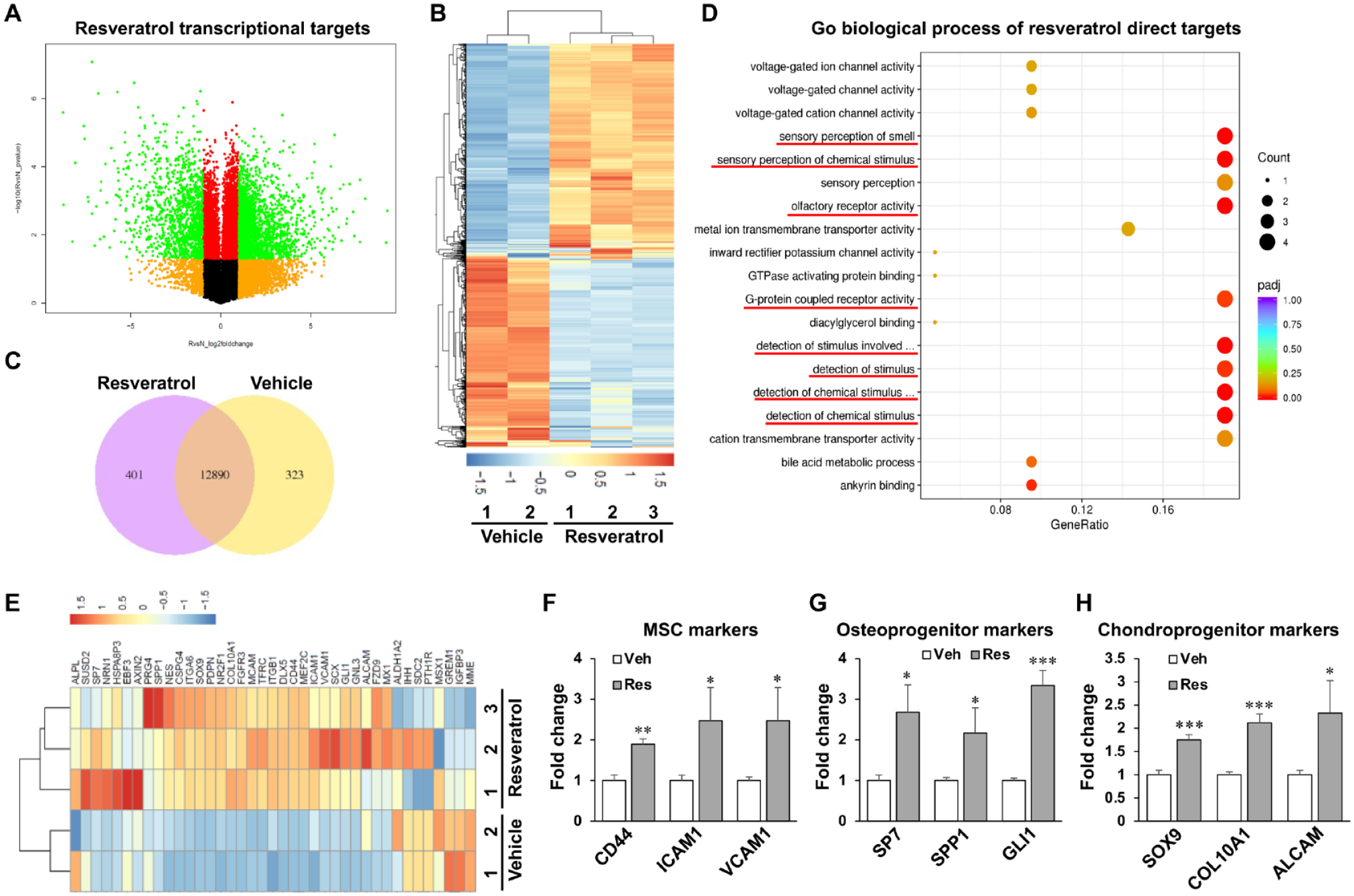
Transcriptome analysis revealed resveratrol promoted periodontal ligament stem cell (PDLSC) stemness. (
Resveratrol Promotes PDLSC Proliferation and Differentiation Capacities
Next, we examined whether resveratrol could increase PDLSC proliferation and differentiation capacities. We then performed GO analysis to focus on cell proliferation pathways and found resveratrol highly activated cell cycle categories, particularly cell cycle, DNA replication, chromosome segregation, and DNA repair/cell cycle checkpoint (Fig. 2A). These results were further confirmed by gene set enrichment analysis (GSEA) to determine the prior gene set is significantly different between 2 biological states. Our results showed that cell proliferation–related gene sets, such as DNA replication, RNA polymerase, and biosynthesis of amino acids, were highly enriched after resveratrol treatment in PDLSCs (Fig. 2B). We next performed immunofluorescence (IF) staining using cell proliferation marker Ki67 to show that resveratrol greatly enhanced Ki67+ PDLSC percentage compared to vehicle control (Fig. 2C). In addition, we measured PDLSC metabolic activity by the MTT assay to show resveratrol largely activated PDLSC viability (Fig. 2D). To further explore the molecular regulation in cell cycle after resveratrol treatment, we performed qPCR to determine that cell cycle suppressor genes p21 and p53, but not p16, were significantly inhibited by resveratrol treatment (Fig. 2E), suggesting resveratrol elevated PDLSC proliferation through inhibition of cell cycle arrest/apoptosis.
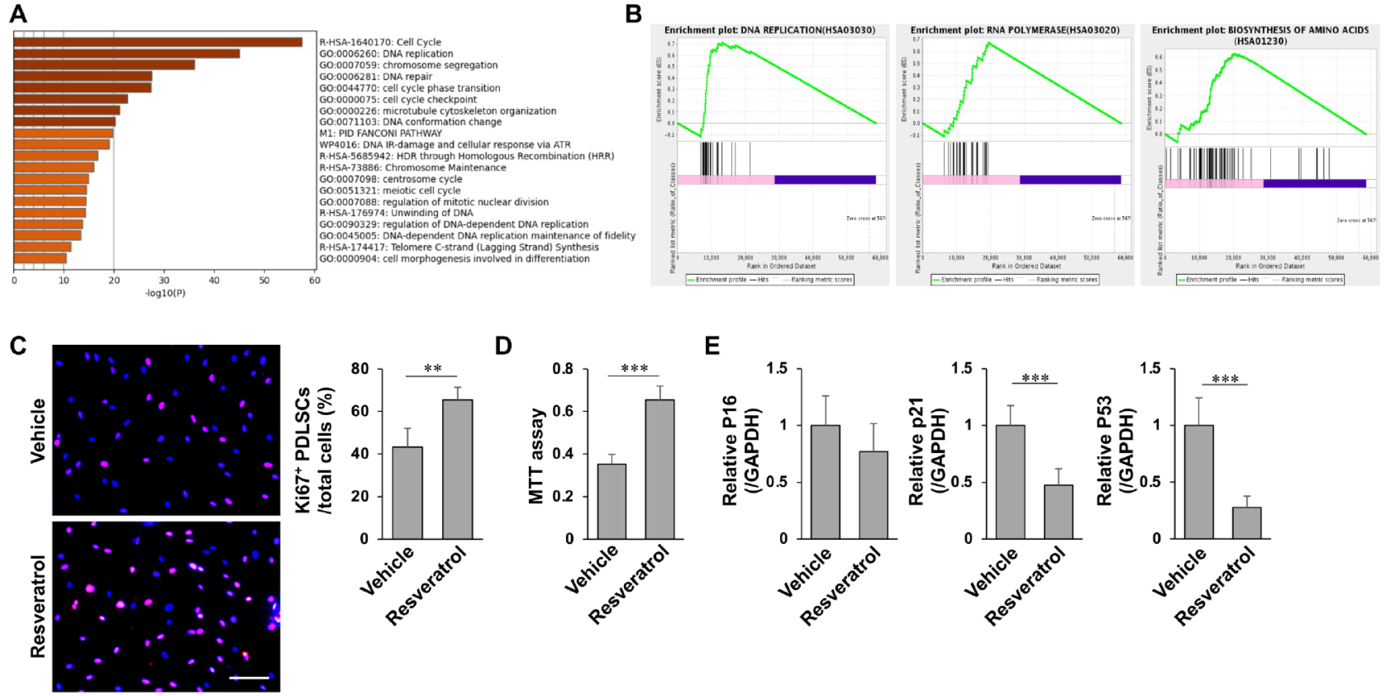
Resveratrol elevated periodontal ligament stem cell (PDLSC) proliferation. (
As RNA-seq showed resveratrol can promote osteoprogenitor and chondroprogenitor markers (Fig. 1F), we then examined the capacities of osteogenesis and chondrogenesis in PDLSCs with resveratrol treatment. Under osteogenic inductions, resveratrol-treated PDLSCs showed superior osteogenesis, as indicated by increased mineralized nodule formation and expression of the osteogenic genes runt-related transcription factor 2 (RUNX2) and alkaline phosphatase (ALP), respectively (Appendix Fig. 1A, B). We next showed that resveratrol-treated PDLSCs generated more new bone than vehicle-treated control at 8 wk postimplantation using an established in vivo MSC implantation assay, in which 4 × 10⁶ PDLSCs with hydroxyapatite tricalcium phosphate (HA/TCP) particles as a carrier were subcutaneously implanted into immunocompromised mice (Appendix Fig. 1C). Parallel studies showed an increased capacity of resveratrol-treated PDLSCs to differentiate into chondrocytes under chondrogenic inductive conditions, associated with the elevated expression of aggrecan (ACAN) and SOX9 by IF staining (Appendix Fig. 1D). To further confirm the effect of resveratrol in chondrogenesis in vivo, we showed resveratrol-treated PDLSCs generated more cartilage-like structures with increased COL2+ chondrocytes than a control group at 8 wk postimplantation using an in vivo chondrogenic implantation assay, in which 4 × 10⁶ PDLSCs with gelfoam-hydrogel as a carrier were subcutaneously implanted into immunocompromised mice (Appendix Fig. 1E). Taken together, these findings indicated that resveratrol promotes PDLSC stem cell properties.
Resveratrol Regulates ERK-WNT Crosstalk through Activation of Olfactory and Growth Factor Pathways
We next aimed to identify the molecular signaling targets of resveratrol in PDLSCs. To confirm our findings in Figure 1D, we performed GSEA analysis to show olfactory-related GPCR gene sets, such as olfactory pathways, olfactory receptor (OR) activity, and sensory receptor of smell, were highly enriched after resveratrol treatment (Fig. 3A). In addition to olfactory signaling, we also identified several growth factors, linked to platelet-derived growth factor (PDGF), vascular endothelial growth factor (VEGF), and epidermal growth factor (EGF) pathways, were highly activated in resveratrol-treated PDLSCs (Fig. 3B). We then examined the expression levels of ORs and growth factors by qPCR to show that ORs including OR52N4, OR10A3, OR2A7, OR2AG2, and OR2A42 and growth factors including PDGFD, EGF, PDGFRL, VEGFA, VEGFC, and PDGFC were highly activated by resveratrol treatment in PDLSCs (Fig. 3C). As the downstream target of both olfactory-GPCR (Mykytyn and Askwith 2017) and growth factor pathways (Bruggemann et al. 2021) is ERK mitogen-activated protein kinase signaling, we then showed that resveratrol significantly activated ERK signaling through phosphorylation of ERK (p-ERK) in PDLSCs (Fig. 3D). The crosstalk between ERK and WNT signaling has been shown to promote MSC lineage commitment toward osteogenesis through stimulation of RUNX2 activity (Cervenka et al. 2011). Our data further revealed that resveratrol was able to enhance the level of active β-catenin in PDLSCs (Fig. 3D). To examine whether WNT/β-catenin signaling acts as a downstream target of the ERK pathway, we used small interfering RNA (siRNA) to knock down ERK in resveratrol-treated PDLSCs. Western blot indicated that siERK treatment downregulated both p-ERK and active β-catenin levels, suggesting ERK signaling controlled WNT/β-catenin cascades in PDLSCs (Fig. 3D). To further explore the functional role of ERK/WNT crosstalk in PDLSCs, siERK treatment greatly blocked osteogenic capacity induced by resveratrol, as indicated by decreased mineralized nodule formation and reduced levels of RUNX2 and ALP (Fig. 3E, F). Collectively, these findings reveal that resveratrol-mediated olfactory and growth factor pathways activate downstream ERK/WNT crosstalk to promote PDLSC stemness.
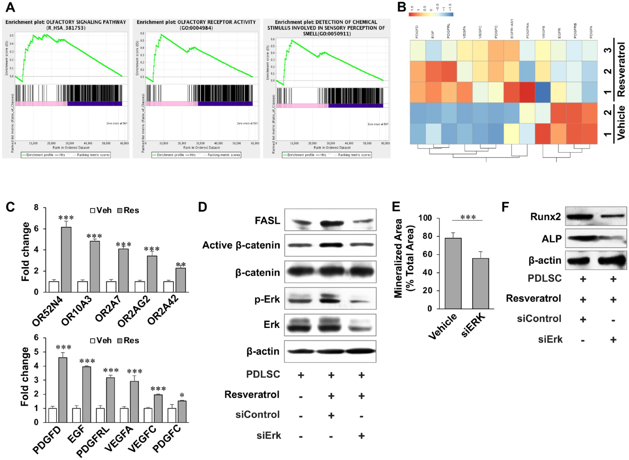
Resveratrol-activated ERK/WNT crosstalk through olfactory and growth factor pathways. (
Resveratrol Is Associated with PDLSC-Mediated Immunomodulation
Since immunomodulatory properties were recently identified as an important characteristic of PDLSCs, which has led to their systemic infusion to treat a variety of immune diseases, we next examined whether resveratrol regulates PDLSC-mediated immunomodulation. We first used a PDLSC/T-cell coculture system to show resveratrol-treated PDLSCs had significantly increased capacity to induce Annexin-V+ apoptotic T cells when compared to the vehicle-treated PDLSCs (Fig. 4A). In order to assess the therapeutic mechanism of resveratrol-treated PDLSCs, we used an inductive experimental colitis model (Alex et al. 2009) to evaluate the therapeutic effect of allogenic PDLSC transplantation (PDLSC-T) at day 3 after dextran sulfate sodium (DSS) induction. Treatment with 0.2 × 10⁶ PDLSCs (positive control group) is considered a standard dosage to elicit a therapeutic response (Chen et al. 2014). Therefore, we infused 10% (0.1×) of that amount (0.02 × 10⁶ of resveratrol-treated PDLSCs) into colitis mice to examine whether resveratrol pretreatment could reduce the dosage of PDLSCs in immunotherapy (Fig. 4B). The body weight of mice with induced colitis was significantly reduced compared to control C57BL6 mice from day 8 to 12 post-DSS induction. After normal, resveratrol-treated or 0.1× resveratrol-treated PDLSC-T, the body weight was partially restored (Fig. 4C). The disease activity index (DAI), including body weight loss, diarrhea, and bleeding, was significantly elevated in the colitis mice. After normal, resveratrol-treated or 0.1× resveratrol-treated PDLSC-T, the DAI score was decreased (Fig. 4D). Furthermore, colon tissue from each group was analyzed. Both the absence of the epithelial layer and inflammatory cell infiltration were observed in the colon of induced colitis compared to the control group. Normal, resveratrol-treated or 0.1× resveratrol-treated PDLSC-T recovered epithelial structure and eliminated inflammatory cells in colitis mice (Fig. 4E). Histological activity index (Alex et al. 2009) confirmed that normal, resveratrol-treated or 0.1× resveratrol-treated PDLSC-T reduced the DAI (Fig. 4G). In addition, IF staining showed significantly increased CD4+IL17+ T-helper 17 (Th17) infiltration in the colon of induced colitis. Transplantation of normal, resveratrol-treated or 0.1× resveratrol-treated PDLSCs dramatically reduced the infiltrated Th17 in colitis mice (Fig. 4F, H). Flow cytometry analysis further showed that elevated Th17 cells were observed in the colitis mice. Normal, resveratrol-treated or 0.1× resveratrol-treated PDLSC-T significantly downregulated Th17 (Fig. 4I). The data therefore suggest that the number of PDLSCs used for immunotherapy could be dramatically reduced to induce T-cell apoptosis and offer a potential treatment for colitis mice. FAS ligand (FASL) is a type II transmembrane protein that binds with FAS to form the death-inducing signaling complex in many cell types (Zhang et al. 2008). To examine whether resveratrol induces PDLSC immunomodulation via ERK-mediated FASL activation, Western blot showed resveratrol treatment significantly elevated the level of FASL in PDLSCs (Fig. 3D). Knockdown of ERK by siRNA dramatically reduced the level of FASL, indicating FASL is required for resveratrol-induced immunotherapy in PDLSCs through activation of ERK signaling (Fig. 3D). As immune components can also target MSCs through death pathways (Liu et al. 2011), we next examined whether resveratrol treatment is able to protect PDLSC survival after cocultured with activated T cells in a PDLSC/T-cell coculture system. Toluidine blue staining showed activated T cells caused PDLSC, but not resveratrol-treated PDLSC, death (Fig. 4J). Collectively, our results revealed that resveratrol can promote PDLSC immunomodulation through activation of FASL-mediated T-cell apoptosis, as well as protection of PDLSC survival.
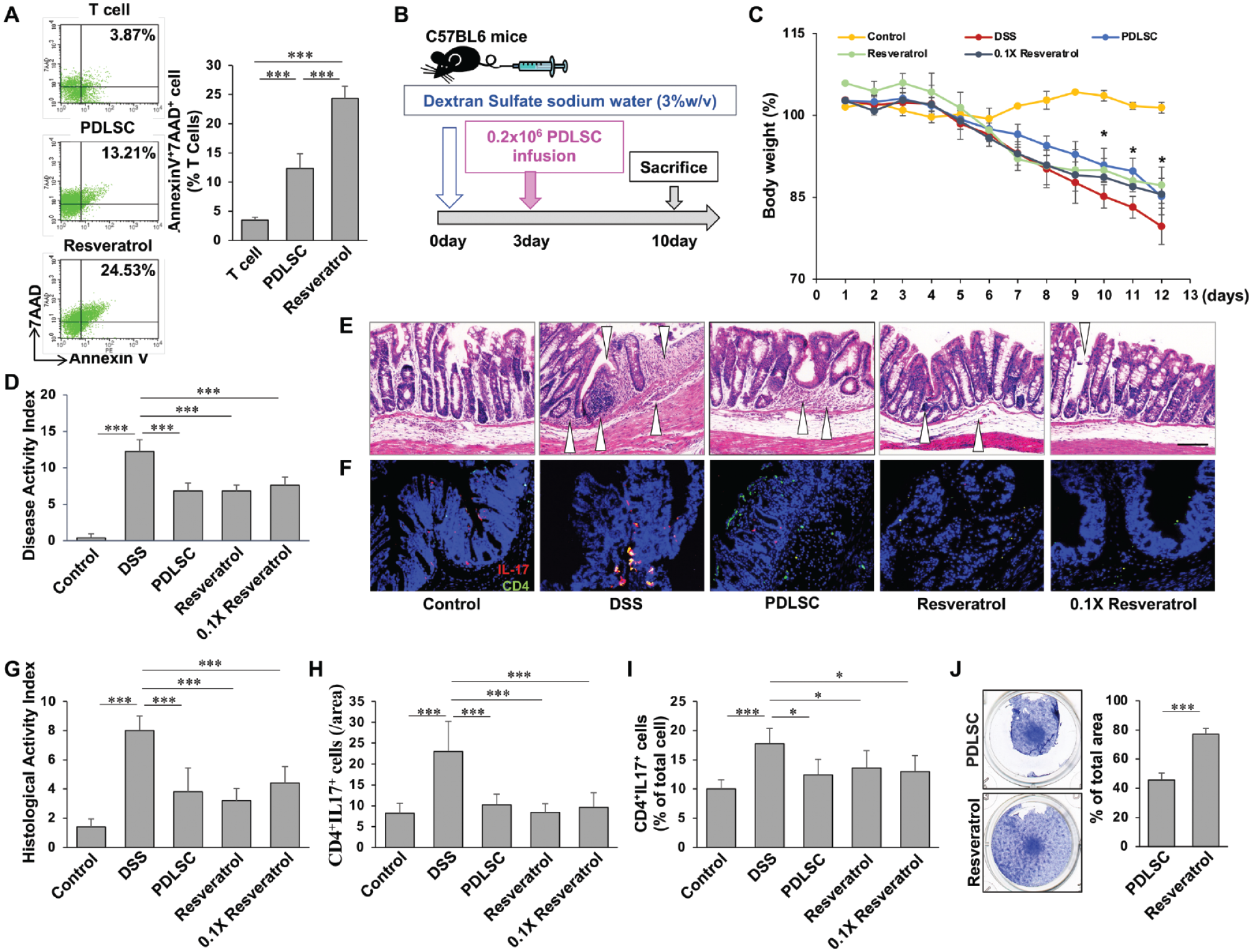
Resveratrol improved periodontal ligament stem cell (PDLSC) immunomodulation in vitro and in vivo in a DSS-induced experimental colitis mouse model. (
Resveratrol Ameliorates Periodontitis Phenotypes through Activating Stemness and Immunomodulation of Endogenous Stem Cells
Based on the recent advances in stem cell biology, immunology, and material sciences, autotherapies, a novel concept for tissue regenerative medicine, have been proposed to optimize endogenous tissue responses and microenvironment for somatic stem cell activation (Lumelsky et al. 2018; Yui et al. 2018). As we find that resveratrol can promote stemness and immunomodulation of PDLSCs, we then asked whether resveratrol could activate endogenous stem cells for tissue regeneration through regulation of inflammatory microenvironment. To this end, we employed a ligature-induced periodontitis model, by which severe alveolar bone loss with an activated proinflammatory microenvironment was established (Marchesan et al. 2018). Local injection of resveratrol was performed at day 0 and day 4 after ligation (Fig. 5A). At day 7 postligation, severe alveolar bone loss was observed around the ligated second molar compared to the control group. In contrast, distance from cementoenamel junction (CEJ) to alveolar bone (AB) crest in the resveratrol-treated group decreased to approximately half of the ligated mice with a clear statistical trend (Fig. 5B). These data prompted us to further examine whether resveratrol treatment activated endogenous stem cells and inhibited proinflammatory T cells in periodontitis mice. IF staining showed the number of costained Ki67 and the MSC marker SCA-1 was dramatically decreased in the ligated mice, while resveratrol treatment significantly elevated the number of Ki67+SCA-1+ cells in vivo (Fig. 5C). In addition, our results showed resveratrol treatment decreased the number of CD4+TNFα+ proinflammatory T cells when compared to ligated mice, in which CD4+TNFα+ T cells were highly expanded after ligation (Fig. 5D). To investigate whether resveratrol treatment can rescue periodontal pathogenic bacteria such as Porphyromonas gingivalis (PS)–induced endogenous stem cell repression, we established an in vitro periodontitis model using PG-lipopolysaccharide (LPS)–treated PDLSCs to show that treatment with LPS significantly inhibited viability and increased cell apoptosis in a dose-dependent manner (Fig. 5E, F). Resveratrol treatment partially elevated PDLSC viability and decreased cell apoptosis under 10 μg/mL LPS stimulation (Fig. 5G, H). Overall, resveratrol treatment promotes endogenous stem cell activation and inhibits proinflammatory microenvironment for autotherapies in the periodontitis mice.
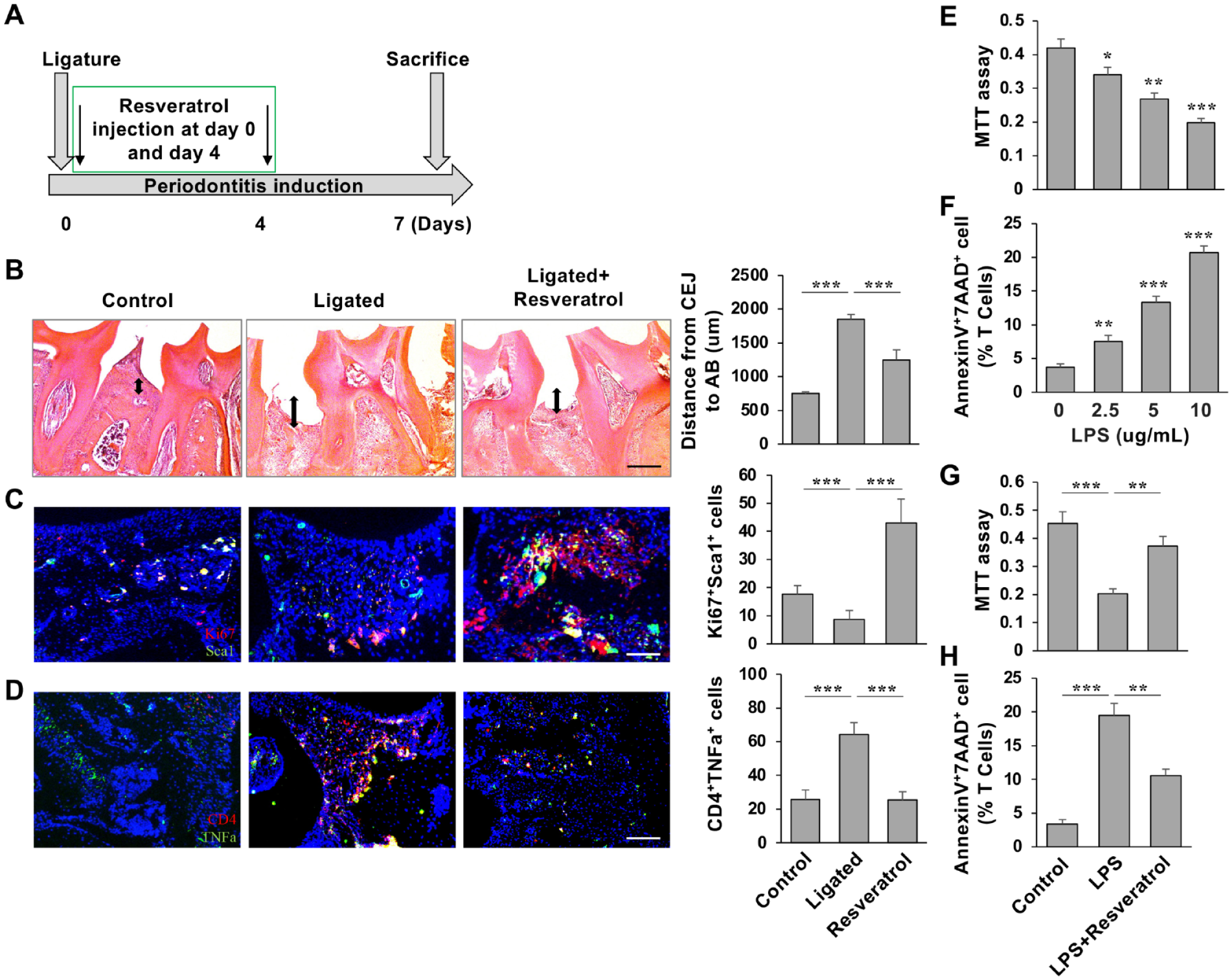
Resveratrol treatment partially rescued disease phenotypes in a periodontitis mouse model. (
Discussion
MSC-based regenerative medicine is a promising approach for tissue reconstruction and disease management, by which MSCs can regulate tissue repair/homeostasis and interact with immune cells for immunomodulation (Prockop 1997; Akiyama et al. 2012; Chen et al. 2017). The increased research indicated that natural products could regulate the immune response with few adverse side effects, which offer new avenues for immunomodulation and can be promising agents in preventing chronic diseases (Kishore et al. 2019). Resveratrol is a promising pharmacological target in regulation of cell viability, proliferation, anti-inflammation, and osteogenesis of MSCs (Wang et al. 2014; Li et al. 2019). In this study, we reveal that resveratrol treatment significantly improves stemness of PDLSCs through activation of stem cell markers, elevation of self-renewal and multipotent differentiation abilities, and upregulation of immunomodulatory capabilities. Furthermore, activation of endogenous stem cells by a pharmacological approach, such as in vivo resveratrol treatment, can markedly improve tissue regeneration and rescue disease phenotypes in a periodontitis mouse model. This study provides experimental evidence that links resveratrol to PDLSC-mediated tissue regeneration and demonstrates the potential to improve autotherapies through activation of endogenous stem cells.
Profiling the transcriptional level of stem cells at a defined condition using RNA-seq is a promising analysis to identify and prioritize genetic variants in the altered expression levels (Schlieben et al. 2021). By using RNA-seq analysis, olfactory and growth factor pathways are identified as downstream targets of resveratrol in PDLSCs. In a recent study, olfactory receptors have been linked to bone metabolism in an osteoporotic condition, suggesting that ORs are expressed in MSC-osteoblast lineage cells and required to maintain skeletal homeostasis (Zhu et al. 2018). Our data reveal that several ORs are expressed in PDLSCs, and resveratrol treatment dramatically increases osteogenic capacity through elevating more than 20 ORs, implying ORs may play a critical role in PDLSC-mediated bone regeneration. Growth factors are well known to be key mediators in supporting MSC survival, proliferation, and differentiation, which are the drivers of regenerative medicine (Nie et al. 2020). Resveratrol significantly activates the expression levels of growth factors, further indicating growth factor and OR pathways may synergistically promote PDLSC stemness.
ERK signaling, the downstream target of both OR-GPCR and growth factor signaling, is required for bone formation during skeletal development and plays an important role in maintaining bone tissue homeostasis (Liu, Zhao et al. 2018). Several natural compounds act as activators of ERK signaling, which is the key driver in osteogenesis for defeating skeletal disorders (Liu, Zhao et al. 2018; Liu et al. 2019). WNT/β-catenin signaling has been shown as a downstream kinase of ERK and is also essential for the self-renewal and multipotent differentiation of MSCs and regulating bone tissue homeostasis (Cervenka et al. 2011; Lin et al. 2019). Our mechanism studies determine that ERK/WNT crosstalk mediates resveratrol-induced PDLSC activation, by which ERK activates downstream β-catenin through OR-GPCR and growth factor signaling. Since resveratrol has been shown to be involved in activation of the WNT/β-catenin cascades to promote osteogenic differentiation (Zhao et al. 2018), collectively, the present study explores the novel targets of resveratrol to activate ERK/WNT crosstalk in PDLSC-mediated tissue regeneration.
MSCs exhibit immunomodulatory properties by mediating the proliferation, migration, and differentiation of several major types of immune cells, and systemic infusion of MSCs or MSC-derived cellular components has been shown to yield therapeutic benefits for a variety of immune-related disorders (Le Blanc et al. 2004; Chen et al. 2015; Liu, Kou et al. 2018). Our previous studies showed that MSCs induce T-cell apoptosis via upregulation of the FASL-mediated FAS death pathway to achieve immune tolerance, by which β-catenin directly binds to the Fasl promoter to drive gene expression at the transcriptional level (Akiyama et al. 2012; Chen et al. 2014). Here, we showed that resveratrol improves PDLSC immunomodulation through FASL activation, which is regulated by ERK/WNT crosstalk. Taken together, our data reveal an ERK/WNT/FASL cascade in the regulation of PDLSC immunomodulation and explore the potential to improve PDLSC-based clinical therapies with reduced cell dosage.
The new paradigm of tissue regeneration shifts to manipulate/activate an endogenous tissue microenvironment to minimize invasive approaches for regenerative medicine, which is termed autotherapies. To achieve autotherapies, preconditioning a regenerative microenvironment, creating a specific stem cell niche, and activating transcription factors for lineage reprogramming are the key components for enhancing endogenous tissue regeneration (Heber-Katz 2017; Lumelsky et al. 2018). Periodontitis is an inflammatory disease, characterized by inflammatory response and alveolar bone loss (Hajishengallis 2015). MSC-based therapy has been widely studied for the treatment of periodontal disease because of its effects on bone regeneration and immunomodulation (Shang et al. 2017). By using an in vivo periodontitis model, we showed resveratrol partially rescues disease phenotypes through activation of endogenous stem cells and inhibition of immune cell infiltration. In summary, this translational study substantially extends current knowledge about stem cell–based autotherapies. We also reveal an ERK/WNT crosstalk mechanism to boost PDLSC stemness and immunomodulation for tissue regeneration.
Author Contributions
W. Li, contributed to design, data acquisition, analysis and interpretation, drafted and critically revised the manuscript; X. Huang, contributed to design, data acquisition, analysis, and interpretation, drafted manuscript; W. Yu, contributed to design, data acquisition, and analysis, critically revised the manuscript; Y. Xu, R. Huang, J. Park, contributed to data acquisition and analysis, critically revised the manuscript; A. Moshaverinia, contributed to data analysis and interpretation, critically revised the manuscript; P. Arora, contributed to conception, data analysis, and interpretation, critically revised the manuscript; C. Chen, contributed to conception, design, data acquisition, analysis, and interpretation, drafted and critically revised the manuscript. All authors gave final approval and agree to be accountable for all aspects of the work.
Supplemental Material
sj-docx-1-jdr-10.1177_00220345211070222 – Supplemental material for Activation of Functional Somatic Stem Cells Promotes Endogenous Tissue Regeneration
Supplemental material, sj-docx-1-jdr-10.1177_00220345211070222 for Activation of Functional Somatic Stem Cells Promotes Endogenous Tissue Regeneration by W. Li, X. Huang, W. Yu, Y. Xu, R. Huang, J. Park, A. Moshaverinia, P. Arora and C. Chen in Journal of Dental Research
Footnotes
A supplemental appendix to this article is available online.
Declaration of Conflicting Interests
The authors declared no potential conflicts of interest with respect to the research, authorship, and/or publication of this article.
Funding
The authors disclosed receipt of the following financial support for the research, authorship, and/or publication of this article: This work was supported by grants from National Institute of Dental and Craniofacial Research, National Institutes of Health, Department of Health and Human Services (R00DE025915 and R03DE028026 to C. Chen), and a Colgate Palmolive Grant (A-2019-590-OC) to C. Chen. Colgate-Palmolive provided support for the in vitro studies shown in this manuscript. Colgate-Palmolive did not provide any support for animal work shown in this manuscript. All the animal studies shown in the manuscript were supported by R00 and R03 grants from NIDCR as cited in the article.
References
Supplementary Material
Please find the following supplemental material available below.
For Open Access articles published under a Creative Commons License, all supplemental material carries the same license as the article it is associated with.
For non-Open Access articles published, all supplemental material carries a non-exclusive license, and permission requests for re-use of supplemental material or any part of supplemental material shall be sent directly to the copyright owner as specified in the copyright notice associated with the article.