






Abstract
Osteoclasts play a key role in the regulation of bone mass and are highly active metabolically. Here we show that a metabolic reprogramming toward the hexosamine biosynthetic pathway (HBP) is required not only for osteoclast differentiation but also to determine the bone resorption mode during physiological and pathological bone remodeling. We found that pharmacological inhibition of O-GlcNAc transferase (OGT) significantly reduced protein O-GlcNAcylation and osteoclast differentiation. Accordingly, genetic deletion of OGT also inhibited osteoclast formation and downregulated critical markers related to osteoclasts differentiation and function (NFATc1, αvintegrin, cathepsin K). Indeed, cells treated with OSMI-1, an OGT inhibitor, also reduced nuclear translocation of NFATc1. Furthermore, the addition of exogenous N-acetylglucosamine (GlcNAc) strongly increased osteoclast formation and demineralization ability. Strikingly, our data show for the first time that O-GlcNAcylation facilitates an aggressive trench resorption mode in human cells. The incubation of osteoclasts with exogenous GlcNAc increases the percentage of erosion by trench while having no effect on pit resorption mode. Through time-lapse recording, we documented that osteoclasts making trenches moving across the bone surface are sensitive to GlcNAcylation. Finally, osteoclast-specific Ogt-deficient mice show increased bone density and reduced inflammation-induced bone loss during apical periodontitis model. We show that osteoclast-specific Ogt-deficient mice are less susceptible to develop bacterial-induced periapical lesion. Consistent with this, Ogt-deleted mice showed a decreased number of tartrate-resistant acid phosphatase-positive cells lining the apical periodontitis site. In summary, here we describe a hitherto undiscovered role of the HBP/O-GlcNAcylation axis tuning resorption mode and dictating bone resorption outcome.
Introduction
Osteoclasts are multinucleated cells originating from hematopoietic progenitors of the myeloid lineage, which requires a series of linked events to differentiate (Boyle et al. 2003; Soe et al. 2015; Ikeda and Takeshita 2016). The binding in myeloid progenitor cells of receptor activator of nuclear factor–κB (RANK) ligand to RANK results in the activation of tumor necrosis factor receptor–associated factor 6 (TRAF-6). This activation stimulates downstream intracellular events, including nuclear factor κB (NF-κB) and nuclear factor of activated T cells, cytoplasmic 1 (NFATc1). NFATc1, a master transcription factor, orchestrates osteoclast differentiation by transcribing various osteoclast-specific genes, such as TRAP, cathepsin K, and DC-STAMP (Asagiri et al. 2005; Asagiri and Takayanagi 2007).
Cellular metabolism is a dynamic process that supplies energy and enables the biosynthesis of macromolecules demanded by biological processes, including proliferation, differentiation, and effector functions (Hart 2019; Park-Min 2019). However, despite the upregulation of metabolic enzymes and nutrient transporters observed during osteoclastogenesis (Czupalla et al. 2005), how metabolic reprogramming regulates osteoclast function and, consequently, bone remodeling remains elusive. Although mature osteoclasts are known to contain an abundant number of mitochondria, it has been shown that glucose and glutamine are critical nutrients required for osteoclastogenesis and bone resorption activity (Kim et al. 2007; Indo et al. 2013). Notably, the hexosamine biosynthetic pathway (HBP) converts glutamine and fructose-6-phosphate, a glucose metabolite, into the end-product uridine diphosphate-N-acetylglucosamine (UDP-GlcNAc) (Love and Hanover 2005). This nucleotide-sugar is the donor substrate for installing O-linked N-acetylglucosamine (O-GlcNAc), a posttranslational protein modification (PTM), on serine or threonine residues of intracellular proteins located in the cytoplasm, nucleus, and mitochondria. O-GlcNAcylation is a reversible PTM that is regulated by 2 enzymes: O-GlcNAc transferase (OGT) that catalyzes its addition and a β-N-acetylglucosaminidase, known as O-GlcNAc hydrolase (OGA), that removes the O-GlcNAc moiety (Hart et al. 2007; Zachara 2018).
Similar to phosphorylation, O-GlcNAcylation of nuclear and cytosolic proteins coordinates protein–protein interactions, stability, and activity, controlling several cellular processes, including transcription, metabolism, signal transduction, and survival (O’Donnell et al. 2004). Dysregulation of O-GlcNAcylation by genetic deficiency of Ogt or Oga causes early embryonic or perinatal lethality in mice, respectively (O’Donnell et al. 2004; Yang et al. 2012). Moreover, the deregulation of O-GlcNAcylation is linked to the pathophysiology of various human diseases such as diabetes, cancers, neurodegenerative disorders, and cardiovascular diseases (Bond and Hanover 2015). Here, we evaluated how an alteration in the HBP/O-GlcNAcylation metabolic axis affects osteoclast resorption mode and, consequently, the effects on bone remodeling under homeostatic and pathological conditions.
Materials and Methods
Osteoclast Differentiation
C57BL/6, LysM-Cre, Cathepsin K–Cre (Ctsk-Cre) Ogt-flox mice were maintained in the animal facility of the School of Pharmaceutical Sciences in agreement with our animal ethics committee and complying with the ARRIVE (Animal Research: Reporting of In Vivo Experiments) checklist. Bone marrow cells isolated from femurs and tibias were cultured in α-MEM supplemented with 1% penicillin/streptomycin and 10% fetal bovine serum (FBS) in the presence of murine M-CSF (30 ng/mL). After 3 d, adherent cells were seeded with M-CSF (30 ng/mL) and murine RANKL (10 ng/mL). The medium was changed every 3 d. Both GlcNAc and OGT inhibitors (5SGlcNAc or OSMI-1) purchased from Sigma-Aldrich were prepared as previously described (Gloster et al. 2011). Cells were treated and collected according to each set of experiments.
After 5 d, murine osteoclasts were stained and TRAP+ cells containing 3 or more nuclei/cell were quantified, and no distinction was made between large and small osteoclasts. Total mature osteoclasts were counted and represented as TRAP+ cells/well.
For human osteoclast differentiation, CD14+ monocytes were isolated by positive selection using magnetic separation from blood donations of healthy female volunteers in accordance with Danish legislation (all donors gave written informed consent for the use of surplus material from the donation). Cells were seeded in culture flasks in α-MEM containing 10% FBS and recombinant human M-CSF (25 ng/mL). The cells were reseeded in 96-well plates with 25 ng/mL M-CSF and RANKL, and 3 d later, GlcNAc and OSMI-1 (solubilized in dimethyl sulfoxide) were added in the fresh medium in the presence of M-CSF and RANKL. The vehicle was added at the same amount to the OSMI-1 (control group). After 7 d of differentiation, the osteoclasts were stained with Giemsa for cell counts. For human osteoclasts, the number of osteoclasts (cells with 2 nuclei or more) and the nuclei were counted in randomized 6 fields per well, given by a random number generator, and using 20× magnification (Olympikus IX71).
Resorption Assay
Murine adherent cells were seeded in 96-well hydroxyapatite-coated plates (OsteoAssay-Corning) with osteoclastogenic media as described above. After 4 d, cells were removed, and the demineralized area was measured using a magnifying glass (Leica MZ6).
For the human resorption assay, CD14+ monocytes were seeded in culture flasks in α-MEM containing recombinant human M-CSF (25 ng/mL) and RANKL (25 ng/mL). After 5 d, mature osteoclasts were detached and reseeded on bone disks (IDS Nordic) in the presence of M-CSF, RANKL, and GlcNAc or OSMI-1. After 3 d, osteoclasts were removed from the bone slices, and resorption events were stained with toluidine blue. The percentage of ES/BS was analyzed by light microscopy using a 100-point grid (Borggaard et al. 2020), and all resorption events were distinguished into pits or trenches. Pits were defined as an excavation, circular in appearance, and where the ratio between length and width of the excavation did not exceed 2. Trenches were defined as an elongated and continuous excavation and at least 2 times longer than its width, as described by Soe and Delaisse 2010; Merrild et al. 2015. The percentage of each type of resorption was determined on all bone slice surfaces. The investigator was blinded during all analyses. Examples of resorption excavation (pits and trenches) can be seen in Figure 3E.
Time-Lapse Recording of Bone-Resorbing OCs
Time lapse recordings were acquired to monitor the resorbing behavior of osteoclasts. Mature osteoclasts were stained with 100 nM SiR-actin and 10 µM verapamil (Spirochrome) and placed at a density of 1 × 10⁵ cells in a rhodamine (NHS-Rhodamine; Thermo Scientific)–coated bone slice (Boneslices.com) placed inside a chambered cover-glass well, as described by Borggaard et al. 2020. The chambered cover-glass was placed in a confocal microscope (Olympus Fluoview FV10i) at 37°C in 5% CO2 in a humidified atmosphere. Osteoclasts from 3 areas for each bone slice were recorded using a 10× magnification and confocal aperture of 1.9. The set interval between frame recording was approximately of 20 min, and the total recording was approximately 72 h.
Apical Periodontitis Induction Model
The present study employed the classical protocol for periapical lesion induction (Fukada et al. 2008). Briefly, the anesthetized mice were positioned on a jaw retraction board, and the mandibular first molar was opened using a low-speed handpiece with a 1/4 carbide round bur under a stereomicroscope. The pulp was left exposed to the oral environment, and the contralateral tooth was considered the control. After 14 d of infection, jaw samples were prepared for histological analysis (hematoxylin and eosin) and TRAP staining. The TRAP-positive cell-lining periapical lesions were counted and expressed as the number per mm2 of bone, as previously described by Fukada et al. (2008).
Further information is provided in the Appendix. All data from this study are available in the institutional repositories.
Results
HBP/O-GlcNAcylation Metabolic Axis Is Required for Osteoclast Differentiation and Effector Function
Previous data have shown increased protein O-GlcNAcylation and the essential role of the HBP axis for osteoclastogenesis (Kim et al. 2021). Employing pharmacological tools, we confirmed that the OGT inhibitors (5SGlcNAc and OSMI-1) reduce protein O-GlcNAcylation and abrogate osteoclast differentiation as evidenced by reduced TRAcP+ cells (Fig. 1A, B and Appendix Fig. 1a–b). To functionally confirm that it was the loss of OGT function that inhibited osteoclast differentiation, we generated LysM-Cre Ogtfl/fl mice, which have a conditional deletion of Ogt in myeloid lineage cells (Appendix Fig. 2). LysM-Cre mice were used as control. Similar to pharmacological inhibition, Ogt deficiency impaired osteoclast formation (Fig. 1C, D) and downregulated NFATc1 (Fig. 1E), late osteoclast differentiation marker αv integrin, and cathepsin K expression (Fig. 1F). The genetic deletion of Ogt also led to a reduction of demineralized area (Fig. 1G). The concordance of these combined pharmacological and genetic approaches supports that OGT is involved in osteoclast differentiation and function.

O-GlcNAc modification of proteins is involved in osteoclast differentiation. Isolated murine bone marrow–derived macrophages (BMMs) were stimulated with macrophage colony-stimulating factor (M-CSF) (30 ng/mL) and receptor activator of nuclear factor–κB ligand (RANKL) (10 ng/mL) for the indicated times. (
We then explored if the OGT-suppressing osteoclastogenesis could be attributed to NFATc1. Our data show that pharmacological inhibition of OGT abrogated receptor activator of nuclear factor–κB (RANKL)–induced nuclear translocation of NFATc1 in mature osteoclasts (Fig. 1H).
Excessive O-GlcNAcylation Enhances Osteoclastogenesis
Given that exogenous supplementation of N-acetyl-D-glucosamine (GlcNAc) is a useful tool to activate HBP and increase intracellular UDP-GlcNAc level (Allison et al. 2012), we performed osteoclast differentiation in the presence of GlcNAc, as a gain-of-function approach. Although exogenous GlcNAc did not affect TRAP+ cell number, it stimulated the formation of bigger osteoclasts (i.e., with more than 10 nuclei), which can be evidenced by increased area and higher number of nuclei per osteoclast (OC) (Fig. 2A–C). The supply of GlcNAc marked increased cathepsin K expression at day 3 (Fig. 2D) and stimulated osteoclast demineralization function (Fig. 2E).
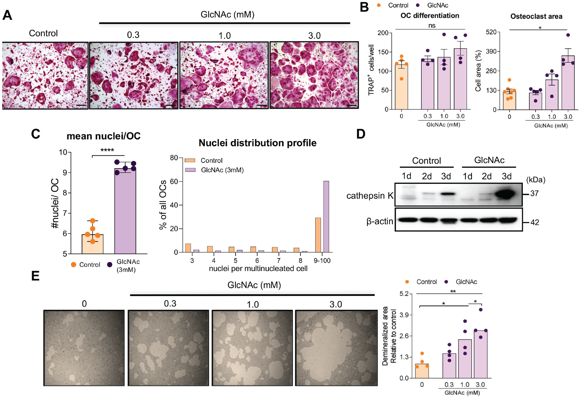
Changes in O-GlcNAc level affects osteoclast formation and function. Isolated murine bone marrow–derived macrophages (BMMs) were stimulated with macrophage colony-stimulating factor (M-CSF) and receptor activator of nuclear factor–κB ligand (RANKL). (
O-GlcNAcylation Dictates Osteoclast Behavior
Previous reports have demonstrated the existence of heterogeneous osteoclast populations with distinct sets of resorption behavior and pit- or trench-forming osteoclasts (Soe and Delaisse 2010, 2017; Merrild et al. 2015). Evaluating these 2 resorption modes can give mechanistic insights on how O-GlcNAcylation affects bone resorption.
For this, we first confirmed that protein O-GlcNAcylation also has an effect on human osteoclast differentiation and bone resorption. Similar to murine osteoclasts, exogenous GlcNAc increased the number of human osteoclasts induced by macrophage colony-stimulating factor (M-CSF) and RANK ligand (RANKL) (Fig. 3A, B). Our data are supported by the fact that exogenous GlcNAc increased the number of nuclei/OC in a dose-dependent manner and upregulated TRAcP activity (Appendix Fig. 3a, b). OGT inhibition by OSMI-1 impaired human osteoclast differentiation (Fig. 3C, D) and led to reduction of the number of nuclei/OC and TRAcP activity, which is supported by a dose-dependent effect (Appendix Fig. 3d, e). Finally, we explored the behavior of mature osteoclasts onto bone slices under GlcNAc or OSMI-1 (Fig. 3E, I). The total eroded surface per bone surface (ES/BS percentage) was affected in opposite ways by GlcNAc and OSMI-1 (Fig. 3F, J). Interestingly, supplementation of GlcNAc specifically led to increased resorption in trench mode while it did not affect pit resorption (Fig. 3G, H; Appendix Fig. 3c). Conversely, OSMI-1 reduced osteoclasts’ bone-resorptive activity, specifically by inhibiting the formation of trenches while not affecting osteoclasts making pits (Fig. 3K, L; Appendix Fig. 3f). The time-lapse recording images of Figure 4, extracted from Appendix Movie 1 and Appendix Movie 2, show osteoclasts (green) resorbing the bone (red). They illustrate the excavation displayed by mature osteoclasts (dark area) in the control group and confirm that O-GlcNAcylation modulates the shape of the excavations performed by osteoclasts stimulating the trench resorption mode (Fig. 4A, Appendix Movie 1). They also show the effect of OSMI-1 reducing the trench excavation mode by osteoclasts (Fig. 4B, Appendix Movie 2) compared to respective control. Overall, we document that the level of O-GlcNAcylation controls the osteoclast fate toward resorbing in a more aggressive trench mode.

Increased osteoclast fusion and bone resorption activity induced by O-GlcNAcylation. CD14+ monocytes and mature osteoclasts were cultured in the presence of macrophage colony-stimulating factor (M-CSF) (25 ng/mL) and receptor activator of nuclear factor–κB ligand (RANKL) (25 ng/mL). (

O-GlcNAcylation regulates osteoclast resorption mode. Illustrative images are selected snapshots of the time-lapse recordings, at indicated time points, from Appendix Movies. Osteoclasts can be identified as green cells, bone slices are stained in red, and dark area represents the eroded pit and trench excavation surface induce by mature osteoclasts from (
Ogt Deficiency in Osteoclasts Reduces Physiological and Inflammatory Bone Loss
To establish the in vivo relevance of our findings, we investigated how the loss of Ogt in osteoclasts would affect homeostatic and pathological bone remodeling. For this, we used genetic approaches by generating mice lacking Ogt in myeloid-expressing cells (LysM-Cre Ogtfl/fl mice) and cathepsin K–expressing cells (Ctsk-Cre Ogtfl/fl mice) (Nakamura et al. 2007); both are relevant tools for assessing Ogt deletion in vivo. Micro–computed tomography (CT) scans revealed that Ogt deficiency in osteoclasts resulted in significantly improved bone microarchitecture parameters assessed in the distal femur relative to the littermate controls (Fig. 5A). The data showed a marked increase in bone volume (BV/TV) and trabecular thickness (Tb.Th.) but no effect on bone mineral density (BMD) in Ctsk-Cre Ogtfl/fl mice (Fig. 5B). The lack of Ogt in osteoclasts also resulted in a significant difference in cortical parameters of the femoral mid-diaphysis, evidenced by increased bone volume (BV/TV), bone thickness, and reduced porosity (PO) on cortical bone (Fig. 5C). Confirming this result, we found that LysM-Cre Ogtfl/fl mice also exhibited a similar bone mass phenotype (Appendix Fig. 4).

Lack of Ogt in osteoclasts induces an osteopetrotic phenotype and reduces inflammation-induced bone loss in a periodontitis model. (
To check whether mice lacking Ogt deficiency in osteoclasts impaired osteoclastogenesis, we cultured bone marrow–derived macrophages (BMMs) from Ctsk-Cre Ogtfl/fl mice under osteoclastogenic conditions. Ogt deficiency resulted in fewer osteoclast numbers and with fewer nuclei (Fig. 5D, E).
To further explore the effect of Ogt deficiency on bone resorption in a pathological chronic inflammatory condition, we employed an apical periodontitis model (Fukada et al. 2008). Briefly, the exposition of dental pulp to the oral microenvironment led to a chronic inflammatory lesion, characterized by a disrupted periodontal ligament, and a blunted inflammatory infiltrate (Fig. 5F). It is important to note that periradicular tissue of Ctsk-Cre and Ctsk-Cre Ogtfl/fl mice with no lesion (control) was histologically similar (Fig. 5F). Ogt deficiency resulted in a smaller periapical lesion as compared to Ctsk-Cre (Fig. 5G).
Our data confirm that mice lacking Ogt in osteoclasts presented a decreased number of multinucleated osteoclasts lining the alveolar bone and tooth root in the periradicular area (Fig. 5H, I). Altogether, our results demonstrate that the deficiency of Ogt in osteoclasts leads to less severe periapical lesion formation associated with a reduced number of osteoclasts.
Discussion
The present study demonstrates a hitherto undiscovered role of the HBP/O-GlcNAcylation metabolic axis stimulating an aggressive osteoclast resorption mode, controlling physiological and pathological bone remodeling. We functionally show that this metabolic pathway is critical for switching osteoclasts to a more aggressive resorption mode. Moreover, using osteoclast-specific Ogt-deficient mice, we demonstrate that the lack of O-GlcNAcylation precisely in osteoclasts disrupts bone homeostasis by increasing bone mass and reducing inflammation-induced bone loss in an apical periodontitis model.
The interplay between cellular metabolism and intracellular signaling pathways is an exciting theme in biological research, and it remains unclear how metabolic reprogramming controls osteoclast behavior. Several studies have highlighted the importance of glucose uptake and consumption in regulating differentiation and bone-resorptive activity of osteoclasts (Williams et al. 1997; Kim et al. 2007; Indo et al. 2013). However, the energy required for osteoclastogenesis mainly derives from mitochondrial oxidative metabolism (Lemma et al. 2016). It has been shown that the expression of glutamine transporter, Slc1a5, is increased in the early stage of osteoclastogenesis, and depletion of L-glutamine or pharmacological inhibition of the Slc1a5 transporter also suppresses differentiation and function (Indo et al. 2013), suggesting that alternative pathways of glucose metabolism must be involved in osteoclastogenesis. A recent published study has shown that RANKL induces increased levels of O-GlcNAcylated proteins throughout osteoclast differentiation and that pharmacological O-GlcNAcylation inhibition reduced osteoclast differentiation in vitro, supporting the role of HBP on osteoclastogenesis (Kim et al. 2021). Here, we expand the knowledge about the impact of O-GlcNAcylation on osteoclast function. Large osteoclasts with a high number of nuclei exhibit a higher bone-resorbing activity. Our study provides new mechanistic insights into the metabolic reprogramming of osteoclasts stimulating osteoclast fusion and dictating osteoclast resorption mode.
O-GlcNAcylation has emerged as a posttranslational protein modification that integrates cell metabolism with several cellular processes (Hart 2019). In bone biology, an increase of O-GlcNAcylation of proteins during osteoblast differentiation was first shown, and the inhibition of OGT reduced the expression of some osteogenic genes in vitro (Nagel and Ball 2014; Koyama and Kamemura 2015). However, the relevance of O-GlcNAcylation on physiological and pathological bone remodeling remained unknown. Here, using genetic and pharmacological gain- and loss-of-function approaches, we confirmed that O-GlcNAcylation is a critical event for murine and human osteoclast differentiation and function. Pharmacological inhibition or genetic deficiency of OGT impaired osteoclast formation, which affected demineralization activity, and the supplementation of osteoclast cultures with GlcNAc, a precursor of the OGT substrate UDP-GlcNAc, boosted osteoclastogenesis and coordinated demineralization activity. However, it has to be mentioned that a previous study showed that GlcNAc, at a high concentration (20 mM), suppressed differentiation of the RAW264.7 macrophage cell line into osteoclasts (Takeuchi et al. 2016, 2017). Plausible explanations for these inconsistent results are the difference in the concentration of GlcNAc and the cell type used. Of note, proteomic analysis revealed that several proteins involved in the cell cycle, metabolism, and cytoskeleton organization are differentially regulated between BMMs and RAW264.7 cells during osteoclast differentiation (Ng et al. 2018). Hence, RAW264.7 cannot be considered fully differentiated osteoclasts since they maintain a large proportion of the macrophage phenotype (Ng et al. 2018).
RANK/RANKL signaling induces the expression and activation of NFATc1, which regulates the expression of genes required to drive myeloid cells toward an osteoclast fate (Ishida et al. 2002; Takayanagi et al. 2002). Here, we demonstrate that genetic deficiency of Ogt reduces the expression of NFATc1. Notably, we also found that OGT inhibitor reduced nuclear translocation of NFATc1 in mature osteoclasts. In support of this, it has been shown that OSMI-1 inhibits the O-GlcNAcylation of NF-κB p65 and NFATc1, affecting their translocation to the nucleus and possibly interfering with osteoclast transcription programs (Allison et al. 2012; Kim et al. 2021). In fact, it was previously shown that OGT interacts with and induces the O-GlcNAcylation of NFATc1 in lymphocytes upon activation, and silencing of OGT impairs the transcriptional activity of NFATc1 (Golks et al. 2007). O-GlcNAcylation is also required for NFATc3 activation and its nuclear translocation in cardiomyocytes (Facundo et al. 2012).
Osteoclasts solubilize 2 main constituents of the bone matrix, mineral and collagen, through 2 distinct bone resorption modes, pit and trench mode (Merrild et al. 2015; Soe and Delaisse 2017). The pit mode is formed by osteoclasts that generate round excavation events separated by migration periods, and trench resorption mode is made by continuous resorption and movement (Merrild et al. 2015; Soe and Delaisse 2017). However, the mechanism of switching the resorptive mode remains unknown. Here we show for the first time that bone-resorptive behavior of mature osteoclasts is sensitive to the O-GlcNAcylation level. Consistently, the supplementation of mature osteoclasts with GlcNAc specifically increased the rate of trench excavation, while OGT inhibition not only reduced their resorption activity but specifically inhibited osteoclasts making trenches. Notably, in contrast to osteoclasts making pits, those making trenches are dependent on the levels of active cathepsin K activity (Borggaard et al. 2020). In support, our data show that modulation of O-GlcNAcylation in osteoclasts increases cathepsin K levels, which coordinates osteoclasts’ resorption aggressiveness. Interestingly, mice lacking Ogt in osteoclasts developed reduced periapical lesions size, which was associated with a decreased number of osteoclasts lining the bone and the apex of the tooth root. Although the mice model presents limitations, as far as we are aware, this is the first evidence for the role of O-GlcNAcylation on osteoclast-induced bone remodeling during periapical lesion formation. We would argue that we have discovered a mechanistic action of O-GlcNAcylation on osteoclastic bone resorption—increased levels of active cathepsin K facilitating an aggressive bone resorption.
In summary, our findings have unveiled a bidirectional link between O-GlcNAcylation, osteoclast resorption behavior, and bone remodeling. Our data show a new mechanism for the bone loss and fragility mediated by excessive osteoclast activity in physiopathological processes uncovering novel avenues for targeting OGT in osteoclasts as a therapeutic intervention in osteolytic diseases.
Author Contributions
T.M. Taira, E.S. Ramos-Junior, K. Søe, S.Y. Fukada, contributed to conception and design, data analysis, drafted and critically revised the manuscript; P.H. Melo, C.C. Costa-Silva, contributed to conception and design, data analysis, critically revised the manuscript; M.G. Alteen, D.J. Vocadlo, W.B. Dias, F.Q. Cunha, J.C. Alves-Filho, contributed to data analysis, critically revised the manuscript. All authors gave final approval and agree to be accountable for all aspects of the work.
Supplemental Material
sj-docx-1-jdr-10.1177_00220345221141043 – Supplemental material for HBP/O-GlcNAcylation Metabolic Axis Regulates Bone Resorption Outcome
Supplemental material, sj-docx-1-jdr-10.1177_00220345221141043 for HBP/O-GlcNAcylation Metabolic Axis Regulates Bone Resorption Outcome by T.M. Taira, E.S. Ramos-Junior, P.H. Melo, C.C. Costa-Silva, M.G. Alteen, D.J. Vocadlo, W.B. Dias, F.Q. Cunha, J.C. Alves-Filho, K. Søe and S.Y. Fukada in Journal of Dental Research
Footnotes
Acknowledgements
We thank Mayara Santos Gomes and Juliana Aparecida Vercesi de Macedo for their technical support and Dr. Shigeaki Kato for the CtsKCre mice.
A supplemental appendix to this article is available online.
Declaration of Conflicting Interests
The authors declared no potential conflicts of interest with respect to the research, authorship, and/or publication of this article. Canadian Institutes of Health Research (PJT-156202). D.J.V. thanks the Canada Research Chairs program for support as a Tier I CRC in Chemical Biology.
Funding
The authors disclosed receipt of the following financial support for the research, authorship, and/or publication of this article: This study was supported by grants from FAPESP (2013/08216-2 and 2017/24073-8) and scholarships from FAPESP (2017/23264-4, 2019/15582-1).

References
Supplementary Material
Please find the following supplemental material available below.
For Open Access articles published under a Creative Commons License, all supplemental material carries the same license as the article it is associated with.
For non-Open Access articles published, all supplemental material carries a non-exclusive license, and permission requests for re-use of supplemental material or any part of supplemental material shall be sent directly to the copyright owner as specified in the copyright notice associated with the article.
