
Abstract
Growing evidence demonstrates the relationship between periodontitis and atherosclerotic cardiovascular diseases. The periodontal pathogen Porphyromonas gingivalis (Pg) has been shown to contribute to the progression of atherosclerosis. Cyclic diadenylate monophosphate (c-di-AMP) has been widely studied as an immune adjuvant for tumor immunotherapy, given its ability to activate the stimulator of interferon genes (STING) and regulate trained immunity. This study sought to elucidate the role of c-di-AMP in Pg-associated atherosclerosis. Periodontitis and atherosclerosis mouse models were established by ligature application around maxillary second molars and feeding ApoE knockout mice with a high-fat diet. We found that periodontitis and atherosclerosis were more severe in mice exposed to Pg than mice that underwent ligature placement only, while prophylactic treatment with c-di-AMP activated trained immunity and elicited significant alleviation of alveolar bone resorption, as well as reduced blood lipid levels and atherosclerotic plaque accumulation. After 3 mo of intervention, c-di-AMP limited the elevation of cytokines interleukin (IL)–6, IL-1β, tumor necrosis factor α, and interferon β; extracellular matrix remodeling enzymes MMP-2 and MMP-9; and adhesion molecules ICAM-1 and VCAM-1 gene expression. The mechanism underlying Pg-aggravated atherosclerosis may be attributed to changes in microbiota composition in oral and aortic plaques and excess inflammatory response, whereas c-di-AMP could prevent the effects of Pg infection due to its potential ability to activate trained immunity and regulate microecological balance. Our findings suggest a positive role of c-di-AMP in alleviating Pg-aggravated atherosclerosis by regulating the immune response and influencing the local microenvironment.
Keywords
Introduction
Periodontitis is a multifactorial inflammatory disease caused by microorganisms accumulating in dental plaques below the gingiva, involving the formation of periodontal pockets and progressive alveolar bone resorption, resulting in clinical attachment loss (Slots 2017). Overwhelming evidence substantiates that pathogens, especially gram-negative bacteria such as Porphyromonas gingivalis (Pg), are associated with the development of periodontitis (Schenkein et al. 2020). These subgingival bacteria invade the bloodstream through the injured gingival connective tissue caused by periodontitis or oral clinical treatments and circulate to reach distant organs (Schenkein et al. 2020). In recent years, the relationship between periodontitis and systemic diseases, such as cardiovascular diseases (CVDs), has been extensively studied (Peng et al. 2022).
CVDs are predominantly caused by atherosclerosis and account for high death and disability rates and disease burden globally. Indeed, besides common risk factors, inflammation has gradually gained increasing attention (Kivimäki and Steptoe 2018). Ample evidence emphasizes the role of inflammation triggered by periodontal bacteria as a bridge between periodontitis and CVDs. Such bacteria can be found in the aortic valves, atherosclerotic plaques, aortic aneurysms, and aortic intima (Brun et al. 2020; Xie et al. 2020; Joshi et al. 2021), indicating the potential role of the oral microbiota in the pathogenesis of atherosclerosis.
Cyclic dimeric adenosine 3′,5′-monophosphate (c-di-AMP, cdA) is a novel second messenger first identified in Bacillus subtilis (Witte et al. 2008), which has been recognized as a significant regulatory molecule in diverse bacteria and archaea (Bai et al. 2013; Whiteley et al. 2015; Moradali et al. 2022). As an agonist of stimulator of interferon genes (STING), c-di-AMP can be recognized by the host’s immune system, thereby triggering a series of subsequent immune responses (Yin et al. 2020). It is now well understood that c-di-AMP enhances trained immunity levels, which play a critical role in tumor and infection-associated immune response (Leventhal et al. 2020; Van Hauwermeiren et al. 2022).
Notwithstanding that c-di-AMP is widely thought of as an immune adjuvant in tumors or during bacterial infection due to its potential ability to activate trained immunity (Quintana et al. 2018; Singh et al. 2022), little has been done to determine whether c-di-AMP could protect against inflammatory diseases like periodontitis or provide cardioprotective effects. Therefore, we aimed to examine whether c-di-AMP could boost trained immunity and improve treatment efficacy against periodontitis and atherosclerosis. We reveal that alveolar bone resorption, lipid metabolism disorder, atherosclerotic plaque accumulation, and inflammatory cytokines expression in mice could be significantly limited by prophylactic use of c-di-AMP. Overall, the study’s findings support the topical application of c-di-AMP in preventing periodontitis or atherosclerosis.
In a nutshell, our study provides hitherto undocumented evidence of a potential mechanism by which c-di-AMP activates trained immunity and hence confers protection against periodontitis and atherosclerosis, providing the foothold for future studies on immunotherapeutic approaches against systematic inflammatory diseases associated with periodontitis.
Materials and Methods
Porphyromonas gingivalis Culture
The growing conditions for Porphyromonas gingivalis strains are listed in the Appendix.
Mice Models and Diets
Six-week-old male ApoE−/− mice based on C57BL/6J genetic background were given silk ligatures around the second maxillary molars and fed a high-fat diet to induce both periodontitis and atherosclerosis. The mice were divided into 3 groups of 6 each to measure levels of trained immunity, with the groups receiving topical applications of phosphate-buffered saline (PBS), 10 µM c-di-AMP, or 100 µM c-di-AMP (3 times a week for 4 wk). Another set of 48 mice were divided into 4 groups of 12 each, to observe the development of periodontitis and atherosclerosis. These groups received topical applications of PBS, 10 µM c-di-AMP, and 100 µM c-di-AMP followed by Pg resuspended in sodium carboxymethyl cellulose (CMC) applied around ligature site or PBS followed by CMC application (3 times a week for 4 wk and 8 wk, respectively) (Fig. 1A). All mice were kept in a standard environment with controlled temperature, humidity, and a 12-h light/dark cycle under specific pathogen–free conditions. Experiments were conducted under conditions authorized by the Institutional Ethical Committee on Experimental Animals (WCHSIRB-D-2020-031) and followed the Animal Research: Reporting of In Vivo Experiments 2.0 (ARRIVE 2.0) guidelines. More details are provided in the Appendix.
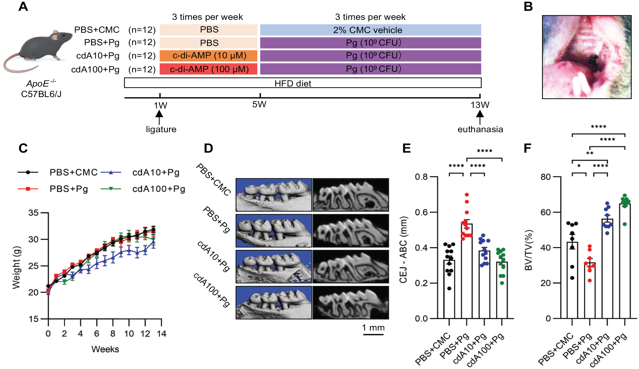
Cyclic dimeric adenosine 3′,5′-monophosphate (c-di-AMP) relieved periodontitis induced by ligature placement with or without Porphyromonas gingivalis infection in ApoE−/− mice. (
Tissue Collection and Preparation
Following the successful establishment of the models, the mice were euthanized and sampled. More details are provided in the Appendix.
Micro-Computed Tomography Analysis
Sagittal views of whole fixed alveolar bones were obtained by micro–computed tomography (µCT) scanning as previously described (Jia et al. 2021). More details are provided in the Appendix.
En Face Staining
Lesion development of aortas was determined using the en face method. More details are provided in the Appendix.
Oil Red O and Hematoxylin and Eosin Staining
To assess atherosclerotic lesions in aortic roots, cryosections of the aortic sinus were prepared and stained with Oil Red O and hematoxylin and eosin (H&E) staining. More details are provided in the Appendix.
RNA Isolation and Quantitative Real-Time Polymerase Chain Reaction
Total RNA extracted and purified from aorta tissue was used for subsequent Fast TB Green quantitative polymerase chain reactions (PCRs), and relative quantitative analysis was performed with the 2−ΔΔCT method. More details are provided in the Appendix.
Immunohistochemistry and Imaging
ICAM-1 and VCAM-1 gene expression and localization were detected by immunohistochemical staining. More details are provided in the Appendix.
Blood Lipids Measurements and the Enzyme-Linked Immunosorbent Assay of Proinflammatory Cytokines
An automatic biochemical instrument was used to measure the levels of blood lipids. The plasma levels of proinflammatory cytokines were assayed by enzyme-linked immunosorbent assay (ELISA) kits according to the manufacturer’s instructions. More details are provided in the Appendix.
16S rRNA Sequencing and Analysis
The microbiomes of silk ligatures and aortic plaques underwent 16S ribosomal RNA (rRNA) analysis, and results were validated by quantitative PCR. More details are provided in the Appendix.
Statistical Analysis
All graphs were generated using GraphPad Prism software (GraphPad Software), and statistical analysis was calculated using GraphPad Prism 9. Data obtained from 16S rRNA sequencing, including α and β diversity, were analyzed with the Kruskal–Wallis H test followed by post hoc uncorrected Welch’s test among 4 groups and the Wilcoxon rank-sum test between 2 groups. Results were presented as median with an interquartile range. For multiple comparisons, a 1-way analysis of variance (ANOVA) test was used. A P value less than 0.05 was statistically significant. Results were presented as mean ± standard error of the mean (SEM) or median ± interquartile range.
Results
c-di-AMP Relieved Periodontitis Induced by Ligature Placement with or without Porphyromonas gingivalis Infection in ApoE−/− Mice
Mice alveolar bones were sampled to assess periodontitis severity by measuring the distance between the cementoenamel junction (CEJ) and the alveolar bone crest (ABC). When the mice were euthanized at 13 wk, the silk ligatures were still left in place, the gingival tissue was red and swollen around the ligature sites (Fig. 1B), and no significant difference in body weight was found (Fig. 1C).
Significant alveolar bone resorption was observed in all mice (Fig. 1D). Compared with mice in the PBS + CMC group, µCT analysis yielded more severe alveolar bone loss in mice that underwent ligature and Pg infection (Fig. 1E, F). Interestingly, prophylactic use of c-di-AMP in mice could attenuate periodontitis, with significantly reduced alveolar bone resorption and enhanced bone density. No significant differences were observed with different doses of c-di-AMP (Fig. 1E, F).
c-di-AMP Reduced Plasma Lipids and Alleviated Aortic Plaque Formation in ApoE−/− Mice Fed with a High-Fat Diet
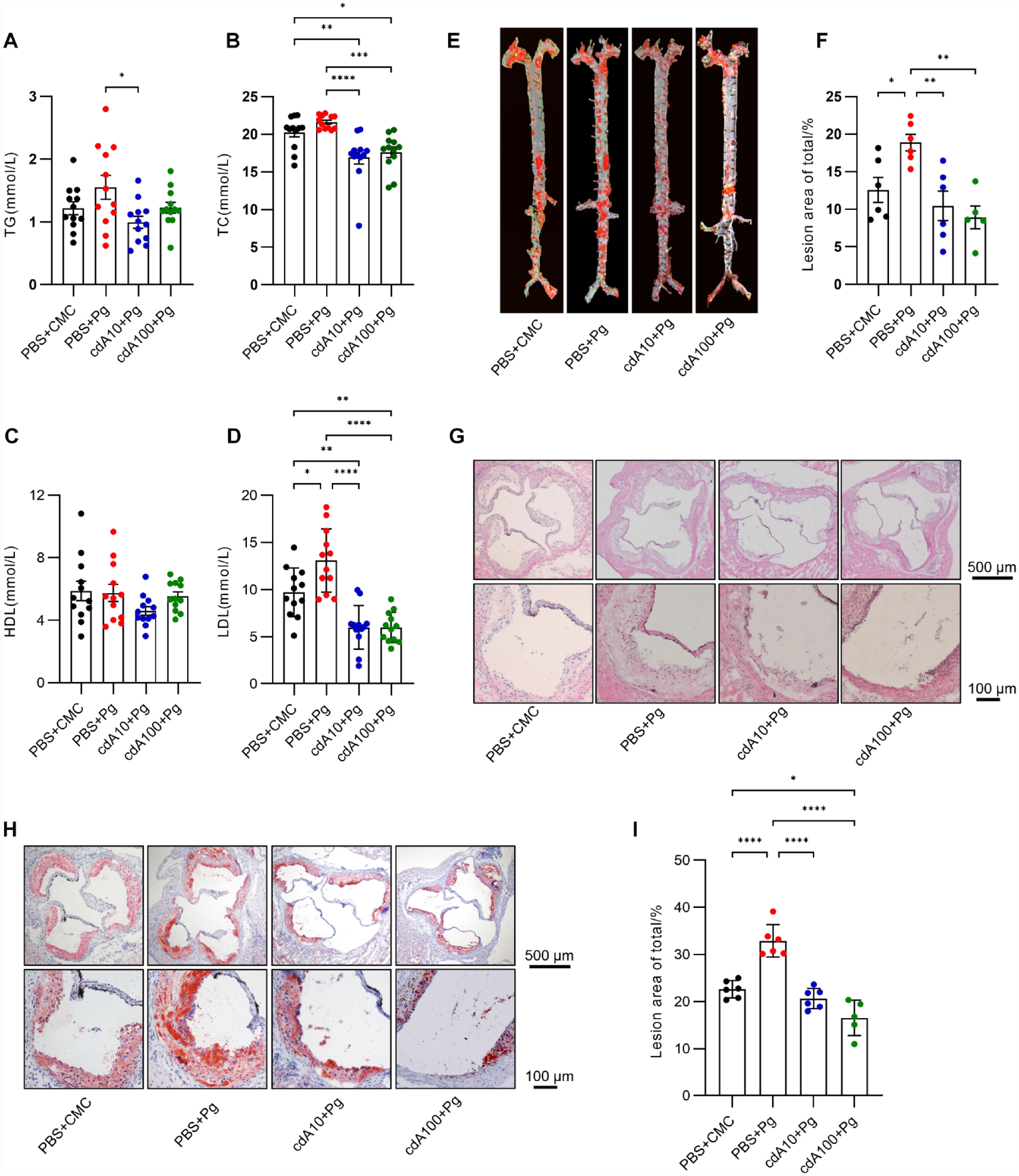
Lipid metabolism disorder and aortic plaque accumulation in the arterial wall are characteristics of advanced atherosclerosis (Bennett et al. 2016). The aortic tissue and plasma of mice were collected after they were euthanized to assess whether c-di-AMP could attenuate the development and progression of atherosclerosis induced by ligature placement with or without Pg infection.
As expected, we observed lipid elevation and plaque formation in all mice (Fig. 2). Compared with the PBS + CMC group, exposure to Pg increased plasma lipids, including total cholesterol (TC), triglycerides (TG), high-density lipoprotein (HDL), and low-density lipoprotein (LDL) (Fig. 2A–D). However, a significant increase was observed only for TC and LDL, previously documented as the most atherogenic lipids. Prophylactic c-di-AMP treatment for a month could significantly decrease the levels of plasma lipids, especially TG, TC, and LDL (Fig. 2A–D).
Cyclic dimeric adenosine 3′,5′-monophosphate (c-di-AMP) reduced plasma lipids and alleviated aortic plaque formation in ApoE−/− mice fed with a high-fat diet (HFD). Plasma levels (mmol/L) of (
Next, the atherosclerotic plaques of the entire aorta were first observed with Oil Red O staining. We found that infection with Pg could promote the aggregation of plaques, while c-di-AMP could partially prevent this effect (Fig. 2E, F). Oil Red O– and H&E-stained histological cross sections of aortic valves exhibited similar results to en face lesions. Pg infection resulted in a 1.5-fold increase in plaque size compared with the PBS + CMC group, whereas fewer lesions were observed in the cdA10 + Pg and cdA100 + Pg groups (Fig. 2G–I), suggesting that c-di-AMP could attenuate Pg-aggravated atherosclerosis.
c-di-AMP Decreased Aortic Tissue Expression of Adhesion Molecules ICAM-1 and VCAM-1 in ApoE−/− Mice
The endothelial function of the aorta can be evaluated by the expression of intercellular adhesion molecule 1 (ICAM-1) and vascular cell adhesion molecule 1 (VCAM-1). In the present study, the aortas of mice treated with Pg showed upregulated gene expression of ICAM-1 and VCAM-1, whereas prophylactic c-di-AMP use significantly downregulated transcriptional levels of ICAM-1 and VCAM-1 (Fig. 3A, B).
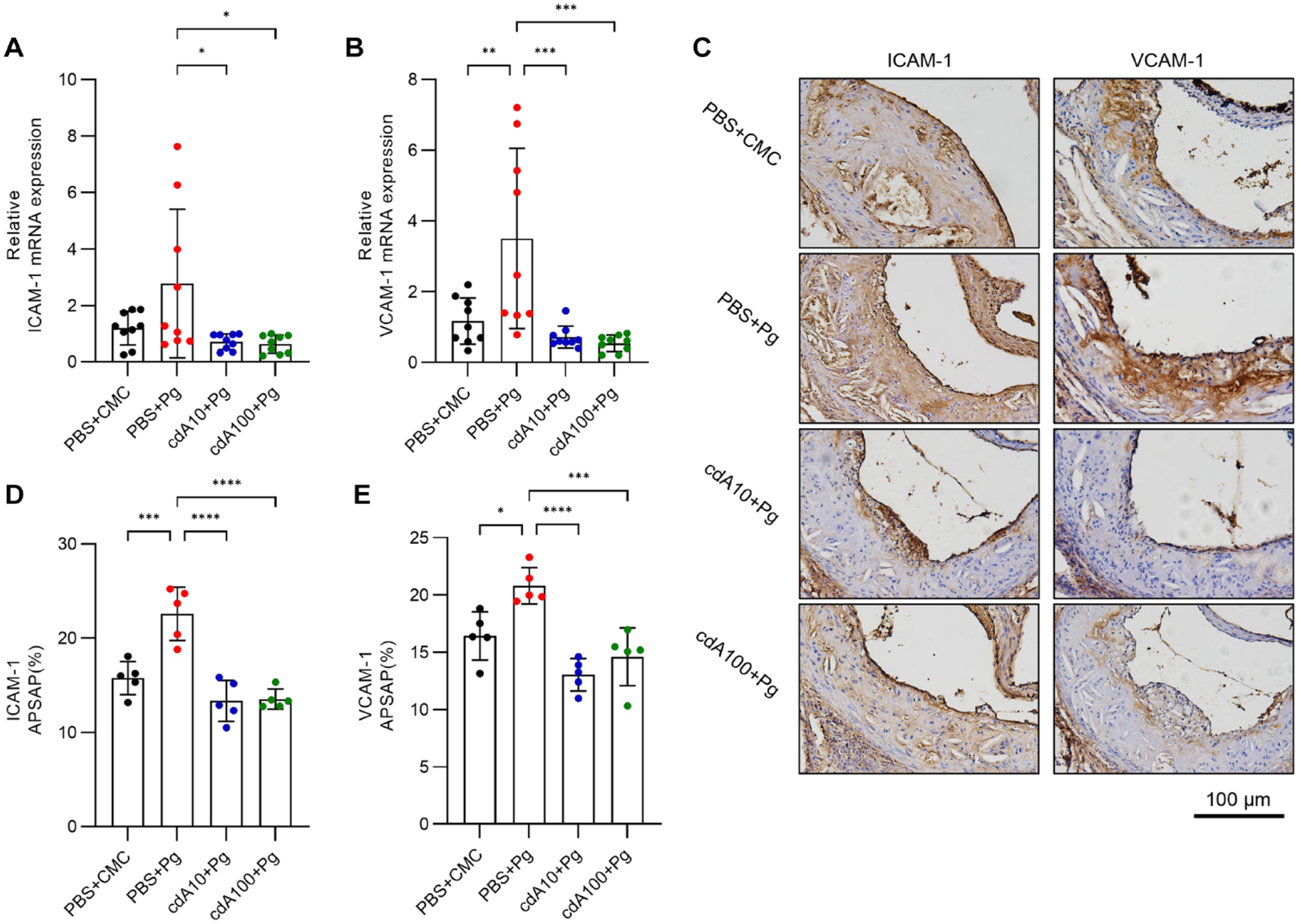
Cyclic dimeric adenosine 3′,5′-monophosphate (c-di-AMP) decreased the expression of adhesion molecules ICAM-1 and VCAM-1 of aorta tissue in ApoE−/− mice. The relative expression levels of adhesion molecules (
We further sought to localize the 2 intercellular adhesion molecules in atherosclerotic lesions by immunohistochemistry staining (Fig. 3C). We found that ICAM-1 and VCAM-1 were mainly localized on the surface of endothelium cells, with low expression inside the plaques and the adventitia. It was found that sections from aortic valves of the PBS + Pg group mice exhibited increased staining for ICAM-1 and VCAM-1, whereas a corresponding decrease in the 2 adhesion molecules was observed in the cdA-treated groups (Fig. 3D-E), substantiating the potent proatherogenic influence of Pg and atheroprotective effects of c-di-AMP.
c-di-AMP Activated Trained Immunity in ApoE−/− Mice
Given that interleukin 6 (IL-6), interleukin 1β (IL-1β), and tumor necrosis factor α (TNF-α) have been highly associated with atherosclerosis, we examined these inflammatory cytokines in mice plasma using ELISA. The results showed lower levels of proinflammatory cytokines in the cdA-treated groups (Fig. 4A–C). Identical results were obtained for interferon β (IFN-β), the major effector molecule of c-di-AMP (Fig. 4D).
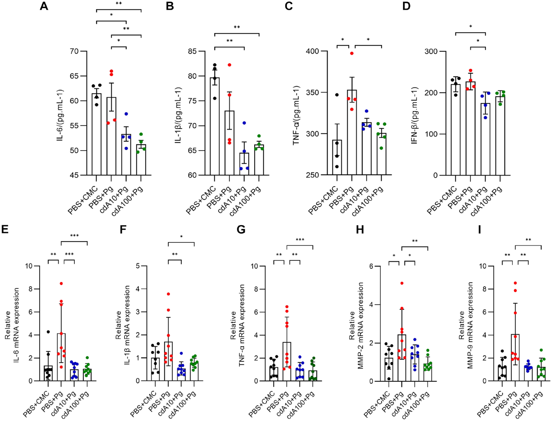
Cyclic dimeric adenosine 3′,5′-monophosphate (c-di-AMP) activated trained immunity in ApoE−/− mice. Expression of (
Next, we examined the expression of inflammatory cytokines using real-time quantitative PCR to confirm whether c-di-AMP could trigger trained immunity and modulate inflammatory response in the development of atherosclerosis. We found that c-di-AMP upregulated the expression of IL-6, IL-1β, TNF-α, and IFN-γ, as well as chemokines MCP-1, CXCL-9, CXCL-10, and CXCL-22 after 1 mo (Appendix Fig. 3). We found decreased inflammatory cytokines at 3 mo in the cdA + Pg groups compared with the PBS + Pg group (Fig. 4E–G).
Similarly, we quantified MMP-2 and MMP-9 gene expression in 3-mo samples. The results showed a 2-fold increase of MMP-2 and a nearly 5-fold increase of MMP-9 between the PBS + Pg group and other groups, which were statistically significant (Fig. 4H, I).
Prophylactic Use of c-di-AMP Prevented Changes in Oral Microbiota Composition Induced by P. gingivalis Infection in ApoE−/− Mice
To investigate the effects of Pg and c-di-AMP on the microenvironment of oral microbiota, the silk ligatures were sampled and underwent 16S rRNA sequencing. Although the Shannon index showed no significant differences (Fig. 5A), the β diversity of the Principal Co-ordinates Analysis (PCoA) exhibited significant differences among groups (Fig. 5B). Analysis of Similarities (ANOSIM) also suggested a significantly different composition of bacteria among the 4 groups (Appendix Table 3). The top 15 most abundant bacteria at the genus levels are shown in Figure 5C. The species composition analysis showed there were mainly 6 phyla of oral bacteria (Appendix Fig. 1a). The dominant phylum of the PBS + CMC group and the cdA groups was Firmicutes (Fig. 5C, Appendix Fig. 1b). However, the composition of the PBS + Pg group was significantly different from the other 3 groups, whose dominant phylum was Proteobacteria (Appendix Fig. 1a). Moreover, Kruskal–Wallis H test showed significant differences for the top 15 genera of bacteria among the 4 groups (Appendix Fig. 1c). Then, we quantified Bacteroidetes and Firmicutes at the phylum levels and Porphyromonas and Pseudomonas at the genus levels. Interestingly, a nearly 4-fold decrease of Firmicutes was observed in the PBS + Pg group. At the genus levels, Porphyromonas and Pseudomonas were increased by 2-fold and 4-fold in the PBS + Pg group compared with the other 3 groups (Fig. 5D–G). The sequencing data results were validated by quantitative PCR (Appendix Fig. 4a, b). Sequencing data of Pg on species levels are shown in Appendix Table 5. Intriguingly, low levels of Pg were observed in the cdA + Pg groups, suggesting a potential inhibition effect toward Pg colonization or growth of c-di-AMP.
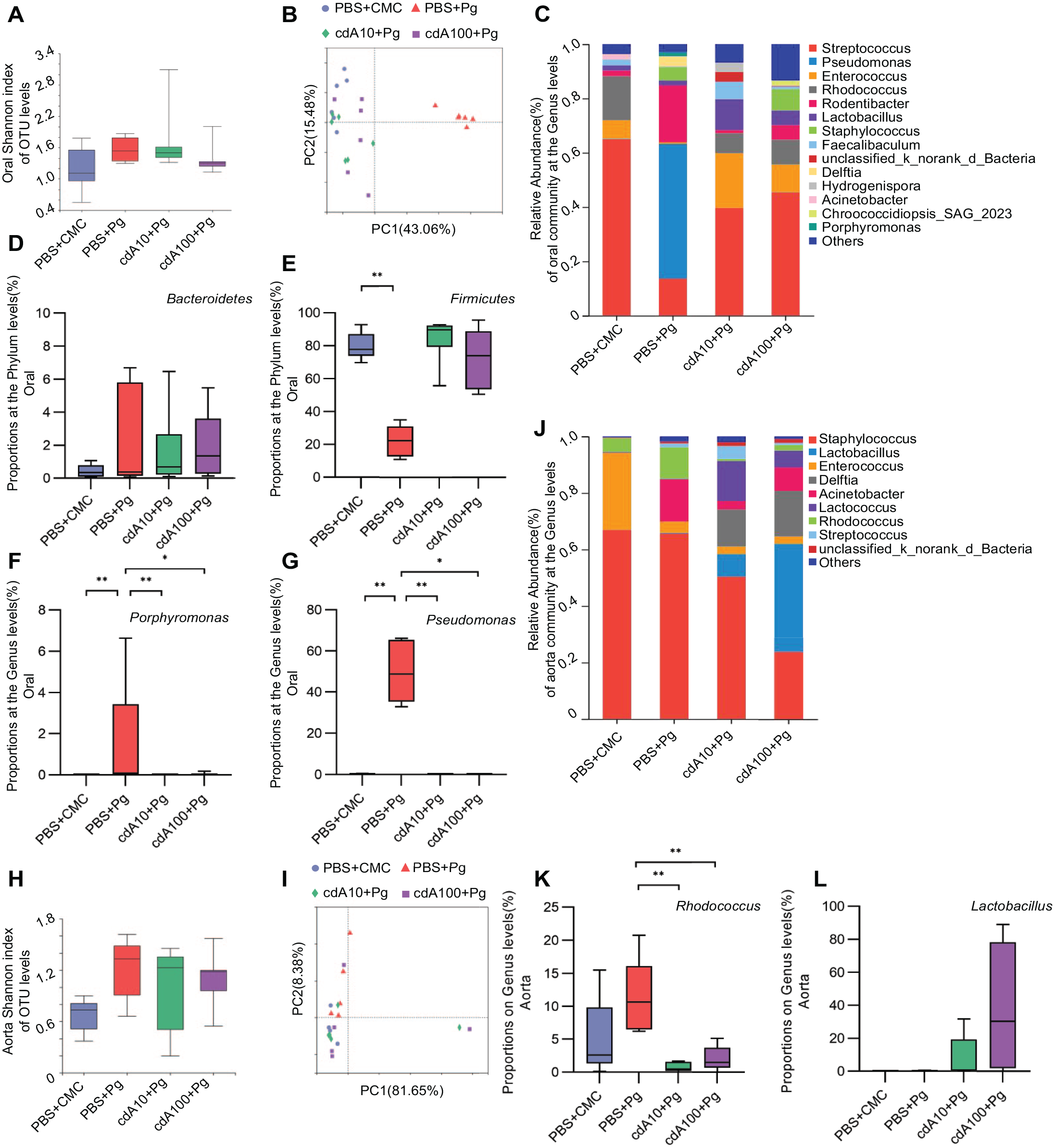
Prophylactic use of cyclic dimeric adenosine 3′,5′-monophosphate (c-di-AMP) reversed the change in oral microbiota composition induced by Porphyromonas gingivalis infection and influenced the bacterial profile of atherosclerotic plaques in ApoE−/− mice. (
Prophylactic Use of c-di-AMP Influenced the Bacterial Profile of Atherosclerotic Plaques in ApoE−/− Mice
The 16S rRNA gene sequencing demonstrated that Pg infection or c-di-AMP use could change the oral bacteria composition. Therefore, we hypothesized that bacteria and/or metabolites produced by bacteria could enter the blood circulation and finally influence the aorta’s microbiota profile. Although no significant difference in the Shannon index (Fig. 5H) was observed, a difference in β diversity was found (Fig. 5I). A comparison of the 2 groups is presented in Appendix Table 4. The species composition analysis showed that 4 phyla of bacteria could be found in atherosclerotic plaques (Appendix Fig. 2a), consistent with oral bacteria analysis results. Firmicutes constituted the majority of bacteria community at the phylum level in all 4 groups (Fig. 5J, Appendix Fig. 2b). Among the top 15 genera, Lactobacillus, Enterococcus, Acinetobacter, Rhodococcus, and Pseudomonas showed significant differences (Appendix Fig. 2c). At the genus level, Rhodococcus were significantly enriched in the PBS + Pg group compared with the cdA-pretreated groups, while a slight elevation in Lactobacillus was observed in the cdA-pretreated groups, although no significant difference was found (Fig. 5K, L). The sequencing data results were subsequently validated by quantitative PCR (Appendix Fig. 4C, D).
Discussion
Over the past few decades, numerous studies have demonstrated a link between periodontitis and atherosclerosis (Zhang et al. 2021). The present study provided compelling evidence that Pg exposure exacerbated periodontitis and atherosclerosis by triggering an inflammatory response, leading to lipid metabolic disorders and endothelial dysfunction of the aorta (Lalla et al. 2003; Miyauchi et al. 2012). Moreover, microbiota-derived STING agonist c-di-AMP could activate trained immunity and attenuate periodontitis and Pg-aggravated atherosclerosis. Finally, Pg infection triggered significant microbial composition change in the oral cavity and accumulation of atherosclerotic plaques in mice, which could be prevented by c-di-AMP pretreatment.
It has been established that one of the earliest detectable indicators of vascular damage is endothelial dysfunction (Pereira et al. 2011; Chiesa et al. 2019). In response to Pg exposure, the levels of proinflammatory cytokines are increased, such as TNF-α, IL-1β, IL-6, and IFN-β, suggesting that Pg infection may induce a durable inflammatory response involving endothelial cells (ECs) lining the intima of the aorta (Miyauchi et al. 2012; Ramalingam et al. 2020).
Serving as a novel pathogen-associated molecular pattern (PAMP), c-di-AMP can be recognized by pattern recognition receptors (PRRs) in the host immune system, which in turn activate STING and trigger type I IFN expression during bacterial infection, closely related to the concept of trained immunity (Devaux et al. 2018). Trained immunity refers to the long-term functional reprogramming of innate immune cells caused by exogenous or endogenous insults, which results in an altered response to a second challenge (Netea et al. 2020). Trained immunity is less specific and of shorter duration than adaptive immune memory (Mulder et al. 2019). Researchers have demonstrated that trained immunity could be induced by c-di-AMP (Singh et al. 2022), consistent with our findings. Cytokine activation in ApoE−/− mice treated with c-di-AMP for 1 mo resulted in an enhanced response to subsequent Pg infection with long-term effects such as downregulation of inflammatory cytokines and remission of diseases. Therefore, c-di-AMP is widely recognized as an immune adjuvant in cancer immunotherapy and treatment of microbiota infections (Fisher et al. 2017; Jiang et al. 2020; Leventhal et al. 2020; Ning et al. 2022; Singh et al. 2022). Although the therapeutic potential of c-di-AMP in tumor immunity has been extensively studied, the potential application and underlying mechanisms of c-di-AMP in atherosclerosis treatment remain to be determined.
Low-density lipoprotein levels represent an important risk factor for atherosclerosis (Xu et al. 2015). Current evidence suggests that excessive LDL accumulates in the vessel wall and is taken up by macrophages, leading to foam cell formation and atherosclerosis (Joo et al. 2017; Kazemian et al. 2020). Moreover, our study found significantly increased gene expression of ICAM-1 and VCAM-1 in the Pg infection group. Adhesion molecules ICAM-1 and VCAM-1 were overexpressed at sites of inflammation, with little or no expression in healthy vessel tissue (Yun et al. 2016), suggesting they play a critical role in regulating immune cells’ accumulation at sites of inflammation, such as recruiting monocytes and macrophages to lesion sites and eventually breaking the endothelial barrier, aggravating vascular inflammatory response (Yun et al. 2016). Additionally, we observed elevated aortic tissue expression of MMP-2 and MMP-9 genes, which have been implicated in vascular remodeling (Schenewerk et al. 2014).
Based on our findings, using c-di-AMP to activate trained immunity can effectively protect the host against Pg infection and exert beneficial effects in alleviating periodontitis and CVDs. However, no significant differences were observed between groups with different c-di-AMP concentrations. Accordingly, more studies are warranted to explore the underlying mechanisms.
Additionally, periodontitis is induced by microbial dysbiosis, characterized by a shift of opportunistic bacteria to a more pathogenic form (Xu et al. 2018). In a bacterial infection model, we found that Pg could change the oral microenvironment and alter microbial community composition, whereas c-di-AMP could prevent the effects of Pg infection, even slightly enriching the microbial community. Studies found that Pg infection could cause an increase in Bacteroidetes and a decrease in Firmicutes (Wade 2013), causing severe immune inflammation in a short period, leading to a sharp increase in the levels of bacteremia and serum inflammatory factors (Peng et al. 2022). We also noted a significant decrease in pathogenic Pseudomonas and Porphyromonas abundance of mice in the cdA-treated groups. Although studies have shown that intracellular c-di-AMP is beneficial for Pg growth and bacterial–bacterial interactions (Moradali et al. 2022), the different roles of extracellular c-di-AMP in bacterial infection have attracted more interest (Louie et al. 2020). Our results demonstrate the inhibitory effects of c-di-AMP against bacteria, suggesting there might be another way for c-di-AMP to influence the immune microenvironment rather than directly impacting bacteria. Further studies will be required to resolve these inconsistencies. Unfortunately, despite different bacterial compositions, we could not detect Porphyromonas in aorta tissue, contrary to previous studies (Velsko et al. 2014; Xie et al. 2020).
The study provides hitherto undocumented evidence on the role of c-di-AMP in periodontitis-aggravated atherosclerosis. Nevertheless, mechanical induction by ligature may cause more severe periodontitis compared with Pg infection, which is considered a chronic process. In addition, our findings were limited by the lack of animal groups without periodontitis and atherosclerosis. In a noninflammatory disease state, the function and mechanism of c-di-AMP in activating trained immunity warrants further research. Besides, although studies over the past decades have raised awareness of the broad benefits of trained immunity toward host defense, the dual role of immune response also showed potentially detrimental outcomes in immune-mediated and chronic inflammatory diseases (Netea et al. 2020). Accordingly, further studies should focus on finding the optimal concentration of c-di-AMP.
In conclusion, we revealed that P. gingivalis infection aggravated the development of periodontitis and subsequent atherosclerosis by triggering the host inflammation response and changes in the microbial community composition in ApoE−/− mice. These effects could be prevented by pretreatment with c-di-AMP due to trained immunity activation. Importantly, c-di-AMP could maintain the microecological balance of the oral cavity and the aorta and could be harnessed as a novel immune adjuvant for clinical use against periodontitis and cardiovascular diseases.
Author Contributions
Q. Wu, X. Peng, X. Zhou, contributed to conception and design, data acquisition, analysis, and interpretation, drafted and critically revised the manuscript; Z. Li, Y. Zhang, K. Luo, X. Xu, J. Li, contributed to data acquisition, analysis, and interpretation, critically revised the manuscript. All authors gave final approval and agree to be accountable for all aspects of the work.
Supplemental Material
sj-docx-1-jdr-10.1177_00220345231162344 – Supplemental material for Cyclic di-AMP Rescues Porphyromonas gingivalis–Aggravated Atherosclerosis
Supplemental material, sj-docx-1-jdr-10.1177_00220345231162344 for Cyclic di-AMP Rescues Porphyromonas gingivalis–Aggravated Atherosclerosis by Q. Wu, Z. Li, Y. Zhang, K. Luo, X. Xu, J. Li, X. Peng and X. Zhou in Journal of Dental Research
Footnotes
A supplemental appendix to this article is available online.
Declaration of Conflicting Interests
The authors declared no potential conflicts of interest with respect to the research, authorship, and/or publication of this article.
Funding
The authors disclosed receipt of the following financial support for the research, authorship, and/or publication of this article: This study was supported by the National Natural Science Foundation of China (32070120, 81991500, 81991501) and Natural Science Foundation of Sichuan Province (2023NSFSC0558).
References
Supplementary Material
Please find the following supplemental material available below.
For Open Access articles published under a Creative Commons License, all supplemental material carries the same license as the article it is associated with.
For non-Open Access articles published, all supplemental material carries a non-exclusive license, and permission requests for re-use of supplemental material or any part of supplemental material shall be sent directly to the copyright owner as specified in the copyright notice associated with the article.