
Abstract
Titanium (Ti)–based biomaterials lack inherent antimicrobial activities, and the dental plaque formed on the implant surface is one of the main risk factors for implant infections. Construction of an antibacterial surface can effectively prevent implant infections and enhance implant success. Silver nanoparticles (AgNPs) exhibit broad antibacterial activity and a low tendency to induce drug resistance, but AgNPs easily self-aggregate in the aqueous environment, which significantly impairs their antibacterial activity. In this study, UiO-66/AgNP (U/A) nanocomposite was prepared, where zirconium metal–organic frameworks (UiO-66) were employed as the confinement matrix to control the particle size and prevent aggregation of AgNPs. The bactericidal activity of U/A against methicillin-resistant Staphylococcus aureus and Escherichia coli increased nearly 75.51 and 484.50 times compared with individually synthesized Ag. The antibacterial mechanism can be attributed to the enhanced membrane rupture caused by the ultrafine AgNPs on UiO-66, leading to protein leakage and generation of intracellular reactive oxygen species. Then, U/A was loaded onto Ti substrates (Ti-U/A) by using self-assembly deposition methods to construct an antibacterial surface coating. Ti-U/A exhibited excellent antibacterial activities and desired biocompatibility both in vitro and in vivo. The U/A nanocomposite coating technique is thus expected to be used as a promising surface modification strategy for Ti-based dental implants for preventing dental implant infections.
Keywords
Introduction
Titanium (Ti) and its alloys are the most commonly used dental implant materials. However, Ti-based biomaterials do not exhibit outstanding antibacterial activity (Rosa et al. 2021). The adhesion and accumulation of dental plaque is recognized as one of the major leading cause of peri-implantitis, which may induce soft tissue inflammatory lesions and alveolar bone resorption, ultimately resulting in implant failure (Berglundh et al. 2018). The mainstream clinical treatments rely on mechanical debridement and antibiotic therapy (Mahato et al. 2016). Although the above methods have achieved positive therapeutic effects, mechanical debridement may cause scratches on the implant surface (Munakata et al. 2022), and abuse of antibiotics may induce bacterial drug resistance (Wang et al. 2020). Therefore, construction of an antibacterial dental implant surface is of crucial significance in preventing implant infection (Chen, Wang, et al. 2021).
Generally, the current research focuses on improving the antibacterial properties by means of construction of a specific micro-nano surface texture (Yang et al. 2022), introduction of functional coatings to change surface properties (He et al. 2023), or application of materials with intrinsic antibacterial properties (Liu et al. 2022). Silver nanoparticles (AgNPs) have been considered as a promising alternate for antibiotics (Vargas-Reus et al. 2012) due to their outstanding antibacterial activity, broad antibacterial activity, and low tendency to induce drug resistance. AgNP-based coatings can be deposited onto the dental implant surface via polydopamine (Choi et al. 2019), chitosan–heparin (Li et al. 2019), or keratin nanofibers (Cochis et al. 2017). However, the antibacterial activity of AgNPs is usually influenced by its dispersion stability. AgNPs tend to spontaneously aggregate into large particles due to the high surface energy and strong cohesiveness of the ultrafine particle size, resulting in inferior antibacterial potency (Tang and Zheng 2018). In order to prevent self-aggregation and maintain biological performance, application of various capping agents (e.g., sodium citrate, polyvinylpyrrolidone, or branched polyethyleneimine) has been shown to successfully confine the size of AgNPs (Badawy et al. 2010; Wu et al. 2019).
Metal-organic frameworks (MOFs) are a class of porous crystalline materials that build from metal ions and organic linkers via coordination bonds (Chen, Li et al. 2021). The uniform submicronic size, large surface area, and regular 3-dimensional (3D) porous structure make MOFs an ideal confinement matrix for AgNPs to enhance their antibacterial performance, such as employing z Zeolite imidazole framework or cyclodextrin MOFs as a template to synthesize ultrafine AgNPs (Shakya et al. 2019; Joo et al. 2023). However, bacterial infection usually creates an acidic environment, which may cause accelerated disruption of MOFs, resulting in AgNP reaggregation and increased local metal ion concentration (Taheri et al. 2021). Among the abundant number of MOFs, the zirconium metal–organic framework UiO-66 possesses excellent chemical stability due to the strong coordination force between Zr6O4(OH)4 nodes and 1,4-benzenedicarboxylate (BDC) ligands (Ahmadijokani et al. 2020). Based on the superior acid resistance, UiO-66 frameworks have been widely used as a delivery vehicle for a series of drugs or metal nanoparticles for antibacterial applications. Therefore, we hypothesized that UiO-66 could be used as the synthesis template and confinement matrix for AgNPs.
Herein, we describe an antibacterial nanocomposite coating for the Ti dental implant. Ultrafine AgNPs were immobilized on UiO-66 via in situ reduction to prepare the UiO-66/AgNPs (U/A) nanocomposite. In order to verify the confinement effect of UiO-66 in the enhancement of dispersible stability and biological performance of AgNPs, the physicochemical and antibacterial properties of U/A were investigated and compared to that of individually synthesized AgNPs. Chitosan (CS)–sodium alginate (SA) self-assembly deposition technology was then employed to load U/A onto a titanium substrate to prepare the antibacterial surface coating. Both in vitro and in vivo antibacterial performance and biocompatibility of U/A nanocomposite coating were investigated, aiming to verify its safety and efficacy in the prevention of implant infections.
Materials and Methods
The detailed materials and methods, including preparation of UiO-66, individually synthesized Ag, U/A and nanocomposite coatings (Ti-UiO-66, Ti-Ag, and Ti-U/A), and sample characterizations, are supplied in the Appendix.
In Vitro Antibacterial Experiments
Methicillin-resistant Staphylococcus aureus (MRSA) ATCC 43300 and Escherichia coli ATCC 25922 were chosen as model gram-positive and gram-negative bacteria, using Ti-U/A as the experimental groups, with Ti substrate Ti-UiO-66 and Ti-Ag as controls and minocycline hydrochloride ointment (Periocline; Sunstar INC) as a positive control. The detailed procedures are supplied in the Appendix.
Biocompatibility Assays
The cytotoxicity of surface coatings toward mouse embryonic osteoblast precursor cells (MC3T3-E1) was assessed by an indirect contact experiment. The detailed procedures are supplied in the Appendix.
In Vivo Antibacterial Experiments
The in vivo animal study was conducted via a subcutaneous infection model, using Ti-U/A as the experimental groups and Ti substrate as controls. The animal experiments were approved by the Experimental Animal Welfare Ethics Branch of Peking University Biomedical Ethics Committee (approval number: PUIRB-LA2023093). We complied with the ARRIVE (Animal Research: Reporting of In Vivo Experiments) 2.0 guidelines. The detailed procedures are demonstrated in the Appendix.
Statistical Analysis
All the experiments were carried out in triplicate. One-way analysis of variance (ANOVA) or Student’s t test was used to measure the statistically significant difference (P) among groups. The data are expressed as the mean ± standard deviation in relevant figures and tables. For in vivo experiments, 6 rats were allocated to each group to ensure an appropriate group size for statistical analysis. Statistical significance was determined as *0.01 < P < 0.05, **0.001 < P < 0.01, ***P < 0.001, ns P > 0.05.
Results
Preparation and Characterization of U/A and Nanocomposite Coatings
As shown in Figure 1A–C, U/A exhibited similar morphology as octahedral-like UiO-66 and uniformly distributed ultrafine AgNPs. With the introduction of UiO-66 in the reduction process, AgNPs were immobilized onto the UiO-66 framework rather than aggregation to large particles (Fig. 1D). The hydrodynamic diameter of UiO-66, Ag, and U/A was about 227 ± 31.38 nm, 495.8 ± 90.68 nm, and 222 ± 46.30 nm, respectively (Fig. 1E). The loading of AgNPs onto UiO-66 caused a decrease in zeta-potential from 7.24 ± 0.28 mV to −6.12 ± 0.60 mV, indicating that U/A has been elaborately synthesized (Fig. 1F).
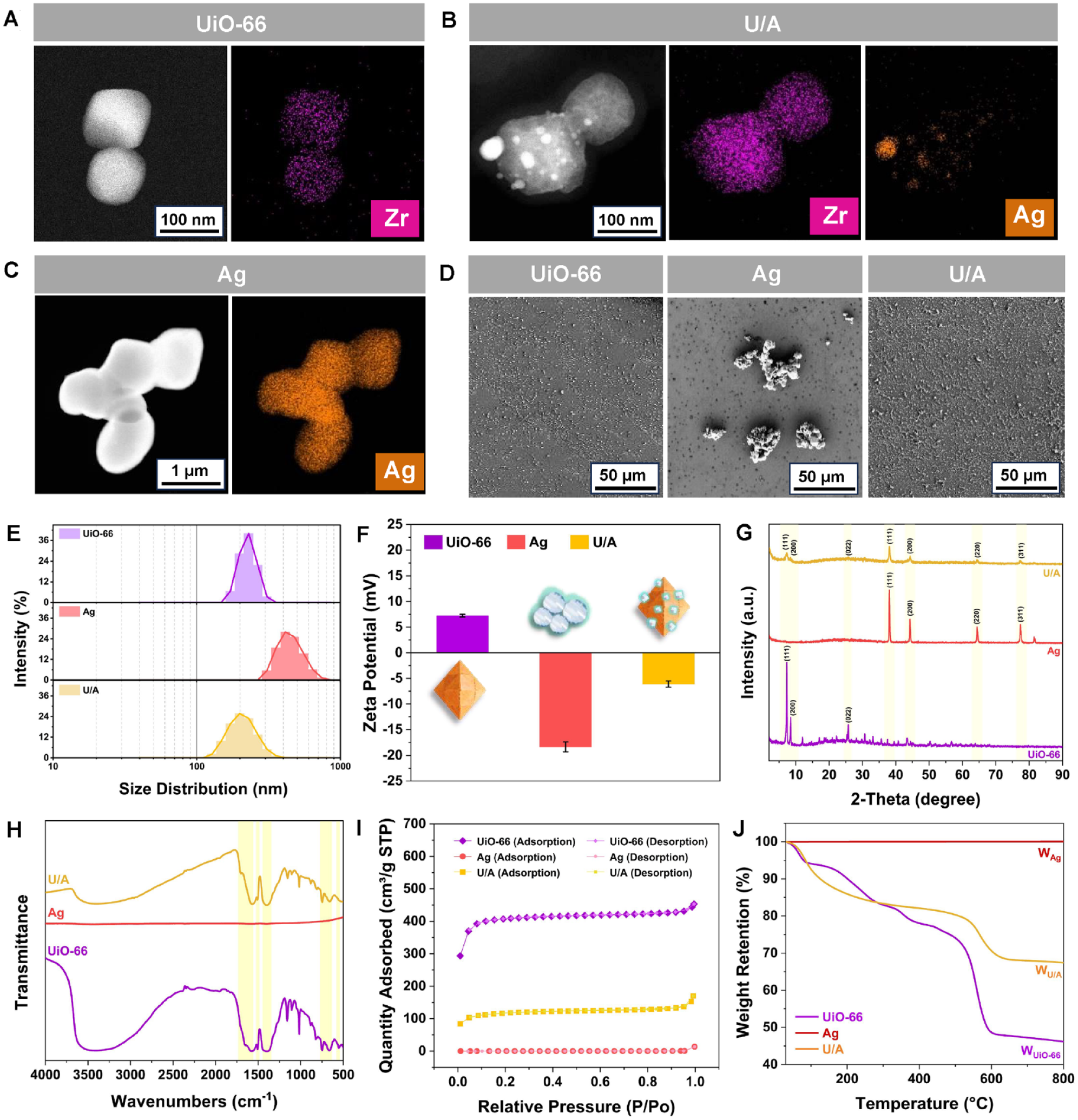
Characterizations of UiO-66/AgNPs (U/A) nanocomposite. (
The crystal structure and homogeneity of different nanoparticles were characterized by X-ray diffraction (XRD) (Fig. 1G). The diffraction peaks at 7.32°, 8.39°, and 25.77° were attributed to the (111), (002), and (022) crystal planes of UiO-66. In contrast, new peaks at 38.01°, 44.35°, 64.44°, and 77.40° appeared on U/A corresponding to the (111), (200), (220), and (311) crystal plane of Ag (Ma et al. 2021). The Fourier-transform infrared spectroscopy (FT-IR) spectrums of the 3 samples are shown in Figure 1H. The peaks at 1,550 to 1,630 cm–1 corresponded to the symmetric and asymmetric stretching vibrations of the carboxyl group connected to Zr4+ in UiO-66 frameworks, a small peak at 1,507 cm–1 referred to stretching vibration of the C=C unit presented in the benzene ring of BDC, a strong peak near 1,400 cm–1 was attributed to the C-O bond of the C-OH group of carboxylic acid of BDC, and the peaks at around 556 cm–1, 667 cm–1, and 749 cm–1 were ascribed to asymmetric stretching vibration of Zr-(OC) (Abid et al. 2012).
According to BET analysis illustrated in Figure 1I and Appendix Figure 1, both UiO-66 and U/A possessed a large specific surface area and exhibited type I isotherms due to their microporous structures. On the contrary, due to the self-aggregation effects, individually synthesized Ag possessed a low specific area only at about 0.71 m²/g. The loading of AgNPs onto UiO-66 decreased the specific surface from 1,222.95 to 430.74 m²/g, but the specific surface area of U/A was still much larger than that of individually synthesized Ag (Appendix Table 1). The TGA profiles of different samples are shown in Figure 1J. For both UiO-66 and U/A, the mass loss that occurred below 200°C was attributed to the evaporation of volatile solvent molecules (methanol, water, and DMF), the mass loss in the range of 200°C to 520°C was caused by decomposition of terephthalic acid while the coordination bonds between Zr and the ligands started to be broken, and the mass loss below 580°C was attributed to the collapse of the 3D framework structure, and beyond this temperature, complete decomposition occurred and the mass loss curve reached equilibrium. On the contrary, the mass loss of Ag was rather negligible. Quantitatively, it could be calculated that the loading content of AgNPs onto UiO-66 was about 0.46 wt% (WU/A/WUiO-66 − 1).
Preparation and Characterization of U/A Nanocomposite Coatings
UiO-66, Ag, or U/A was separately loaded onto a titanium surface via CS-SA self-assembly deposition technology, with the detailed synthesized routine illustrated in Figure 2A. The surface functionalization and grafting of CS barely affected the pristine morphologies and roughness of titanium substrates (Appendix Fig. 2). With the proceeding treatments of H2SO4/H2O2, APTES, GA, and grafting of CS, the C, O, and N contents of Ti-OH, Ti-NH2, Ti-GA, and Ti-CS increased, which indicated successful modification at each step (Appendix Fig. 3). After being coated with the SA layer containing UiO-66, Ag, or U/A, even-distributed nanoparticles were observed on Ti-UiO-66 and Ti-U/A, but random agglomeration of large Ag particles appeared on the surface of Ti-Ag (Fig. 2B, C). The obvious presence of Zr 3d at 245.03 eV, 426.38 eV and Ag 3d at 374.32 eV, 368.28 eV demonstrated the successful deposition of U/A nanocomposite coating onto titanium substrate (Fig. 2D, E). Ti-OH exhibited a more hydrophilic nature compared with pristine Ti due to the abundant hydroxyl groups after treatment with H2SO4/H2O2. The hydrophilicity of Ti-NH2 decreased due to the existing alkyl chains of APTES. After grafting of GA and CS, the surface wettability gradually increased because of the hydrophilic nature of polysaccharides. Further deposition of SA containing UiO-66, Ag, or U/A caused a slight decrease in surface hydrophilicity; the respective water contact angle of Ti-UiO-66, Ti-Ag, and Ti-U/A was around 23.09° ± 4.19°, 25.67° ± 3.80°, and 26.35° ± 2.12°, respectively; and the wettability tendency of 3 surface coatings corresponded to that of UiO-66 nanocrystals, individually synthesized Ag, and U/A nanocomposites (Fig. 2F and Appendix Fig. 4). The release of Ag+ from Ti-U/A increased rapidly within the initial 24 h, and then the release profile of Ag+ tended to flatten. The cumulative release amount of Ag+ within 336 h in pH 7.5 and 5.0 was around 3.40 mg/L and 2.90 mg/L (Fig. 2G).
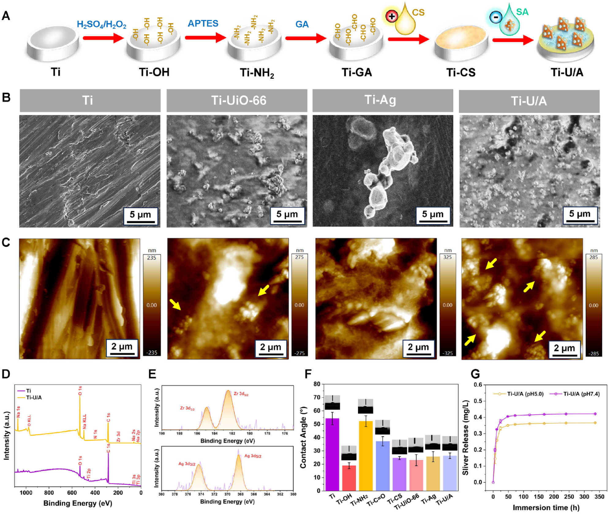
Preparation and characterizations of UiO-66/AgNPs (U/A) nanocomposite coating. (
Investigation of the Antibacterial Mechanisms of U/A Nanocomposite
The minimum bactericidal concentration (MBC) of U/A nanocomposite against MRSA and E. coli was around 22.07 μg/mL and 0.86 μg/mL, which was much lower than that of UiO-66 (MRSA: 2,000.00 μg/mL; E. coli: 1,666.67 μg/mL) and Ag (MRSA: 1,666.67 μg/mL; E. coli: 416.67 μg/mL) (Fig. 3A). For both MRSA and E. coli, the OD570 values for U/A significantly decreased compared to control, UiO-66, and individually synthesized Ag, indicating a more serious damage caused by U/A to the bacterial cell membrane (Fig. 3B). Meanwhile, much higher protein leakage occurred after incubation with U/A rather than other samples (Fig. 3C). The intracellular reactive oxygen species (ROS) levels within bacteria incubated with U/A were much higher (*P < 0.05) than those of UiO-66 and Ag for both MRSA and E. coli (Fig. 3D). Taking these results together, U/A can cause membrane rupture, leading to protein leakage, generation of intracellular ROS, and finally superior antibacterial ability (Fig. 3J).
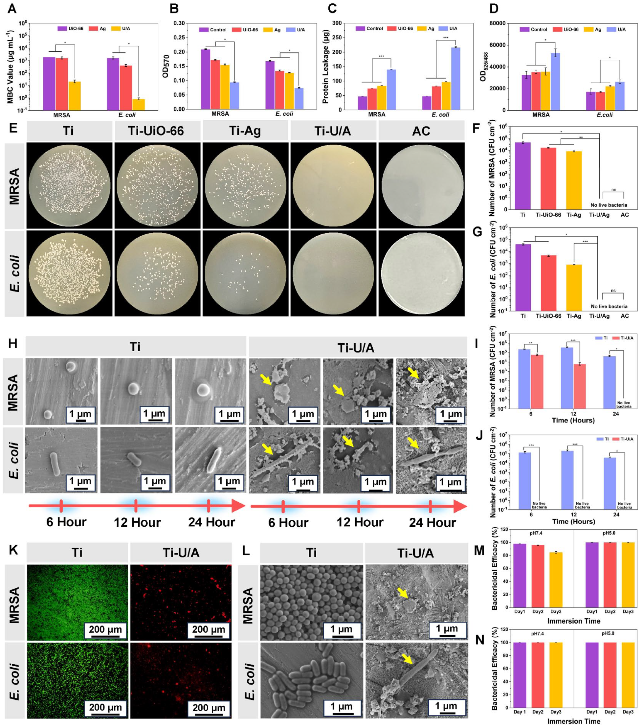
Investigation of the antibacterial performance of UiO-66/AgNPs (U/A) nanocomposite and titanium (Ti)–U/A. (
In Vitro Antibacterial of Performance of Ti-U/A
Ti-U/A exhibited a much stronger inhibitive effect on both MRSA and E. coli than pristine Ti, Ti-UiO-66, and Ti-Ag (Fig. 3E–G). The fluorescence observation further confirmed the superior antibacterial activity of Ti-U/A, which was comparable to that of minocycline hydrochloride (5 mg/mL) (Appendix Fig. 5). The bacteria that adhered on Ti-U/A started to shrink after a 6-h incubation, severe shrinkage and deformation grew over time, and finally, the integrity of the bacteria cell membrane deteriorated (Fig. 3H). The number of bacterial colonies remained almost unchanged on the Ti surface but was gradually decreased on Ti-U/A with the extension of incubation time and was completely eliminated within 24 h (Fig. 3I, J). After being continuously incubated for 24 h, the biofilm of both MRSA and E. coli on Ti was stained green, while only a few with red fluorescence were observed on Ti-U/A (Fig. 3K). This was further confirmed via scanning electron microscopy (SEM) observation; the Ti surface was evenly covered by large amounts of alive bacteria, but only a small number of scattered bacteria with damaged morphologies were observed on Ti-U/A (Fig. 3L). The antibacterial efficiency of Ti-U/A against MRSA and E. coli was still maintained over 80% after a 3-d presoaking whether in a neutral or an acidic environment (Fig. 4M, N, Appendix Fig. 6).
Assessment of cytotoxicity. (
Biocompatibility Assays
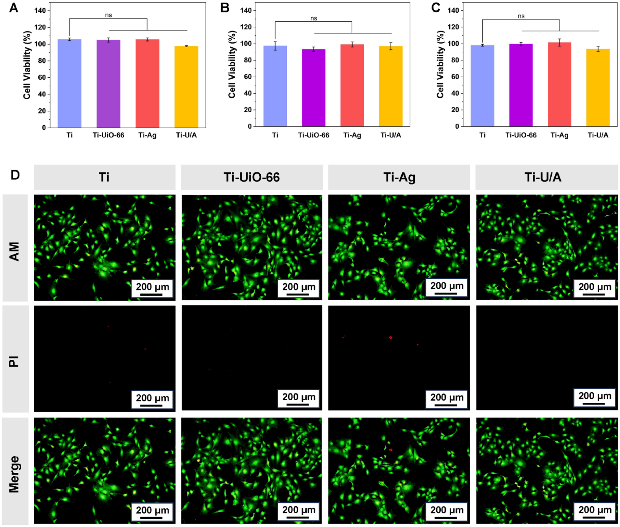
The viability of MC3T3-E1 remained over 90% after being incubated with the extracts of Ti-U/A for 1, 4, and 7 d, respectively (Fig. 4A–C). There was no significant difference between Ti-U/A and Ti (ns P > 0.05). The live/dead staining assay further confirmed the desired cytocompatibility of Ti-U/A, indicating that the deposition of Ti-U/A nanocomposite coating would not cause an adverse effect on cell metabolic activity (Fig. 4D).
In Vivo Examination of the Antibacterial Experiments and Biosafety of Ti-U/A
To evaluate the in vivo antibacterial performance and biosafety, Ti-U/A or Ti was implanted subcutaneously in Sprague–Dawley rats, and MRSA suspension (106 CFU/mL) was injected into the implant site to induce bacterial infection (Fig. 5A). The infection status of wounds was determined by quantification of the viable bacteria extracted from the implant and surrounding tissues, as well as the inflammation of peri-implant tissues. As illustrated in Figure 5B, a large number of living bacteria existed on Ti samples, while few bacteria were observed on Ti-U/A at days 3 and 7. Besides, the U/A nanocomposite coating also exhibited an enhanced inhibitory effect against the bacteria in the peri-implant tissues, which was proved by the significantly decreased bacteria colonies surrounding Ti-U/A at days 3 and 7 (Fig. 5C).
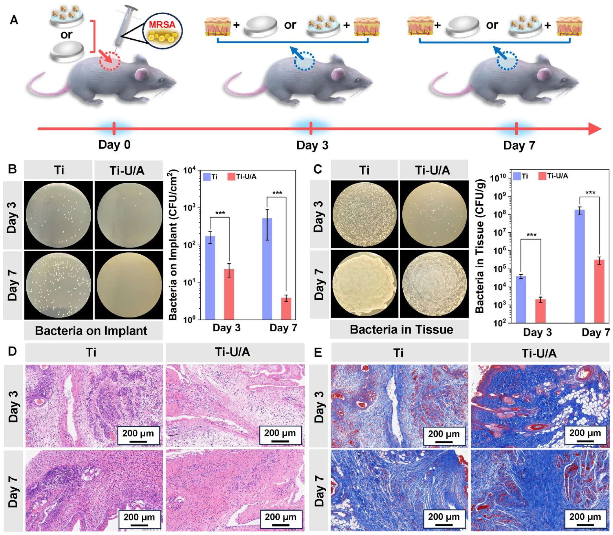
In vivo antibacterial experiments and histological analysis. (
To evaluate the bacterial infection-induced inflammation in the peri-implant tissues, hematoxylin and eosin (H&E) staining (Fig. 5D) was performed. The peri-implant tissues around Ti samples exhibited typical signs of bacterial infections, which were reflected by numerous nuclei into the tissues, corresponding to the colony-forming unit (CFU) enumeration results. However, Ti-U/A exhibited a milder inflammation with reduced inflammatory cells at days 3 and 7. The tissues around pristine Ti samples were seriously infected by MRSA, so the peri-implant tissues exhibited much more inflamed nuclei compared to that of Ti-U/A. The inflammatory cell ratios calculated from H&E staining data for Ti and Ti-U/A were around 27.11% and 14.60% on day 3 (n = 6, ***P < 0.001) and 30.31% and 16.46% on day 7 (n = 6, ***P < 0.001) (Appendix Fig. 7). The number of inflammatory cells and the bacterial infection for Ti-U/A were significantly decreased compared with Ti for days 3 and 7. The collagen distribution around implant sites was analyzed by Masson staining (Fig. 5E), and the destruction of collagen was observed in the Ti control groups due to the mass bacteria survival while collagen degradation significantly decreased in Ti-U/A due to the superior antibacterial activity of U/A. Besides, compared to the blank controls without treatment of MRSA suspension and implantation, no obvious tissue lesion, inflammation, or damage was observed in the rats’ organs (heart, liver, spleen, lung, and kidney) (Appendix Fig. 8). These outcomes demonstrated that the U/A hybrid nanocomposite coating possessed good antibacterial activity and biocompatibility.
Discussion and Conclusion
Ti-based dental implants lack an inhibitory effect on adhered bacteria (Busscher et al. 2010; Do Nascimento et al. 2013). The adhered bacteria will proliferate and develop into sturdy biofilms that firmly attach to the implant surface, which is recognized as one of the main risk factors for peri-implantitis (Berglundh et al. 2018). Construction of the antibacterial surface is of great significance to prevent implant-associated bacterial infections and enhance implant success. AgNPs are well recognized as one of the most effective antimicrobial agents, but the antibacterial activity of AgNPs is closely related to its colloidal stability. Due to the high surface energy and strong cohesiveness of the ultrafine particle size, AgNPs directly reduced by NaBH4 easily self-aggregated to large particles in the aqueous environment, which significantly impaired its antibacterial activities. Porous MOFs can be considered a confinement matrix in the synthesis of AgNPs to prevent self-aggregation and maintain its colloidal stability and biological performance. Thereby, we designed a U/A nanocomposite coating to prevent bacterial infection while simultaneously meeting the biocompatible demand.
The U/A nanocomposite synthesized in this study exhibited superior antibacterial activity compared to published research (Appendix Table 2). This can be attributed to the relatively larger specific surface area of UiO-66 synthesized in our work, which can provide sufficient volume to adsorb more Ag ions for the preparation of AgNPs. This could be further confirmed by XRD patterns. The nanocomposite of UiO-66 and AgNPs synthesized in our work exhibited very strong Ag(111) crystal planes, which has proved to be of critical significance in antibacterial progress (Rizzello and Pompa 2014; Hu et al. 2016). The consensus antimicrobial mechanism of U/A can be attributed to the contact sterilization and ion-mediated bactericidal effects. AgNPs can directly interact with the bacterial cell membrane and interfere with DNA transcription and cellular respiration (Durán et al. 2010; Tang and Zheng 2018). In addition, the released Ag ions can bind to thiol groups (-SH) of proteins, thereby inhibiting the synthesis of respiratory enzymes and leading to ROS generation (Chopra 2007; Taglietti et al. 2014).
There are still negligible obstacles in the clinical application of U/A nanocomposites. The flow and erosion of gingival crevicular fluid may decrease the retention time of U/A on the target site, thereby failing to provide sufficient protection against bacterial infection. Therefore, U/A was deposited onto Ti surfaces via a versatile self-assembly deposition method. As the precursor layer, positively charged CS was covalently grafted onto an aldehyde functionalized titanium surface to ensure the stability of whole-surface coating. Subsequently, Ti-CS was immersed into a solution mixture of SA and U/A, where U/A nanocomposites were deposited onto Ti-CS via an electrostatic interaction between the polycation CS and polyanionic SA. Since it is easy to achieve activation and functionalization of Ti substrates, the synthesis procedure of U/A was relatively mild. The U/A nanocomposite coating technique is thus expected to be used as a promising surface modification strategy for Ti-based implants, such as dental or orthopedical implants, with the aim to effectively prevent bacterial infection. It is worth mentioning that U/A nanocomposites exhibited a negatively charged nature (Fig. 1F), which further ensured the uniform dispersion of nanoparticles in the hybrid nanocomposite surface coating. According to SEM images and energydispersive X-ray spectroscopy (EDX) analysis elemental analysis in Appendix Figure 6, presoaking would not cause noticeable effects on the coating morphology and Ag and Zr contents regardless of the pH changes in the immersion environment, which corresponded to the results obtained by inductively coupled plasma optical emission spectrometer (ICP-OES) results, indicating satisfactory stability of the deposited U/A hybrid nanocomposite coating to fight against an initial bacterial infection. The antibacterial efficacy and biosafety of U/A nanocomposite coatings were further confirmed via in vivo experiments. Therefore, U/A hybrid nanocomposite coating can be considered a promising surface modification strategy for Ti-based dental implants to prevent bacterial infections. Although laboratorial data regarding U/A nanocomposite coatings have significantly advanced, the gap between laboratory research and commercially available products clearly illustrates that investigations still come up against several nonnegligible obstacles before moving forward to a clinical setting. Further efforts should focus on improving coating stability and long-term functionality as well as the evaluation of the potential effects of surface modification on implant–bone integration and immune response.
In conclusion, construction of an antibacterial coating consisting of UiO-66/AgNPs nanocomposite can effectively prevent bacterial infection while simultaneously maintaining excellent biocompatibility both in vitro and in vivo, and it can be considered a promising surface coating for a Ti-based dental implant.
Author Contributions
C. Yu, contributed to design, data acquisition and analysis, drafted and critically revised the manuscript; Y. Yu, contributed to conception, design, data analysis and interpretation, drafted and critically revised the manuscript; Y. Lu, contributed to design, data acquisition and analysis, critically revised the manuscript; K. Quan, contributed to design, data acquisition, drafted the manuscript; Z. Mao, contributed to data acquisition, drafted the manuscript; Y. Zheng, L. Qin, D. Xia, contributed to conception, design, critically revised the manuscript. All authors gave final approval and agree to be accountable for all aspects of the work.
Supplemental Material
sj-docx-1-jdr-10.1177_00220345241229646 – Supplemental material for UiO-66/AgNPs Coating for Dental Implants in Preventing Bacterial Infections
Supplemental material, sj-docx-1-jdr-10.1177_00220345241229646 for UiO-66/AgNPs Coating for Dental Implants in Preventing Bacterial Infections by C. Yu, Y. Yu, Y. Lu, K. Quan, Z. Mao, Y. Zheng, L. Qin and D. Xia in Journal of Dental Research
Footnotes
Declaration of Conflicting Interests
The authors declared no potential conflicts of interest with respect to the research, authorship, and/or publication of this article.
Funding
The authors disclosed receipt of the following financial support for the research, authorship, and/or publication of this article: This study was supported by the National Key Research and Development Program of China (grant 2023YFC2412600), the National Natural Science Foundation of China (grants 52171233, 22376176), the Beijing Natural Science Foundation–Haidian Original Innovation Joint Fund Project (grants L212014, L222030), the Beijing Nova Program (grant 20230484459), the National Clinical Key Discipline Construction Project (grant PKUSSNKP-T202103), and the Research Foundation of Peking University School and Hospital of Stomatology (grant PKUSS20230104).
A supplemental appendix to this article is available online.
References
Supplementary Material
Please find the following supplemental material available below.
For Open Access articles published under a Creative Commons License, all supplemental material carries the same license as the article it is associated with.
For non-Open Access articles published, all supplemental material carries a non-exclusive license, and permission requests for re-use of supplemental material or any part of supplemental material shall be sent directly to the copyright owner as specified in the copyright notice associated with the article.