
Abstract
The addition of ion-releasing particles to experimental adhesives has been evaluated as a strategy to increase the longevity of bonded interfaces. This study evaluated the effect of calcium silicate particles (CaSi) applied as a pretreatment to acid-etched dentin surfaces and/or added (5% by mass) to the bonding resin of a commercial 3-step adhesive system. The mechanical properties of the adhesive layer, hybrid layer, and underlying dentin were measured by nanoindentation after 24 h and 6 and 12 mo of storage in simulated body fluid (SBF) and observed by scanning electron microscopy. Calcium and phosphorus were quantified by energy-dispersive x-ray spectroscopy. Enzymatic activity was estimated by in situ zymography and hydroxyproline assay after 24 h and 12 mo. Microtensile bond strength was determined after 24 h and 12 mo in SBF. Data were analyzed by repeated measures analysis of variance and Tukey test (alpha, 5%). CaSi used as a pretreatment or added to the bonding resin reduced the mechanical properties of the adhesive layer. Notwithstanding, only where CaSi was present did groups show significant increases in mechanical properties for the hybrid layer and the underlying dentin after prolonged storage, suggesting the occurrence of apatite precipitation. This result corroborates the higher calcium content disclosed by energy-dispersive x-ray spectroscopy. Also, CaSi-containing pretreatment and/or the addition of CaSi to the bonding resin inhibited metalloprotease activity, as confirmed in situ and by the reduced hydroxyproline release. However, microtensile bond strength reductions after 12-mo storage in SBF were observed in all groups. The findings suggest that CaSi, whether applied as a pretreatment, added to the bonding resin, or both, promoted mineral precipitation and inhibited enzymatic activity at the bonded interface.
Keywords
Introduction
In adhesive dentistry, the initial stage of the bonding procedure involves dentin acid conditioning with phosphoric acid or acidic monomers present in self-etch systems. The bonded interface quality relies on the micromechanical interlocking between the adhesive monomers and the dentin collagen scaffold (Betancourt et al. 2019). However, adhesive infiltration usually results in partially exposed collagen fibrils, which are susceptible to enzymatic degradation (Mazzoni et al. 2012). Also, the integrity of the bonded interface is further compromised by the hydrolysis of the resin component (Breschi et al. 2018).
Various strategies were proposed to extend the longevity of bonded interfaces. One approach is the incorporation of calcium-releasing particles in experimental or simplified commercial adhesive systems (Rifane et al. 2023) or as a pretreatment applied to acid-etched dentin (Bauer et al. 2019). Calcium ions combined with phosphate ions in the dentinal fluid promote mineral precipitation in water-rich voids at the bonding interface after 24 h of thermocycling storage or after 7 d of nonthermocycling aging (Al-Hamdan et al. 2020), which may increase its mechanical properties, reduce hydrolytic degradation, and inhibit collagenolytic activity (Gu et al. 2010).
Experimental adhesives containing calcium silicate particles (CaSi) particles promoted apatite deposition on mineral-depleted dentin and remineralization of the hybrid layer (HL) and underlying dentin after 30 and 90 d in simulated body fluid (SBF) storage (Sauro, Osorio, Fulgencio, et al. 2013; Sauro, Osorio, Osorio, et al. 2013). Moreover, bond strength was preserved after 10- or 12-mo storage (Profeta 2014a; Bendary et al. 2020). However, previous studies did not explore the effect of adhesives containing CaSi on proteolytic activity. Also, rather than using experimental formulations, it would be interesting to verify if CaSi could improve the performance of commercial adhesive systems.
Ion release from resin-based materials occurs according to diffusional and relaxational processes (Trinca et al. 2024). Consequently, mineral precipitation may not occur fast enough to protect collagen from proteolytic degradation, which is triggered immediately after dentin demineralization (Gao et al. 2024). Therefore, CaSi particles delivered to acid-etched dentin as a pretreatment would lead to faster mineral precipitation and more effective protection of collagen against hydrolytic degradation. Still, there is a paucity of information regarding the benefits of pretreating demineralized dentin with CaSi particles on mineral precipitation and their effects on collagenolytic activity.
Aiming to improve the longevity of resin-bonded interfaces, this study synthesized and characterized CaSi nanostructured agglomerates and evaluated their effect as a dentin pretreatment or as an addition to the bonding resin of a commercial 3-step adhesive system. The mechanical properties and enzymatic activity at the adhesive interface, as well as its microtensile bond strength (µTBS), were tested immediately and after prolonged storage in SBF. The tested null hypothesis was that incorporating CaSi into the bonding procedure, either as a pretreatment or as an addition to the bonding resin of a commercial adhesive system, does not significantly affect µTBS, microstructural characteristics, mechanical properties, or the proteolytic activity at the adhesive interface immediately or after aging.
Materials and Methods
Particle Synthesis and Characterization
CaSi particles were synthesized by the sol-gel technique based on a previously described method (Santana et al. 2011; Fan et al. 2016). Particle morphology was observed under scanning electron microscopy (Quanta FEG600; FEI Co.) and transmission electron microscopy (model JEM-2100; JEOL). Particles were characterized regarding the crystalline phase (XRD, MultiFlex; Rigaku Corp.), size distribution (Mastersizer 2000; Malvern Instruments Ltd.), and surface area (Nova 1200e; Quantachrome Instruments). A detailed description is in the Appendix.
Experimental Groups and Specimen Preparation
CaSi particles (5% by mass) were added to the bonding resin of a 3-step adhesive system (Adper Scotchbond Multipurpose/SBMP; 3M ESPE) and vortexed for 1 min immediately before use (Thermo Fisher Scientific Inc.). The effect of CaSi particles on resin’s degree of conversion, cohesive strength, and Ca2+/Si4+ release was tested.
Six experimental groups were defined according to pretreatment (3 levels) and the presence of CaSi particles in the bonding resin (2 levels). Pretreatment consisted of a 95% ethanol solution containing 5% of CaSi particles: P(+) (percentages by mass). Pretreatment with only ethanol, P(–), and no pretreatment (noP) were also tested. Specimen preparation and tested groups are shown in Appendix Figure 2. A total of 108 sound third molars (Local Ethics Committee, CAAE 8741220.3.0000.0075) had their occlusal surfaces flattened, and the smear layer was standardized with 600-grit sandpaper. The bonding protocol was as follows:
1) Dentin etching with 37% phosphoric acid for 15 s (Condac 37; FGM), followed by deionized water rinsing for 15 s.
2) Removal of water excess by absorbent paper (Kimberly-Clark).
3) For P(–) and P(+) groups, 30 μL of the ethanol solution (without or with CaSi, respectively) was actively applied on the etched surface for 20 s (Point Brush; SDI), followed by air-drying.
4) SBMP primer application (30 µL, 10-s active application), followed by air-drying (5 s).
5) One layer of bonding resin application with CaSi, Ad(+), or without, Ad(–), followed by light curing (40 s, 1000 mW/cm2, Valo Grand; Ultradent).
6) Placement of 2 horizontal increments of composite (1.5 mm, Filtek Z350; 3M ESPE) and light curing (40 s, 1000 mW/cm2, Valo Grand).
Nanoindentation
Nanoindentation (n = 6) was performed in 1.5-mm-thick slabs after 24 h and 6 and 12 mo of storage in SBF (15 mL; Kokubo and Takadama 2006), which was renewed weekly. An ultramicrohardness tester (DUH 211S; Shimadzu Corporation) with a Berkovich indenter (tip radius, 0.1 µm; tip angle, 115º) was used to perform 5 lines of 3 indentations each (5 mN, 5 s) parallel to the bonded interface. The nanohardness (NH; kgf/cm2) and elastic modulus (E; GPa) of the indented areas were calculated per the load/unload versus displacement curves. A detailed description is in the Appendix.
Scanning Electron Microscopy/Energy-Dispersive X-ray Spectroscopy
The specimens tested for nanoindentation after 12 mo were repolished, and the adhesive-dentin interfaces (n = 2) were observed in a scanning electron microscope (Quanta 650 FEG; Thermo Fisher Scientific Inc.). Calcium and phosphorus were quantified (normalized intensity) in a 15-µm line perpendicular to the interface with EDS (Quantax 4030 and Xflash 6-60; Bruker Optics). For 24-h interface analysis, a new set of specimens was prepared as previously described.
In Situ Zymography
In situ zymography was performed at the bonded interfaces after 24 h or 12 mo of storage by quenched fluorescein-conjugated gelatin as MMP substrate (EnzChek Gelatinolytic/Collagenolytic Assay Kit, E-12055; Thermo Fisher Scientific Inc.) as described by Mazzoni et al. (2012). A detailed description is in the Appendix.
Hydroxyproline Assay
Samples containing MMP-2 (25 μmol/L; Sigma-Aldrich Inc.) were incubated in Tris buffer (pH 7.5) with gelatin (10 mg/mL; Sigma-Aldrich Inc.) at 37 °C for 24 h. Furthermore, the following was added to the solutions prior to incubation: 10 μL of the bonding resin, Ad(–) and Ad(+); pretreatments, P(–) and P(+); or their combinations, P(–)Ad(–), P(–)Ad (+), P(+)Ad(–), or P(+)Ad(+). Hydroxyproline (HYP) release was quantified by an enzyme-linked immunosorbent assay kit (Bt Lab) and a spectrophotometer (Molecular Devices) to acquire absorbance values. An incubate, consisting of only gelatin, was used as the intra-assay blank sample. A detailed description is in the Appendix.
Microtensile Bond Strength
Sound molars (n = 5) were prepared as in the Experimental Groups and Specimen Preparation section. After 24-h storage in SBF at 37 °C, beams were obtained (cross-sectional area, 0.64 mm2). Five randomly selected beams from each were tested at a crosshead speed of 1 mm/min (model 5565; Instron Corp.). Bond strength values of the 5 beams from the same specimen were averaged. Another 5 beams were kept in SBF at 37 °C for 12 mo before testing. Fractured interfaces were observed under a stereomicroscope (model SZ61; Olympus) and classified according to their failure mode: adhesive, cohesive (dentin or composite), or mixed.
Statistical Analysis
Data were subjected to normality (Shapiro-Wilk) and homoscedasticity (Levene) tests. NH and E for adhesive layer, HL, and µTBS were analyzed by 3-way analysis of variance. Dentin NH and E were analyzed by 4-way analysis of variance. Storage period was considered a repeated measure. In all cases, a Tukey test was used for multiple comparisons. The global significance level was 5%. In situ zymography and HYP assay results were not subjected to statistical analysis. All analyses are detailed in Appendix Table 1.
Results
Particle Characterization
CaSi presents an irregular morphology (Fig. 1A), which at higher magnification was revealed to be aggregates of submicrometric particles (Fig. 1B). Transmission electron microscopy images (Fig. 1C, D) revealed coalescence areas (electron-dense areas) distributed inside these basic structural units. The diffractogram (Fig. 1E) shows the presence of peaks characteristic of dicalcium silicate (β-Ca2SiO4/belite) and tricalcium silicate (Ca3SiO5/alite). Particle size distribution (Fig. 1F) indicates a median size (D50) of 5 µm. Particle mean surface area was 55 m2/g.
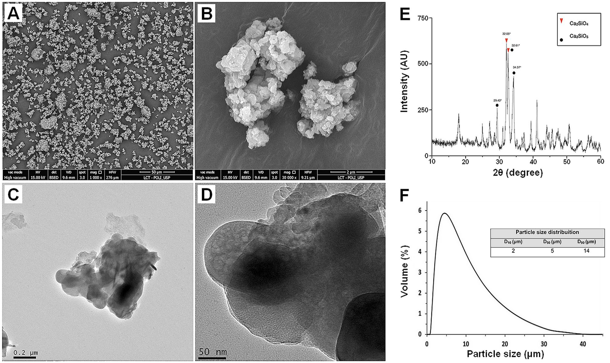
CaSi particle characterization. Low- and high-magnification images for milled CaSi clusters synthesized: (
Nanoindentation
Adhesive Layer
Means and standard deviations for adhesive layer NH and E are shown in Figure 2A and B, respectively. Statistically significant interactions are detailed in Appendix Table 2. At 24 h, Ad(+) groups presented significantly lower NH as compared with Ad(–) groups. However, after 12 mo, only Ad(+) groups did not show statistically significant reductions in hardness (P < 0.05; Appendix Table 3). The inclusion of CaSi in the adhesive and as a pretreatment consistently reduced the E of the adhesive layer, regardless of the storage duration. Additionally, extended storage (6 and 12 mo) negatively affected the adhesive layer’s E, resulting in statistically significant reductions (Appendix Table 4).
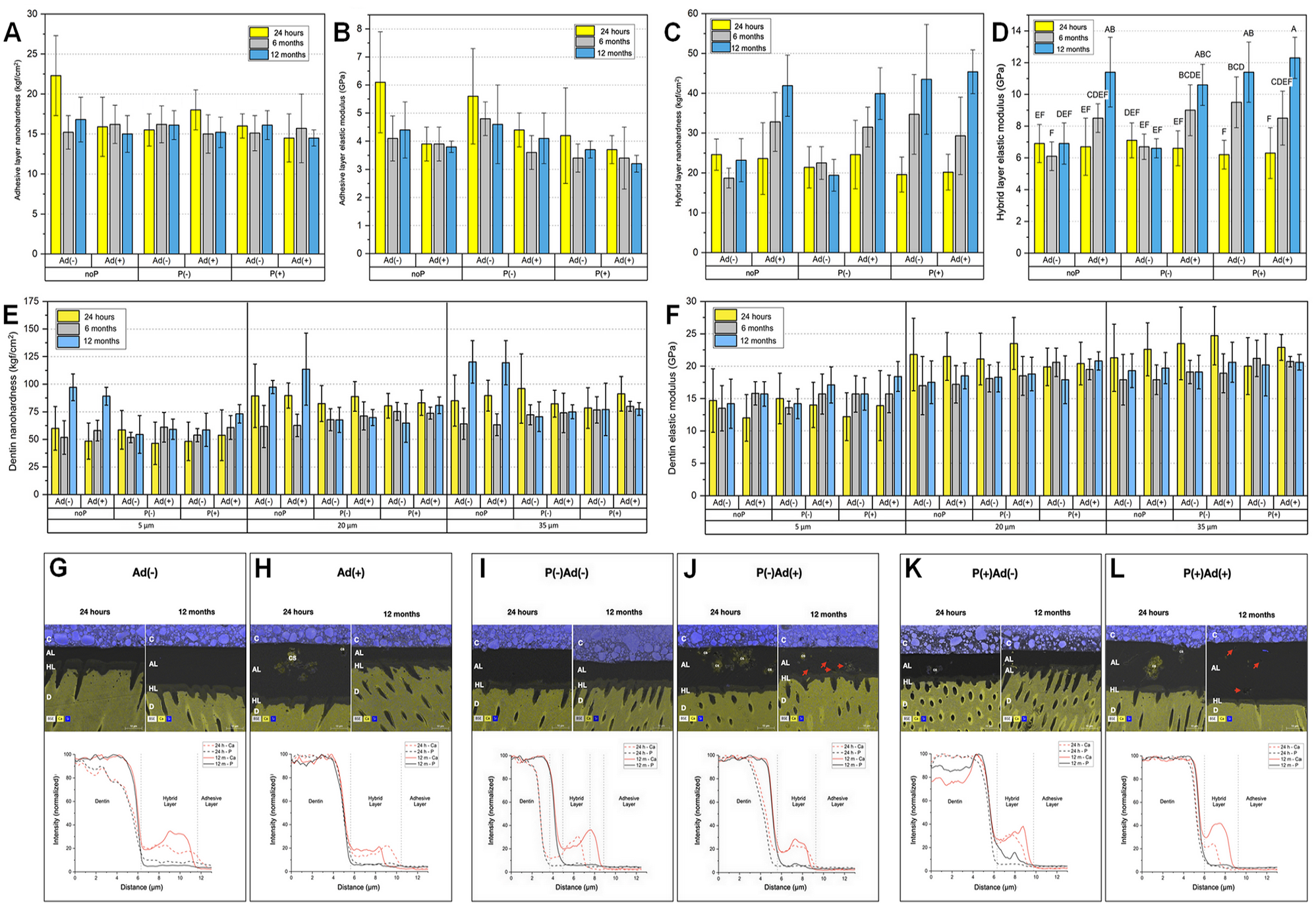
Mechanical properties (NH and E) of adhesive layer, hybrid layer, and dentin and interface characterization (SEM/EDS). Means and standard deviations for (
Hybrid Layer
Statistically significant interactions are shown in the Appendix. Groups containing Ad(+) or P(+) presented a statistically significant increase of NH after 12 mo of storage (P < 0.01; Fig. 2C and Appendix Tables 5 and 6, respectively). For the pretreatment × bonding resin interaction (Appendix Table 7), when noP or P(–) was used, their combination with CaSi-containing bonding resin resulted in statistically higher NH values as compared with the unfilled bonding resin (P < 0.01). When P(+) was used, no statistically significant difference was observed between the filled and unfilled bonding resin.
For E, the bonding resin × pretreatment × storage period interaction was statistically significant (P < 0.01; Fig. 2D). Ad(–) and P(–)Ad(–) groups did not present statistically significant differences after aging. Statistically significant increases between 6 and 12 mo were observed for the Ad(+) and P(+)Ad(+) groups (34% and 45%, respectively). The P(+)Ad(–) group presented a statistically significant increase between 24 h and 6 mo (35%). P(–)Ad(+) also presented a significant increase in E between 24 h and 12 mo (38%).
Dentin
Means and standard deviations for dentin NH and E are shown in Figure 2E and F, respectively. The pretreatment × storage period interaction (Appendix Tables 8 and 9) indicates that the noP and P(–) conditions presented a statistically significant decrease in NH (P < 0.05) and E (P < 0.01) between 24 h and 6 mo, whereas the P(+) groups were able to maintain stable NH and E over time. Furthermore, the statistically significant interaction for distance from the HL × storage period (P < 0.01; Appendix Tables 10 and 11) demonstrates that dentin at the bottom of the HL exhibited statistically lower NH and E as compared with the other 2 depths at 24 h and 6 mo (P < 0.01). Finally, dentin adjacent to the HL showed a statistically significant increase in E after 12 mo, while at the other depths, statistically significant reductions were verified. A similar trend was observed for NH.
Scanning Electron Microscopy/Energy-Dispersive X-ray Spectroscopy
Curves for calcium and phosphorous counts along the dentin-adhesive interface are shown in Figure 2G to L. Overall, after 12 mo, all groups showed increased Ca2+ content at the HL, regardless of the presence of CaSi in the pretreatment and/or bonding resin. However, for CaSi-containing groups, higher Ca2+ content was observed at 24 h and 12 mo, with a greater intensity of phosphorus in the HL after storage.
In Situ Zymography
Representative images of the dentin-adhesive interface after 24 h and 12 mo of storage are displayed in Figure 3. Negative control groups Ad(–) (Fig. 3A) and P(–)Ad(–) (Fig. 3B) showed high levels of gelatinolytic activity. In contrast, groups treated with CaSi, regardless of the protocol or duration, demonstrated a significant reduction in enzymatic activity. After 24 h, groups with CaSi-filled bonding resin alone, Ad(+) (Fig. 3A) and P(–)Ad(+) (Fig. 3B), exhibited a >60% decrease in gelatinolytic activity at the HL and underlying dentin, as shown by 3-dimensonal quantification. After 12 mo, the Ad(+) treatment showed even more pronounced inhibitory effects, with nearly no detectable gelatinolytic activity. Adding CaSi solely to the pretreatment, P(–)Ad(–) (Fig. 3B) versus P(+)Ad(–) (Fig. 3C), also reduced gelatinolytic activity at the HL in both periods. Finally, the P(+)Ad(+) group (Fig. 3C) showed almost complete suppression of gelatinolytic activity at 24 h and 12 mo.
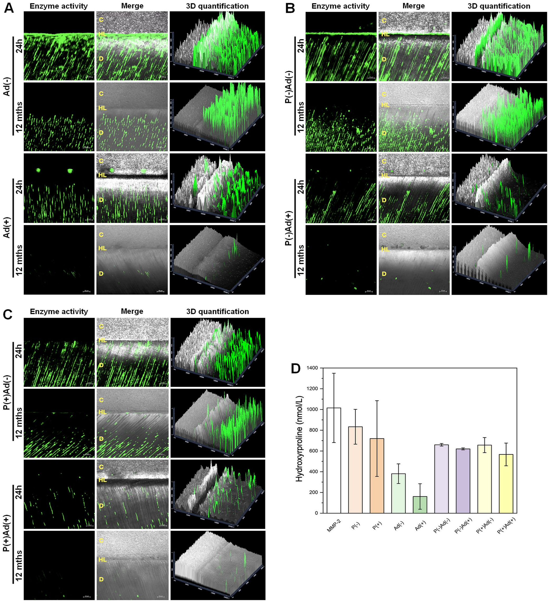
Results for enzymatic assays. (
HYP Assay
The HYP assay (Fig. 3D) showed that samples containing CaSi effectively inhibited collagenolytic activity, regardless of the application method. HYP release was as follows: P(–) 833.52 nmol/L > P(+) 719.83; Ad(–) 380.74 > Ad(+) 161.27; P(–)Ad(–) 659.75 > P(–)Ad(+) 620.16; and P(+)Ad(–) 656.79 > P(+)Ad(+) 566.91. Moreover, relative to full MMP-2 activity, the Ad(+) and P(+)Ad(+) groups demonstrated inhibition rates of 84.1% and 44.2%, respectively, corroborating the in situ zymography results.
Microtensile Bond Strength
Means and standard deviations for µTBS are shown in Figure 4A. Pretreatment and the storage period were statistically significant factors (P < 0.05 and P < 0.01, respectively). Pretreatment pooled data analysis indicated that interfaces restored with CaSi-filled pretreatments exhibited lower µTBS values when compared with the noP groups. Also, a statistically significant reduction of 37% in bond strength was observed between 24 h and 12 mo of storage. The result of failure mode analysis is shown in Figure 4B. Mixed failure mode was the most prevalent, followed by adhesive failures. After 24 h, the occurrence of cohesive failures was relatively low. However, after storage, the trends were reversed, with an increase in composite cohesive failures.
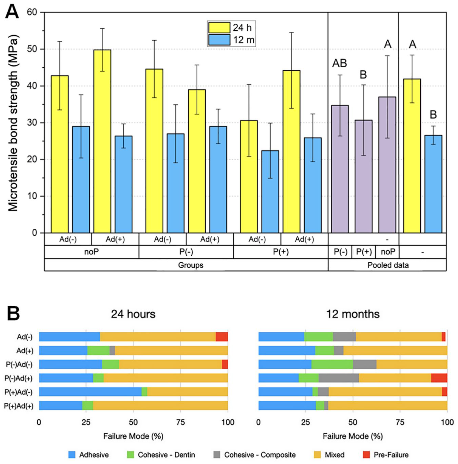
(
Discussion
CaSi particle incorporation during the bonding procedure was proposed as a strategy to deliver calcium ions at the HL, which, in combination with phosphate ions from the physiologic fluid, would lead to mineral precipitation. This approach aimed to enhance the mechanical properties at the bonded interface, reduce hydrolytic degradation, and protect collagen against degradation, ultimately improving the long-term interface performance (Van Meerbeek et al. 2020).
Regardless of the presence of particles, prolonged storage adversely affects the mechanical properties of the adhesive layer and µTBS, likely due to resin component hydrolysis (Sideridou et al. 2003; Malacarne et al. 2006; Yiu et al. 2006; Reis et al. 2007; Marchesi et al. 2010). In CaSi-containing adhesives, particle dissolution and their lack of chemical bonding with the organic matrix facilitate the transit of fluids, which may have accelerated degradation (Chiari et al. 2021). Additionally, particles from the pretreatment can precipitate on the dentin surface and be incorporated into the bonding resin, exacerbating the mentioned phenomena.
However, CaSi enhanced the mechanical properties of the HL and underlying dentin after aging. Similar results were reported for interfaces containing modified CaSi particles (Sauro, Osorio, Fulgencio, et al. 2013; Profeta 2014b; Sauro et al. 2015). The adhesive interface is semipermeable (Tezvergil-Mutluay et al. 2015); as such, Ca2+ ions released from the bonding resin and/or pretreatment are transferred to the voids in the HL, enabling mineral precipitation. These crystallites fill water voids, protecting collagen fibrils from enzymatic attack and making dentin more acid resistant (Gao et al. 2024). Moreover, Ca2+ ions can coordinate and chelate negatively charged groups present in demineralized dentin matrix, such as -R-COO- and PO43-, displacing the confined water between collagen fibrils and improving penetration of hydrophobic monomers. Thus, there is a significant reduction in defects in the HL, which contributes to its longevity (Pan et al. 2023). The negative controls, Ad(–) and P(–)Ad(–), presented some mineral precipitation due to ions prevenient from the SBF. However, it was not sufficient to promote significant recovery in mechanical properties.
The structural conformation of collagen is crucial for guiding mineral nucleation and crystal growth (Carvalho et al. 2023). The decreased gelatinolytic activity in CaSi-containing groups observed by in situ zymography suggests that collagen structural integrity was preserved, which may have contributed to the increase in mineral content of the HL and underlying dentin. A natural reduction of gelatinolytic activity over time is expected, as confirmed by the reductions in fluorescence observed in all groups after aging (Altinci et al. 2019)
The HYP assay indicated a decrease of gelatin degradation when CaSi was added to the incubates, as observed with incorporation of bioactive glass particles in commercial adhesives (Rifane et al. 2023). However, the use of only 1 metalloprotease and gelatin as a substrate makes this evaluation limited. The interactions among demineralized tissue, collagen, collagenases involved in the enzymatic degradation process (MMP-2, -3, -8, -9, -20, and cathepsins) (Nascimento et al. 2011; Mazzoni et al. 2012), and CaSi-based treatments are more reliable in the in situ test.
Despite the positive effects verified in the HL and the evidence of reduced enzymatic activity, long-term µTBS was not improved in groups where CaSi was used, in agreement with previous studies (Sauro et al. 2012; Profeta et al. 2013; Sauro, Osorio, Fulgencio, et al. 2013; Wang et al. 2014; Sauro et al. 2015). It can be hypothesized that the reduction in cohesive strength of the CaSi-containing adhesive (more severe than in the control adhesive; Appendix) offset the improvements observed in the HL and underlying dentin. Therefore, the adhesive layer became the interface’s weakest link (Carrilho et al. 2005; Zhang et al. 2015). As discussed previously, the incorporation of particles from the pretreatment into the bonding resin may explain the similar behavior of groups receiving the CaSi pretreatment. Moreover, the precipitation of CaSi on the dentin surface may obstruct the penetration of monomers into the collagen matrix, potentially resulting in decreased µTBS.
Different mechanisms may be involved to explain Ad(+), P(–)Ad(+), and P(+)Ad(+) improvements in the mechanical properties of the HL and the reduced enzymatic activity. First, apatite precipitation within the voids of the adhesive/HL and underlying dentin protects collagen fibrils from enzymatic degradation by hindering the access of the proteases’ catalytic site to the specific collagen-binding domains found in the organic matrix (Moreira et al. 2021). Second, dentin remineralization enhances its mechanical properties and can lead to the inactivation of MMPs (Kim et al. 2010). Third, the precipitation of a Si2+-rich layer in, and its reaction with, Ca2+ and PO43- ions could create high molecular weight complexes (Ca/P–MMPs), restricting MMP-2 and MMP-9 activity at the HL (Kremer et al. 1998). Finally, the possible release of hydroxyl ions (OH-) from the Ca(OH)2–phase products from hydration, dissociation, and re-precipitation of CaSi can alkalinize the medium, denaturing enzymes and consequently interfering in protease activity (Pashley et al. 2004; Aggarwal and Bhasin 2018).
In contrast with previous studies that have explored various calcium-releasing materials, such as calcium orthophosphates and bioactive glasses, this study specifically investigated the effects of synthesized, nonmodified CaSi particles. This targeted approach allowed for a more in-depth understanding of CaSi’s unique properties and potential benefits. The presented data suggest for the first time that the use of a CaSi-containing pretreatment and/or bonding resin is an effective strategy for enhancing mechanical properties and reducing enzymatic activity at the HL over time. Therefore, the null hypothesis can be rejected. Based on the findings, this study demonstrates that making a minor adjustment to one of the system components or incorporating an additional step in the bonding procedure can enhance the longevity of adhesive restorations. While this early stage of research indicates that the clinical restorative process may take longer due to the added step, it is crucial to continue researching for more efficient dentin-bonding agents.
Author Contributions
M. Damasceno e Souza Chiari, F. Dupart Nascimento, contributed to conception and design, data acquisition, analysis, and interpretation, drafted and critically revised the manuscript; F. Maron Vichi, E. Iraê Almeida Hummel Pimenta Santos, F. Rodrigues de Oliveira, contributed to acquisition, critically revised the manuscript; H. Marques Honório, contributed to analysis and interpretation, critically revised the manuscript; R. Ruggiero Braga, contributed to conception and design, data analysis and interpretation, drafted and critically revised the manuscript. All authors gave final approval and agreed to be accountable for all aspects of the work.
Supplemental Material
sj-docx-1-jdr-10.1177_00220345251336125 – Supplemental material for Calcium Silicate Particles: Biological and Mechanical Role in the Hybrid Layer
Supplemental material, sj-docx-1-jdr-10.1177_00220345251336125 for Calcium Silicate Particles: Biological and Mechanical Role in the Hybrid Layer by M. Damasceno e Souza Chiari, F. Maron Vichi, E. Iraê Almeida Hummel Pimenta Santos, F. Rodrigues de Oliveira, H. Marques Honório, F. Dupart Nascimento and R. Ruggiero Braga in Journal of Dental Research
Footnotes
Declaration of Conflicting Interests
The authors declared no potential conflicts of interest with respect to the research, authorship, and/or publication of this article.
Funding
The authors disclosed receipt of the following financial support for the research, authorship, and/or publication of this article: This study was funded by grants 2020/05986-5, 2023/10291-4, and 2023/18285-3, São Paulo Research Foundation and Coordination for the Improvement of Higher Education Personnel.
A supplemental appendix to this article is available online.
References
Supplementary Material
Please find the following supplemental material available below.
For Open Access articles published under a Creative Commons License, all supplemental material carries the same license as the article it is associated with.
For non-Open Access articles published, all supplemental material carries a non-exclusive license, and permission requests for re-use of supplemental material or any part of supplemental material shall be sent directly to the copyright owner as specified in the copyright notice associated with the article.