
Abstract
Preadhesion mineralization and in situ nano-deposition improving dentin bonding durability are currently explained by divergent paradigms: the former is attributed to collagen strengthening and protease fossilization, while the latter is known for the release of interface-confined water. This study challenges this dichotomy by hypothesizing that preadhesion mineralization fundamentally acts as a dehydration strategy. Two bonding strategies were formulated: a polyacrylic acid–stabilized amorphous calcium fluoride (PAA-ACF) preadhesion mineralization and in situ ACF nano-deposition. Cryo–transmission electron microscopy (TEM) revealed collagen binding by ACF nanoparticles within 30 s, whereas scanning electron microscopy, X-ray diffraction, and TEM showed that PAA‑ACF–mediated intrafibrillar mineralization on etched dentin began within 30 min and reached completion by 3 h. The PAA-ACF 3-h mineralization group showed approximately twice the surface roughness and elastic modulus of the phosphoric acid (PA) and ACF 30-s deposition groups, as measured by atomic force microscopy, whereas its gelatinase activity (assessed by zymography) was substantially lower, only about one-fifth and one-third of the levels in the PA and ACF 30-s groups, respectively. Fourier transform infrared spectroscopy, water absorption, and thermogravimetric analysis confirmed that both strategies effectively released the interface-confined water. Notably, both the nano-deposition and mineralization groups displayed similarly low proteolytic activity in resin–dentin interfaces. After 30,000 thermal cycles, the micro-tensile bond strength of the 3-h group was statistically comparable to that of the ACF 30-s group and exceeded that of conventional PA wet-bonding. Pearson correlation analyses revealed that bonding strength correlated not with mechanical properties or protease activity of preadhered dentin but with the extent of dehydration, which facilitated infiltration of the hydrophobic adhesive monomer (Bis-GMA), as validated by Nile red tracing and micro-Raman analysis. In summary, both strategies enhance adhesive infiltration by releasing the interface-confined water, forming a defect-low hybrid layer. This finding unifies the understanding of dentin bond durability improvement under a common dehydration-based mechanism.
Keywords
Introduction
The longevity of resin–dentin bonding remains a primary clinical challenge. The main cause is from a defective hybrid layer caused by insufficient adhesive infiltration in demineralized dentin matrix (DDM) (Van Meerbeek et al 2020; Bertolo et al 2021). Although there is a consensus that the restriction of water in the DDM to hydrophobic adhesive infiltration causes suboptimal dentin bonding (Spencer et al 2010; Pashley et al 2011), the fragility and enzymatic susceptibility of apatite-depleted, resin-sparse dentin collagen fibrils also compromise the bonding durability (Breschi et al 2008). Therefore, the corresponding strategies to extend restoration lifespan are investigated: reinforcing the collagen network with crosslinkers (Chen et al 2021), inactivating proteases with inhibitors (Tezvergil-Mutluay et al 2012), or displacing water in DDM with ethanol via the ethanol-wet bonding technique (Sadek et al 2010). However, the primary determinant among these adverse factors remains unclear, which confounds the development of optimal strategies.
We identified “interface-confined water” as a critical factor impairing adhesive penetration (Pan et al 2023). This water is derived from the complex 3-dimensional (3D) structure and intrinsic polarity of the DDM, which includes hydrogel-like water bound to hydrated noncollagenous proteins (NCPs), capillary water constrained within the hierarchical porous structure, and water molecularly confined by polar side chains and collagen fibril nanostructures. Subsequent research has therefore explored methods to release this interface-confined water, such as polycation assembly (Lei et al 2023; Shu et al 2025), metal ion coordination (Cheng et al 2025; Li et al 2025), and in situ nano-deposition (Xu et al 2021; Luo et al 2025; Xu et al 2026b). The nano-deposition strategy is that kinetically unstable supersaturated solutions (e.g., calcium fluoride solution, calcium phosphate solution) are used to in situ form nanoparticles (amorphous calcium fluoride [ACF] and amorphous calcium phosphate [ACP]) at negatively charged groups in the DDM. This removes the hydration capacity of the DDM and releases the water confined at the interface, enhancing adhesive infiltration and forming a low-defect hybrid layer.
In parallel, a “preadhesion mineralization” strategy has been recently reported. Bonding improvement was achieved by mineralizing the DDM before adhesive application within minutes, using a rapid mineralizing solution such as polyacrylic acid–stabilized ACF (PAA-ACF) or polyaspartic acid–stabilized ACP (Gao et al 2024; Li et al 2024). They attributed the resultant improved bonding to the physicochemical properties of the mineralized dentin, including increased elastic modulus and the “fossilization” of proteases.
However, this presents a paradox from a composite materials science perspective. The long-term stability of dentin bonding is fundamentally governed by the integrity of the hybrid layer between the DDM and the adhesive resin. The ultimate strength and durability depend on their synergistic interaction, specifically, flawless integration. Considering the mineralization process itself progressively displaces water molecules from the collagen fibrils (Kim et al 2010), we therefore hypothesize that the dehydrating effect of the preadhesion mineralization, which facilitates superior adhesive infiltration, is the unifying mechanism underpinning the improved bonding durability, rather than the physicochemical reinforcement of the dentin matrix itself.
Herein, this study aims to elucidate the key determinant of bonding durability by directly comparing 2 strategies: the rapid in situ ACF nanoparticle deposition and the more protracted PAA-ACF–mediated intrafibrillar remineralization. By comparing their bonding effects and elucidating their respective contributions to dehydration, mechanical reinforcement, and enzyme inhibition, we seek to determine whether the improvement of bonding durability caused by the release of interface-confined water to improve adhesive infiltration is the unifying principle underlying durable dentin adhesion.
Materials and Methods
Details of materials and methods are provided in the Appendix.
ACF Solution and PAA-ACF Mineralization Medium
PAA-ACF mineralization medium was made by rapidly mixing 20 mM NaF (Tris-buffer with 150 mM NaCl) and 10 mM CaCl2·2H2O (Tris-buffer with 300 mM NaCl and 800 μg/mL PAA). ACF solution was prepared similarly but without PAA. Cryo–transmission electron microscopy (TEM) with selective area electron diffraction (SAED) and electrophoretic light scattering was used to characterize size distribution and Zeta potential.
ACF and PAA-ACF Conditioning
Type I rat tail collagen solution was pipetted onto the gold meshes; incubated in ACF for 30 s and PAA-ACF for 10 min, 30 min, and 1 h; and detected by cryo-TEM. High-resolution TEM identified the morphology and lattice fringes.
For the dentin model, groups were named as follows: SD, sound dentin; PA, acid-etched with 37% phosphoric acid for 15 s (according to manufacturer recommendation); 30S, PA-etched dentin conditioned with ACF solution for 30 s; and 10MIN, 1H, 3H, and 6H, PA-etched dentin mineralized in PAA-ACF solution for 10 min, 1 h, 3 h, and 6 h, respectively.
Physiochemical and Proteolytic Activity of Conditioned Dentin
Scanning electron microscopy (SEM), TEM, X-ray diffraction (XRD), ATR–Fourier transform infrared spectroscopy (FTIR), and X-ray photoelectron spectroscopy (XPS) were used. Surface roughness and mechanical properties were demonstrated with atomic force microscopy (AFM). Endogenous gelatinase activity and localization were analyzed by in situ zymography and immunohistochemistry. Exogenous collagenase degradation was assessed by hydroxyproline release.
Water Analysis
Water sorption was determined as the percentage of weight loss after lyophilization, as previously described (Gonulol et al 2015). Thermogravimetric analysis (TGA) was conducted in an airflow with a heating rate of 10 °C/min from 25 °C to 1,000 °C to detect the bound water content and mineral content of 10 mg conditioned DDM powder. ATR-FTIR characterized the types of water. Specifically, 2-mm-thick conditioned dentin slabs were soaked in deionized water and gently wiped with filter paper, and signals were collected.
Adhesive Infiltration and Bonding Performance
Nile red fluorescent staining was used to trace adhesive penetration (Bis-GMA). Dental adhesive (containing 0.01 wt% Nile red) was applied. Resin–dentin slices were examined using confocal laser scanning microscopy (CLSM) with a 63× oil objective (excitation/emission: 540/590 nm).
A micro-Raman spectrometer was employed with a 532-nm laser beam with a 50× lens (power: 50%, exposure time: 60 s). The scanning line (step width: 1 μm) started in dentin, across the hybrid layer, and ended in the adhesive layer. CH2 (1,450 cm-1) and C = O (1,720 cm-1) were present for both HEMA and Bis-GMA; C-O-C (1,113 cm-1) and phenyl (1,609 cm-1) bands were specific to Bis-GMA. The degree of hydrophobic monomer (Bis-GMA) infiltration into the hybrid layer was quantified as the ratio of the integral area of the Bis-GMA–specific 1,113-cm-1 band to that of the reference CH2 band at 1,450 cm-1 using Origin software.
Resin–dentin samples were thermocycled for 0 or 30,000 times and evaluated using micro-tensile bond strength (μTBS) (Single Bond 2 and a self-made adhesive composed of simple components without water or fillers), in situ zymography, and silver nanoleakage staining.
Statistical Analyses
All data were analyzed using GraphPad Prism version 9.1.1 (GraphPad Software), with a significance level set at α = 0.05. Data are expressed as mean ± SD. After verifying normality and homoscedasticity, parametric tests were applied: 1-way analysis of variance (ANOVA) with Šidák post hoc tests for collagenase activity of dentin surface, surface roughness, Derjaguin–Muller–Toropov (DMT) modulus, and water sorption, as well as 2-way ANOVA for μTBS values, silver nanoleakage distribution, and in situ zymography. The relationship between roughness, elastic modulus, gelatinase, in situ zymography, water absorption, adhesive infiltration, and bond strength was evaluated using Pearson multivariable correlation analysis. Correlation coefficients with their 95% confidence intervals and 2-tailed P values were calculated.
Results
PAA-ACF Mineralization and ACF Deposition
ACF solution rapidly aggregated amorphous nanoparticles, while PAA-ACF solution in the form of precursors (78–122 nm) could be stabilized for 6 h (Appendix Fig. 1A–B). Zeta potentials of ACF (+31 ± 1.96 eV) and PAA-ACF (−18.9 ± 1.67 eV) are in Appendix Figure 1C. Cryo-TEM detected ACF nanoparticles that were electrostatically attached to the negatively charged bands of collagen fibrils. PAA-ACF precursors initially infiltrated the gap zones within 10 min and transformed into CaF2 crystals after 30 min; complete mineralization was observed after 1 h in cryo-TEM (Fig. 1A). High-resolution TEM showed the intrafibrillar mineralization and lattice fringe spacings of the (111) and (220) crystal planes of CaF2 (Appendix Fig. 2A, B). Mesoscale mineralized spherules were observed within 3D collagen membranes at 30 min (Fig. 1B) and gradually merged after mineralizing for 1 h (Appendix Fig. 3).
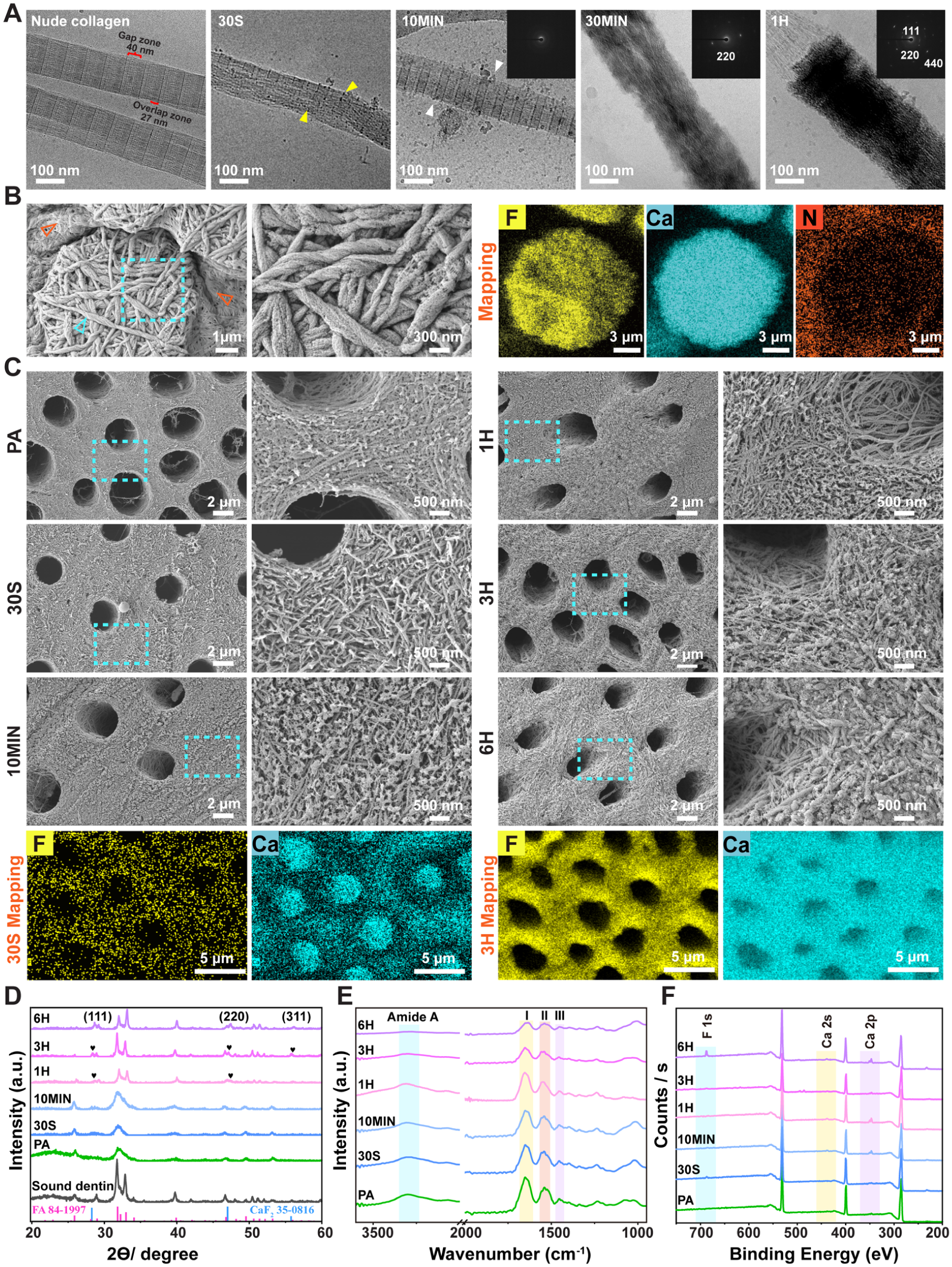
Amorphous calcium fluoride (ACF) and polyacrylic acid–stabilized amorphous calcium fluoride (PAA-ACF)–conditioned collagen and dentin. (
Ultrastructure and chemical properties of dentin samples are shown in Figures 1C–F and 2. For the 30S group, amorphous spherical CaF2 nanoparticles were deposited. For the PAA-ACF groups, no crystals were detected in the 10MIN group, the 500-nm-thick zone at the bottom of the DDM was remineralized with needle-shaped crystals after 1 h, and the 3- to 5-μm-thick region was completely remineralized after 3 h. After 6 h, crystals densely occupied extra- and intrafibrillar spaces. SAED patterns and XRD spectra presented the 2θ = 28.2° (111), 47° (220), 55.7° (311) crystal planes of CaF2. ATR-FTIR spectra (Fig. 1E) revealed that the intensity of amide bands of collagen decreased as the mineralizing time extended. XPS spectra showed that the F1s and Ca 2s, 2p peaks in the ACF- and PAA-ACF–mineralized groups were more pronounced than in the PA group (Fig. 1F).
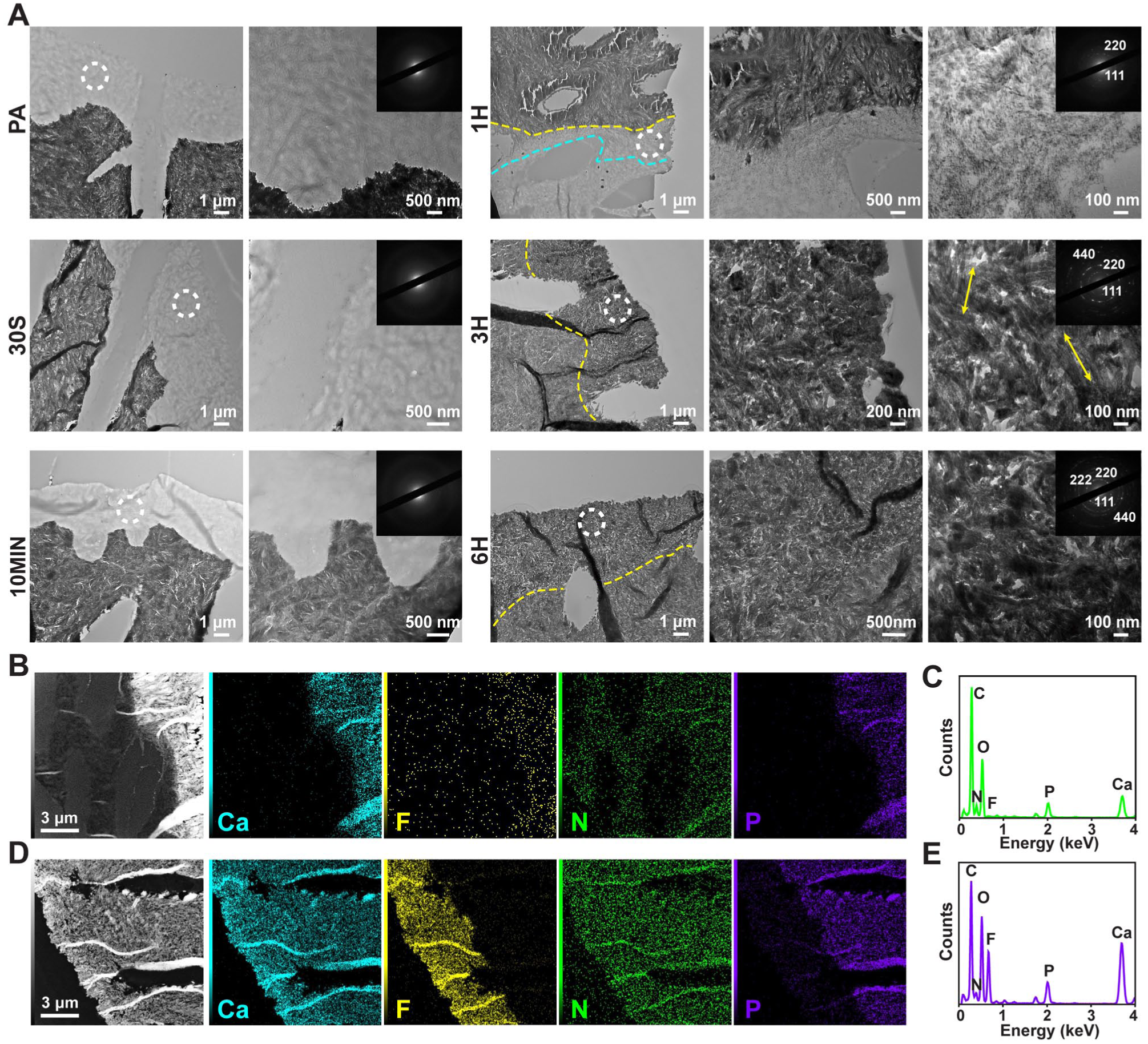
Characterization of amorphous calcium fluoride (ACF)–conditioned and polyacrylic acid–stabilized amorphous calcium fluoride (PAA-ACF)–mineralized dentin. (
Physicochemical Properties
AFM (Fig. 3B, F) revealed that the PA group was the smoothest (20.9 ± 5.9 nm). 10MIN (26.1 ± 6.7 nm), 1H (37.7 ± 12.3 nm), and 3H (56.1 ± 9.5 nm) were rougher than ACF 30S (32.7 ± 13.2 nm) (P < 0.05). DMT modulus (Fig. 3C, G) of 10MIN (14.5 ± 1.9 Gpa) and 1H (16.5 ± 5.2 Gpa) groups increased with mineralization time, and the 3H group (24.6 ± 5.4 Gpa) reached the mechanical property of sound dentin (25.9 ± 4.2 Gpa) and was twice that of ACF 30S (11.9 ± 1.1 Gpa) and PA (9.5 ± 1.9 Gpa).
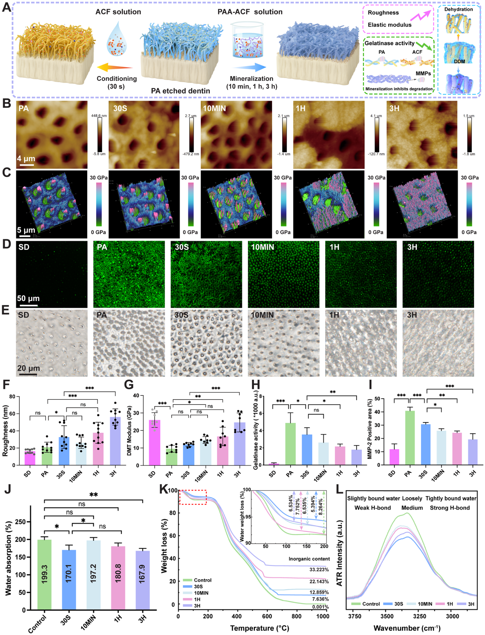
Physicochemical properties of amorphous calcium fluoride (ACF)–conditioned and polyacrylic acid–stabilized amorphous calcium fluoride (PAA-ACF)–mineralized dentin. (
In situ zymography (Fig. 3D, H, Appendix Fig. 4) showed the highest fluorescence in the PA and 30S groups, twice that of the 3H group, and 1.5 times that of the 1H group. Immunohistochemical staining localized MMP-2 primarily to the dentinal tubules and peritubular dentin (Fig. 3E, I, Appendix Fig. 5). The hydroxyproline release after exposure to exogenous collagenase (Appendix Fig. 6) detected no difference between PA (18.86 ± 3.226 μg/mL) and 30S groups (16.42 ± 4.094 μg/mL) (P > 0.05), was higher than the 10MIN group (11.56 ± 1.491 μg/mL), and was twice that of 1H (7.893 ± 2.412 μg/mL) and 3H groups (7.799 ± 3.567 μg/mL).
Water absorption (Fig. 3J) was decreased compared to the control (199 ± 8.2%), with 170% ± 13.7% in the 30S group and 167.9% ± 6.7% in the 3H group (P < 0.05), with no difference between these 2 groups (P > 0.05). TGA (Fig. 3K) revealed that bound water content was reduced in ACF (5.394%), PAA-ACF 10MIN, 1H, and 3H (6.535%, 7.752%, 6.534%) groups relative to control (8.264%) but had higher inorganic remnants in ACF 30S (7.63%) and all the 10MIN, 1H, and 3H (12.85%, 22.14%, 33.22%) groups compared to control (0.001%). ATR-FTIR spectra (Fig. 3L) exhibited a blueshift of the loosely bound water band, and the content of the 3 types of water significantly decreased.
Adhesive Infiltration
Nile red tracing (Fig. 4A–C) showed that the intensity of 30S, 1H, and 3H groups was stronger than that of the PA and 10MIN groups. Raman spectra of adhesive layer, hybrid layer, and dentin were recorded (Fig. 4D, E, Appendix Fig. 7). Based on the intensity at 1,720, 1,450 cm–1 (C = O, CH2, all monomers in the adhesive) and 1,113, 1,609 cm–1 (C-O-C, phenyl, Bis-GMA), Raman analysis showed that the content of Bis-GMA, calculated by the ratio of the 1,113/1,450 integral area, was increased in the hybrid layer of 30S (75.15% ± 4.08%), 1H (65.37% ± 24.5%), and 3H (69.46% ± 18.01%) groups, compared to the PA group (30.86% ± 12.21%). No significant difference was seen in 30S and 3H groups (P > 0.05).
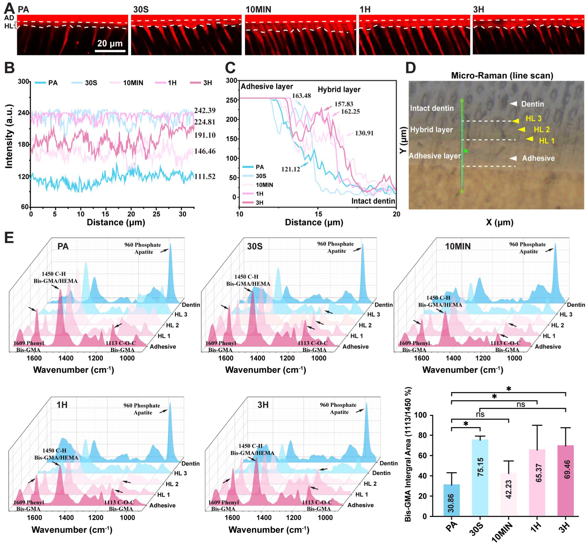
Prompting adhesive infiltration enhanced dentin bonding via water release. (
Bonding Performance
Nanoleakage observed under SEM displayed scattered silver particles along the resin–dentin interface in 30S and mineralizing groups, which were significantly fewer than the PA group in immediate mode (P < 0.05) (Fig. 5A, B). After aging, silver deposition increased in the PA, 10MIN, and 1H groups, exceeding that in the 30S and 3H groups (P < 0.05). In situ zymography (Fig. 5B, C) showed that both ACF and PAA-ACF groups, which did not present a significant difference (P > 0.05), exhibited significantly weaker fluorescence than the PA group in immediate and aging modes (P < 0.05).
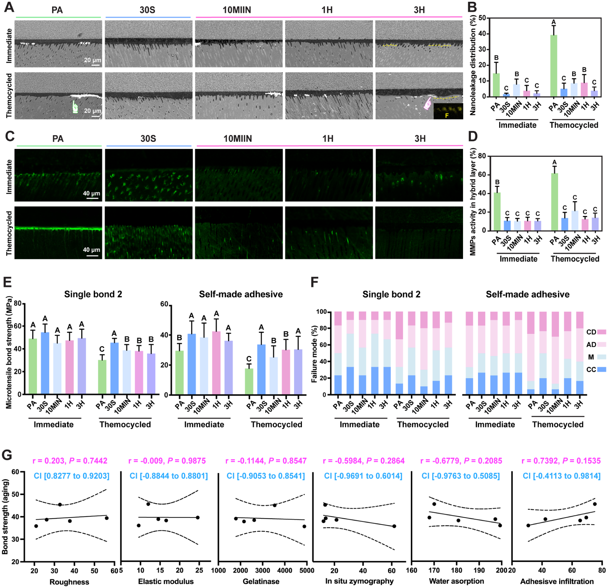
Bonding performance of amorphous calcium fluoride (ACF)–conditioned and polyacrylic acid–stabilized amorphous calcium fluoride (PAA-ACF)–mineralized dentin. (
For Single Bond 2 adhesive, neither conditioning strategy significantly enhanced the immediate bonding strength (Fig. 5D, P > 0.05). After aging, although all groups exhibited a decline in μTBS, the PA, 10MIN, and 1H groups exhibited a significant reduction (P < 0.05). In contrast, the 30S and 3H groups maintained relatively stable bonding performance (P > 0.05). For self-made adhesive, both ACF conditioning and mineralization improved the immediate μTBS values (P < 0.05). After aging, stable bonding performance was maintained only in the 30S and 3H groups (P > 0.05). Failure mode analysis revealed that the percentage of mixed failure and cohesive failure in the composite predominated in the 30S, 1H, and 3H groups before aging. Conversely, the high percentage of adhesive failure and cohesive failure in dentin in the PA and 10MIN groups suggested a high failure at the bottom of the hybrid layer.
Pearson’s correlation analysis (Fig. 5G, Appendix Fig. 8) revealed that no significant correlation was found between μTBS and surface roughness, elastic modulus, or dentin gelatinase activity. Although statistical significance was not reached due to the limited number of groups (n = 5), a negative correlation trend was observed between water sorption and aged bond strength (r = −0.6779). Moreover, a positive correlation trend was found between adhesive infiltration and aged bond strength (r = 0.7392).
Discussion
Recent studies show that elevated calcium and phosphorus concentrations facilitate calcium phosphate (CaP) prenucleation nanocluster aggregation and nucleation, thereby promoting their transformation into the hydroxyapatite phase (Yao et al 2019; Amornkitbamrung et al 2023). Therefore, to accelerate the remineralization process, the present study increased the concentration of calcium, fluoride, and PAA by 2 times compared to the previous reports (Ping et al 2022; Gao et al 2024). Likewise, the concentration of calcium and fluoride of the ACF solution was also increased compared to our previous study (Xu et al 2026b). Notably, cryo-TEM, for the first time, revealed the CaF2-mediated mineralizing and nanoparticle deposition process in a reconstituted collagen model, ensuring a close-to-native state and avoiding dehydration artifacts (Thompson et al 2016; Lei et al 2022). We observed ACF nanoaggregates precipitated on the surface of collagen due to the electrostatic attraction at 30 s. PAA-ACF–mediated intrafibrillar mineralization occurred at 30 min, with complete mineralization after 1 h, involving precursor infiltration of gap zones, phase transformation, and oriented growth, consistent with nonclassical crystallization theory (Gower 2008; Song et al 2019).
However, phosphoric acid–etched dentin is more complex than reconstituted collagen (Gu et al 2011; Shao et al 2018). Although we doubled the concentration of the remineralization medium, the results exhibited slower remineralization rates than those in the preadhesion remineralization reports: no detectable crystals at 10 min, a 300- to 500-nm remineralized region by 1 h, and full mineralization after 3 h. Based on these kinetics, we prepared dentin specimens with varying mineralization levels, designated as 10MIN (nonmineralized), 1H (partially mineralized), and 3H (fully mineralized) groups, for subsequent bonding experiments.
The surface roughness and mechanical properties are often considered contributing factors to micro-tensile bonding strength (Gao et al 2024; Li et al 2024). In this study, AFM revealed that the PAA-ACF 3H group exhibited the highest roughness and elastic modulus, followed by 1H and 10MIN groups. This trend indicated that both intrafibrillar mineralization and the degree of mineralization play critical roles in enhancing collagen’s mechanical properties, which aligns with earlier findings (Kinney et al 2003; Zhong et al 2023; Li et al 2024). Specifically, the roughness and elastic modulus of the 3H group were approximately twice those of the ACF 30S group. However, Pearson correlation analysis indicated no significant relationship between bonding strength and either roughness or elastic modulus.
The activation of endogenous matrix metalloproteinases (MMPs) is known to contribute to the degradation of collagen fibrils within the hybrid layer (Zhang and Kern 2009). MMP-2 immunohistochemistry revealed the localization of gelatinase predominantly within dentin tubules and peritubular dentin, consistent with previous studies (Seseogullari-Dirihan et al 2016; Stape et al 2018). In situ zymography showed that 3H and 1H mineralizing groups exhibited lower gelatinase activity than the PA and ACF 30S groups, which can be attributed to the inhibitory effect of fluoride, owing to its strong electronegativity (Kato et al 2014) and the shielding of protease cleavage sites by mineral encapsulation. Additionally, remineralized dentin exhibited resistance to exogenous collagenase degradation due to the physical barrier provided by minerals, which protected collagen. Strikingly, different from the current understanding that MMP harms the longevity of resin–dentin bonds (Pashley et al 2004), our Pearson correlation analysis revealed that gelatinase activity of dentin before adhesion was not significantly correlated with bonding strength after aging. Intriguingly, it was the gelatinase activity within the hybrid layer that showed a negative correlation with the aged bonding strength. These findings suggest that the enzymatic degradation is associated with the structural integrity of the final hybrid layer, dependent on adhesive infiltration, and the elimination of defects in the hybrid layer can effectively inhibit MMP activation.
Collagen mineralization can enhance its mechanical properties and release intrafibrillar water (Jiao et al 2016). Similarly, ACF deposition has been reported to reduce the DDM interface-confined water (Xu et al 2021). In line with these findings, this study observed a general decrease in water absorption with increasing mineralization time. Notably, the ACF 30S group showed the lowest water adsorption rate, which was not significantly different from the 3H mineralization group. ATR spectra further confirmed a reduction in confined water signal in the ACF 30S group. This decrease may be attributed to the masking of negatively charged groups on NCPs and collagen, which would reduce interfacial polarity and thereby facilitate the release of interface-confined water. The process of mineralization not only released interface-confined water but also progressively replaced bound water with mineral crystals (Figueiredo et al 2010), a phenomenon corroborated by TGA analysis. Collectively, these results demonstrate that both mineralization and amorphous nanoparticle deposition strategies function as effective dehydration methods. Importantly, Pearson analysis revealed that water absorption had a significant negative correlation with aged bonding strength, underscoring the critical role of interface-confined water in the adhesive infiltration that determines the final structure of the hybrid layer.
To evaluate monomer infiltration, Nile red was used to trace the key hydrophobic Bis-GMA (Xu et al 2021, 2026a), a monomer critical for providing mechanical robustness and hydrolysis resistance essential for long-term bonding stability (Van Meerbeek et al 2020). Nile red fluorescence analysis of the resin–dentin interface revealed that the ACF 30S, PAA-ACF 1H, and 3H groups significantly promoted Bis-GMA infiltration compared to the PA group (Fig. 4A–C), and the ACF 30S and PAA-ACF 3H groups exhibited comparable and the most pronounced improvements. These observations were quantitatively corroborated by micro-Raman spectroscopy of Bis-GMA content in the hybrid layer (Fig. 4D, E). The improved adhesive infiltration was associated with fewer structure defects and reduced activity of MMPs within the hybrid layer (Fig. 5). Critically, Pearson correlation analysis demonstrated a positive correlation between the degree of adhesive infiltration and the aged bonding strength. This provides direct evidence that improving the penetration of hydrophobic resin components is a critical factor in achieving durable dentin bonding.
Conclusion
In summary, the 30-s nano-deposition strategy achieved comparable long-term bonding strength to the 3-h preadhesion mineralization bonding strategy. Bonding durability was not primarily determined by surface roughness, elastic modulus, or endogenous MMP activity of the preadhered dentin but rather by the effective management of interface‑confined water. Both strategies share a common mechanism that involves masking polar groups in the DDM and releasing interface‑confined water. This mechanism improves adhesive infiltration and modulates phase interactions between the adhesive resin and the DDM, thereby promoting the formation of a defect‑low hybrid layer essential for durable dentin bonding.
Author Contributions
Q. Zhong, contributed to design, data acquisition, analysis, drafted the manuscript; C. Shu, contributed to design, data acquisition, drafted the manuscript; M. Pan, J. Xu, contributed to acquisition, drafted the manuscript; Q. Luo, contributed to conception, data analysis and interpretation, critically revised the manuscript; X. Li, contributed to conception and design, data interpretation, critically revised the manuscript. All authors gave final approval and agreed to be accountable for all aspects of the work.
Supplemental Material
sj-docx-1-jdr-10.1177_00220345261442125 – Supplemental material for Mineralization/Nano-deposition Boosts Adhesive Infiltration via Dehydration
Supplemental material, sj-docx-1-jdr-10.1177_00220345261442125 for Mineralization/Nano-deposition Boosts Adhesive Infiltration via Dehydration by Q. Zhong, C. Shu, M. Pan, J. Xu, Q. Luo and X. Li in Journal of Dental Research
Footnotes
Acknowledgements
The authors thank Lingyun Wu, Tingyu Liu, and Qin Han at the Center of Cryo-Electron Microscopy (CCEM), Zhejiang University, for their technical assistance on Cryo-TEM and CLSM; Xiaomin Zhang, Xi Zheng, and Dongmei Qi at the Analysis Center of Agriculture, Life and Environment Sciences, Zhejiang University, for their technical assistance with TEM and SEM; Qingyun Lin from the Center of Electron Microscopy of Zhejiang University for her technical assistance on high-resolution TEM; and Xiangqian Wang, the assistant research fellow of the Training Platform of Clean Energy Utilization, Polytechnic Institute of Zhejiang University, for his contribution in carrying through the AFM.
Declaration of Conflicting Interests
The authors declared no potential conflicts of interest with respect to the research, authorship, and/or publication of this article.
Funding
The authors disclosed receipt of the following financial support for the research, authorship, and/or publication of this article: This work was supported by Natural Science Foundation of Zhejiang Province grant (LQ23H140002), National Natural Science Foundation of China grant (32371380, 32271407, 82301118).
Data Availability Statement
A supplemental appendix to this article is available online.
References
Supplementary Material
Please find the following supplemental material available below.
For Open Access articles published under a Creative Commons License, all supplemental material carries the same license as the article it is associated with.
For non-Open Access articles published, all supplemental material carries a non-exclusive license, and permission requests for re-use of supplemental material or any part of supplemental material shall be sent directly to the copyright owner as specified in the copyright notice associated with the article.