
Abstract
The dual route model (DRM) of reading suggests two routes of reading development: the phonological and the orthographic routes. It was proposed that although the two routes are active in the process of reading; the first is more involved at the initial stages of reading acquisition, whereas the latter needs more reading training to mature. A number of studies have shown that deficient phonological processing is a core deficit in developmental dyslexia. According to the DRM, when the Lexical Decision Task (LDT) is performed, the orthographic route should also be involved when decoding words, whereas it is clear that when decoding pseudowords the phonological route should be activated. Previous functional near-infrared spectroscopy (fNIR) studies have suggested that the upper left frontal lobe is involved in decision making in the LDT. The current study used fNIR to compare left frontal lobe activity during LDT performance among three reading-level groups: 12-year-old children, young adult dyslexic readers, and young adult typical readers. Compared to typical readers, the children demonstrated lower activity under the word condition only, whereas the dyslexic readers showed lower activity under the pseudoword condition only. The results provide evidence for upper left frontal lobe involvement in LDT and support the DRM and the phonological deficit theory of dyslexia.
Reading is a complex task conducted in the information processing system, where a number of cognitive factors, ranging from low-level sensory to high-level cognitive processes, are activated. During reading, a complex neural network is activated, involving different brain regions (Berl et al., 2010; Binder et al., 2003; Hofmann et al., 2008; Pugh et al., 2001). Specifically, previous data support the notion that the frontal lobe is involved in the process of reading, providing higher executive functionality such as memory and attention to the process of reading (Binder et al., 2003; Fiebach, Ricker, Friederici, & Jacobs, 2007).
The dual route model (DRM) of reading is a well-established and accepted model that proposes a description for the process of reading (Coltheart, Rastle, Perry, Langdon, & Ziegter, 2001; Fiebach, Frederici, Muller, & von-Cramon, 2002). The model suggests the occurrence of two information processing pathways: the orthographic route and the Phonological Route. These routes are considered to be noncompetitive and rather complementary to each other. Both routes process the written word into its appropriate spoken representation. However, they differ in their complexity, speed of processing, and course of action. The orthographic (lexical) route is more active when identifying a whole pattern representations of the written word from the mental lexicon (the set of rules that defines the written script), finds semantic context, and then identifies the appropriate spoken representation of the written word. The phonological route combines the grapheme (the smallest alphabetic representation of the written word) with the phoneme (its acoustic representation) and then provides phonological representation for the written word. The DRM has been used to study different aspects of reading (Fiebach et al., 2002; Jobard, Crivello, & Tzourio-Mazoyer, 2003), such as developmental reading (Acha & Perea, 2008) and reading disabilities, specifically dyslexia (Bergmann & Wimmer, 2008; Wimmer & Schurz, 2010; Ziegler et al., 2008). It has been shown, for example, that the phonological route is more involved in the early phases of reading acquisition, whereas the orthographic route needs more printed exposure to develop (Ehri, 2002; Share, 1995). In the context of learning disabilities, a deficit within the phonological route has been suggested as one of the underlying causes of dyslexia (Share, 1994; Snowling, 1995; Stanovich, 1988; Temple et al., 2001).
One of the methods to investigate the DRM is the Lexical Decision Task (LDT; Bergmann & Wimmer, 2008; Binder et al., 2003; Carreiras, Mechelli, & Price, 2006; Edwards, Pexman, Goodyear, & Chambers, 2005; Fiebach et al., 2002; Fiebach et al., 2007; Heim et al., 2005; Hofmann et al., 2008; Jacobs, Graf, & Kinder, 2003; Jacobs & Grainger, 1994; Sela et al., 2011). The task requires the participant to decide whether a chain of letters represents a real word or a nonword (or pseudoword). In a typical reader, the identification of a real word is thought to be more a process of the orthographic route where word patterns as a whole are retrieved from the mental lexical storage. As no patterns of pseudowords exist in the human brain, the pseudowords can be identified only by recoding the alphabetic code in the phonological route, where grapheme to phoneme correspondent takes place (Coltheart et al., 2001).
The current study used a neuroimaging device based on functional near-infrared spectroscopy (fNIR) for recording frontal lobe activity during the performance of the LDT. The fNIR is a relatively new, noninvasive brain-imaging technology that allows for the measurement of hemodynamic changes within the brain. It is a portable, affordable, and easy-to-use device that is considered to be more tolerant to movement artifacts as compared to other brain imaging tools such as electroencephalography (EEG) and functional magnetic resonance imaging (fMRI). Because of these many attractive attributes, fNIR has become a commonly used tool in various areas of cognitive research (Ayaz et al., 2012; Hofmann et al., 2008; Holtzer et al., 2011; K. Izzetoglu, Bunce, Onaral, Pourrezaei, & Chance, 2004; Menda et al., 2011; Quaresima, Ferrari, van der Sluijs, Menssen, & Coiler, 2002; Sela et al., 2011). Specifically, several studies have focused on the involvement of the frontal lobe in different aspects of language. For example, fNIR was used to show the effect of aging on the left prefrontal cortex activity during a series of lingual and memory tasks (Sakatani, Lichty, Xie, Li, & Zuo, 1999). In addition, it was used to show the involvement of the left Broca in the process of language translation task (Quaresima et al., 2002) and to show the correlation between language dominance and handedness (Watanabe et al., 1998). A recent study used fNIR to demonstrate the involvement of the left superior and inferior frontal lobe in the performance of LDT among typical readers (Hofmann et al., 2008). It was suggested that this brain region is involved in the process of decision making, where an intralexical positive trigger is sent from parietal regions when a word is identified and an expiration of an extralexical temporal threshold produces a negative response following a pseudoword stimulus (Fiebach et al., 2007). Furthermore, another LDT study where fNIR was used among typical 12-year-old readers compared to young adults (Sela et al., 2011) indicated a lower activity in the upper left frontal lobe under the Word condition among the children as compared to the young adults during the identification of words, but no such activity was noted when pseudowords were identified. It was suggested that based on the DRM model (Coltheart et al., 2001), it is plausible that the orthographic route, which is responsible for word identification, matures later, after years of print exposure, whereas the phonological route remains active at all ages when needed (i.e., for pseudoword identification).
During the normal reading process, both orthographic and phonological routes follow developmental sequence (Stanovich, 1988). However, if dyslexic readers exhibit a deficit within the phonological route (Share, 1994; Snowling, 1995; Stanovich, 1988; Temple et al., 2001), there is a possibility that their brain activity during LDT might exhibit atypical activation compared to both typical adult and child readers.
In the current study, we used fNIR to measure the hemodynamic activity in the frontal lobe during the performance of LDT and compared the results of three groups: typical readers of age 12, young adult dyslexic readers, and young adult typical readers. Based on the DRM and the phonological deficit theory, it was hypothesized that the children’s group would show lower activation in their upper left frontal lobe compared to young adult typical readers under the Word condition (Sela et al., 2011). In contrast, young adult dyslexic readers would show lower activity in their upper left frontal lobe as compared to their typically reading peers under the pseudoword condition.
Method
Participants
Three groups were included in this study: 17 typically reading 7th grade children (age M = 12.70, SD = 0.457; 8 females and 9 males), 17 young adult dyslexic readers (age M = 25.65, SD = 2.668; 9 females and 8 males), and 17 young adult typical readers (age M = 25.06, SD = 2.384; 9 females and 8 males). No significant age difference between the two reading levels of young adults groups was obtained, t(32) = 0.678, p = .503). Reading score was measured by Shatil’s One Minute Test (OMT; Shatil, 1997), in which the participant is required to read aloud as many words as possible from a given list in one minute (Breznitz & Misra, 2003). A significant group effect was found in both word and pseudoword conditions, F(2, 48) = 78.245, p < .001; F(2, 48) = 52.015, p < .001; respectively, stemming from the apparent reading advantage of the young adults typical readers over the other two research groups (Table 1). Fluency was measured by reading connected texts, each containing 264 words, taken from the Reading Test section of the Israeli Psychometric SAT (1994). The texts were read orally by the participants. The number of words read per second was calculated (Table 1). All children and young adult typical readers fell into the criteria of a typically reading definition (0 SD and above), and all young dyslexic readers fell into the criteria of dyslexic reading (–1.5 SD and below) based on the Standard Hebrew Reading Test (MATAL, 2007).
The Groups’ Means and Standard Deviations and Between-Group Comparisons on Baseline Measures.
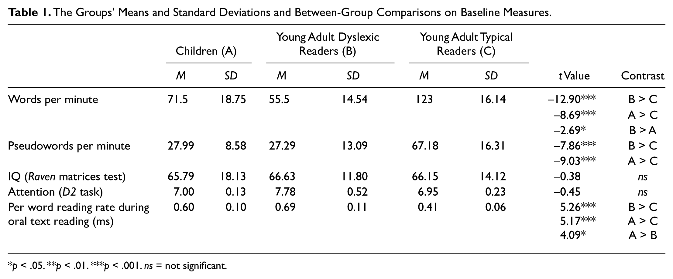
p < .05. **p < .01. ***p < .001. ns = not significant.
All young adult dyslexic readers were recruited through the Student Support Service of the University of Haifa, which assists students with learning disabilities. They were diagnosed as dyslexic during childhood and classified as impaired readers by the Student Support Service. The children were recruited from middle-class secondary schools in the Israeli school system. All participants and their parents provided informed consent prior to inclusion in the study. The young adults were paid volunteers, and the children were compensated with a gift at school. No significant between-group differences were found in nonverbal IQ percentile scores as measured by the Raven Standard Progressive Matrices (Raven, 1960) or in scores on the D2 attention measure (Brickenkamp, 1981; Table 1). All participants were native Hebrew speakers from a middle-class background. All participants were right-handed, displayed normal or corrected-to-normal vision in both eyes, and were screened for normal hearing. None of the participants reported chronic use of medications. Informed consent approved by the University of Haifa ethics committee was obtained prior to each respondent’s participation in the study.
Apparatus
The equipment used in this study included two computer sets. One set was dedicated for the presentation of the LDT stimuli via ePrime software (Psychology Software Tools, Inc.; http://www.pstnet.com). The ePrime application was also used to collect participants’ reaction and reaction times. The second computer set incorporated the fNIRS system (fNIR Devices LLC; http://fnirdevices.com/). The fNIR device was composed of head probe and control box. The flexible head probe held four light sources and 10 light detectors with 2.5 cm light source-detector separation (Figure 1), resulting in 16 different spatial data sources (Channels). The detectors were positioned. The head probe was positioned on the participants’ forehead to image the underlying cortical regions. Data were sampled every 500 ms and streamed into the host computer via the control box. The COBI studio software (Drexel University) was used to store the data and managing data acquisition. The two computer sets were connected with a COM cable to synchronize the two ePrime and fNIRS data sets. MATLAB software (Version 2010a, The Mathworks, Natick, MA) was used for the signal processing and to prepare data for statistical analysis, which was performed using IBM SPSS (Version 19, IBM SPSS Inc., Chicago, IL).
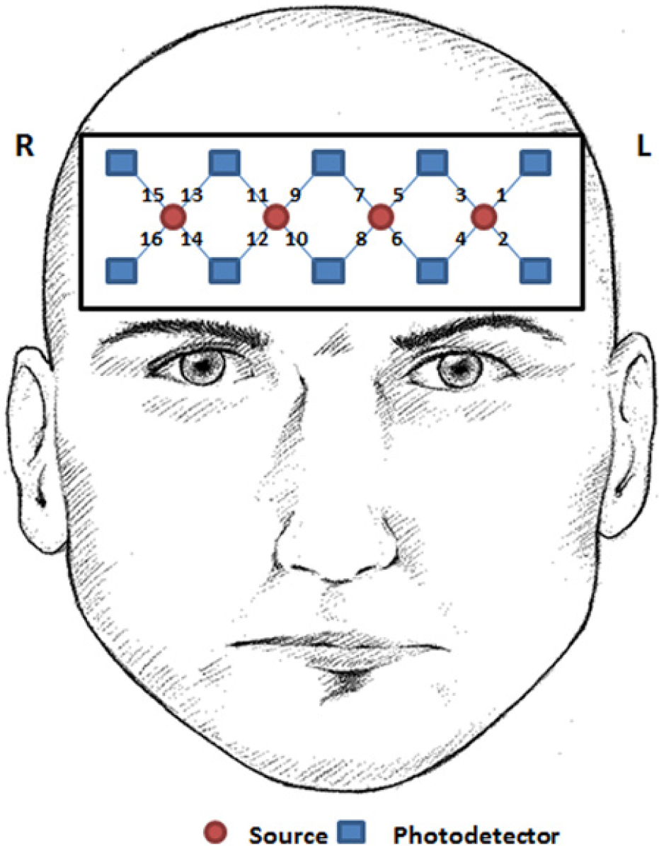
The fNIR Device model 1000.
Task
The LDT (Bergmann & Wimmer, 2008; Binder et al., 2003; Carreiras et al., 2006; Edwards et al., 2005; Fiebach et al., 2002; Fiebach et al., 2007; Heim et al., 2005; Hofmann et al., 2008; Jacobs et al., 2003; Jacobs & Grainger, 1994; Sela et al., 2011) included 96 trials; half (48) were high-frequency words in the Hebrew language (Frost, 2001), and the other 48 trials were pseudowords created from the same letters as the real words. In each trial, the stimulus was presented as white letters on a gray background for 400 ms horizontally in the center of the screen. The participants were asked to sit comfortably in front of a computer screen. They were asked to click with their right hand on 1 if the set of characters represented a word and 2 if a pseudoword. Trials were separated by a 10-s time interval with a jitter of ±4 s to allow sufficient time for the hemodynamic response to fully evolve (M. Izzetoglu, Nioka, Chance, & Onaral, 2005; Miezin, Maccotta, Ollinger, Petersen, & Buckner, 2000). An fNIR data set with a 10-s interval was recorded prior to task initiation and served as a baseline reference for the relative blood oxygenation change computation (M. Izzetoglu, Bunce, Izzetoglu, Onaral, & Pourrezaei, 2007).
Behavioral Data Extractions
The trial’s reaction time was defined as the time between the stimulus onset and the participant’s response. The participants’ reactions and reaction times were extracted from the ePrime output log file. For each participant, the accuracy rate and mean reaction time for words and pseudowords were computed.
fNIRS Data Processing and Feature Extraction
A low-pass filter with a cutoff frequency of 0.14 Hz was used to clean the fNIR data from heart pulsation, respiration and high-frequency noise. The cleaned light intensity signals were converted into relative changes of hemodynamic responses using the modified Beer–Lambert law (M. Izzetoglu et al., 2007). The data conversion resulted in two different signals: the amount of oxygenated (OxyHb) and deoxygenated (DeoxyHb) hemoglobin change. Note that since there is an age difference between the two study groups, an age-dependent correction to the path length factor was integrated in MBLL to accurately extract the hemodynamic signals (Duncan et al., 1999; Quaresima et al., 2002). For each LDT trial, the hemoglobin change data set was segmented from the stimulus onset to 15 s later on. For each of the trials and for each of the two hemodynamic signals, a baseline adjustment was applied by subtracting the mean value of the signal at the 5-s time interval prior to the beginning of the trial. In the final stage of the fNIR data extraction, the mean trial value was extracted for each trial separately. For each of the features, a mean value was computed per individual, Channel (16 voxels; Figure 1), stimulus type (2: word, pseudoword), and reaction accuracy (2: right, wrong responses). Trials with noisy data, mainly caused by movement artifacts, were excluded from the analysis. As the current study focused in the activity within the upper left frontal lobe, and based on previous reports (Hofmann et al., 2008; Sela et al., 2011), the fNIR results presented here are the mean OxyHb measured and calculated based on the data collected from Channel 3 (Figure 1). Because of the ceiling effect obtained in the LDT, only correct reaction trials were used in the statistical analysis.
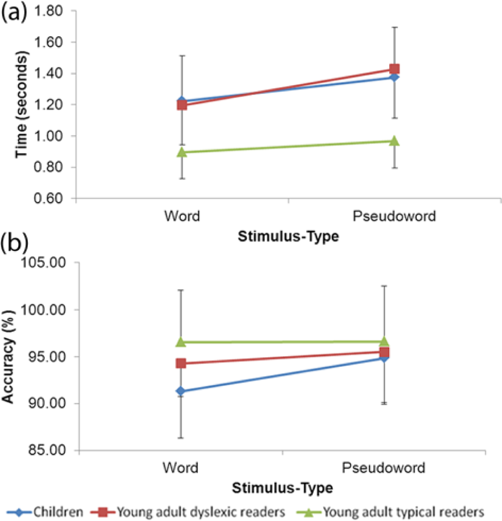
Behavioral results: The mean groups’ (a) reaction time and (b) accuracy, for the children (blue), dyslexic readers (red), and adult typical readers (green).
Statistical Analysis
To test for group (3: children, young adult dyslexic readers, and young adult typical readers) and stimulus type (2: word and pseudoword) effects, a series of 3 × 2 mixed model analysis of variance (ANOVA) tests was applied for each of the current study’s extracted features. In specific cases where two of the study’s groups were compared, 2 × 2 ANOVA tests were applied. Only data extracted from correct response trials were included in the analysis. Greenhouse–Geisser correction for nonsphericity was applied where appropriate. Between-group tests and paired t tests were employed in cases in which significant group or stimulus type effects were obtained to identify the source of difference.
Results
The results of the statistical analysis yielded significant differences between the groups as well as task conditions. Moreover, significant between-group and stimulus type interaction was found in the fNIR data, suggesting that the task conditions affected each of the groups differently.
Behavioral Reaction Time
The mean and standard deviation values of the groups’ reaction times are presented in Figure 2a. The results revealed a significant group effect, F(2, 48) = 15.11, p < .001, as well as significant stimulus type effect, F(1, 48) = 29.939, p < .001, indicating a slower response to the pseudoword as compared to the word. A marginal but nonsignificant group by stimulus type interaction was found, F(2, 48) = 2.969, p = .06. As Figure 2a clearly shows, the adult typical readers were significantly faster in their responses as compared with the group of children and the dyslexic readers group, F(1, 32) = 25.272, p < .001 and F(1, 32) = 27.003, p < .001, respectively. Both latter groups demonstrated a relatively equal speed of response, F(1, 32) = 0.073, p = .789.
Behavioral Accuracy
The analysis of the three reading groups (Figure 2b) revealed significant main effects for both group, F(2, 48) = 3.865, p < .05, and stimulus type, F(1, 48) = 4.861, p < .05. No group by stimulus type interaction was found, F(2, 48) = 2.091, p = .135. The adult typical readers showed the highest accuracy value (nearly 97% under both stimulus type conditions), the dyslexic readers were accurate in 94.9% of the task trials, and the children were accurate in around 92% of the trials. In-depth examination revealed that the children showed a significant difference between the two stimulus types, where their accuracy level under the pseudoword condition was higher than in words, t(16) = −2.285, p < .05). A significant group difference was found under the word condition between the children and the adult typical readers, t(32) = −3.294, p < .01, where the accuracy for the adult typical readers was higher.
Channel 3: Mean OxyHb
The time courses of the OxyHb response in Channel 3 (upper left frontal lobe, Figure 1) for word and pseudoword are presented in Figure 3a and 3b, respectively. Examining the graphs revealed that the young adult typical readers responded for both conditions with an increase in the value of OxyHb. The increase in OxyHb started around 4 s following stimulus presentation and reached the maximum after 8.5 s. It is also evident from the graphs that the group of dyslexic readers tended to respond in a similar manner as compared to the young adult typical readers under the word condition (Figure 3a). However, a significant lower response was observed for the group of dyslexic readers under pseudoword as compared to the young adult typical readers (Figure 3b). Moreover, although the group of children followed a similar time course in hemodynamic response as compared to the young adult typical readers for pseudoword (Figure 3b), their hemodynamic response for word was significantly lower (Figure 3a).
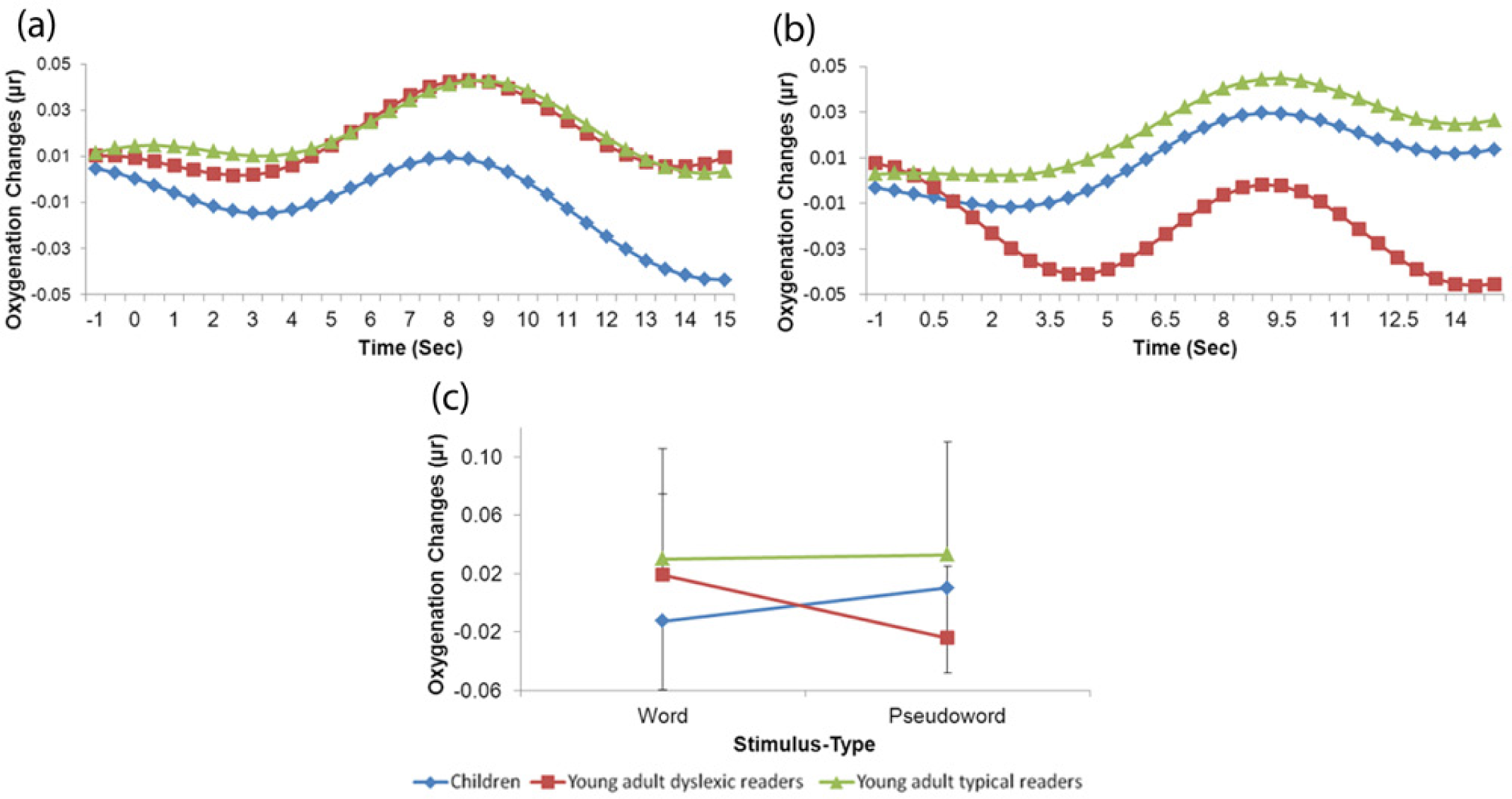
fNIR results: The groups’ mean OxyHb change time-course as a response for (a) word and (b) pseudoword stimulus types as well as (c) the groups’ mean OxyHb changes measured in Channel 3 for the children (blue), dyslexic readers (red), and adult typical readers (green).
The analysis of the mean value of the OxyHb changes in Channel 3 revealed a significant group effect, F(2, 48) = 3.832, p < .05, with no significant stimulus type effect, F(1, 48) = 0.0, p = .845, and a nearly significant group by stimulus type interaction, F(2, 48) = 3.14, p = .052. However, as can be seen in Figure 3c, the adult typical readers demonstrated a similar value of mean OxyHb changes under both stimulus type conditions (~0.03 µr), higher than the children and higher than the dyslexic readers. Moreover, although the dyslexic readers did not differ from the adult typical readers in their mean OxyHb changes under the word condition, t(32) = −0.482, p = .633, there was a significant difference between the adult typical readers and the dyslexic readers under pseudowords, t(32) = −2.564, p = .015. In contrast, when the group of children was compared to the adult typical readers, a significant difference was found under the word condition, t(32) = −2.392, p < .05, but not under the pseudowords, t(32) = −0.833, p = .411. The comparison between the children and the dyslexic readers indicated that both groups did not differ in the overall mean OxyHb changes, F(1, 32) = 0.004, p = .949. However, there was a significant group by stimulus type interaction, F(1, 32) = 7.688, p < .01. Thus, the children and the dyslexic readers were affected differently by the word and pseudoword conditions. Results indicated that the children obtained higher mean OxyHb in the pseudoword condition and the adult dyslexic readers obtained higher mean OxyHb during the word condition.
In sum, the results of the current study indicated that the young adult typical readers showed an ability to perform the LDT faster and more accurately than both the children and the young adult dyslexic readers. In addition, they showed higher values of mean OxyHb changes in Channel 3, which, as has been suggested, reflects higher activity in the sampled voxel. The children and the young adult dyslexic readers demonstrated a significant interaction in the mean OxyHb changes in Channel 3 where the dyslexic readers had lower mean OxyHb changes under the pseudoword condition as opposed to the children who showed lower mean OxyHb changes under the word condition.
Discussion
Overall, the results of the current study support the hypothesis that the group of children would show lower activity in the upper left frontal lobe under the word condition as compared to young adult typical readers and that the young adult dyslexic readers would show lower upper left frontal lobe activity under the pseudoword condition as compared to their young typical reader peers. Thus, the results support the notion that the upper left frontal lobe is involved in LDT and might represent a developmental trend as well as support the phonological deficit hypothesis as an explanation of the dyslexic phenomenon even among adult dyslexic readers who have been exposed for years to printed materials and remedial reading.
According to a widely accepted definition of developmental dyslexia, a dyslexic reader is one who exhibits slow and inaccurate reading performance unrelated to his or her IQ level or educational opportunities (British Psychological Society, 1999; Shaywitz & Shaywitz, 2008). The reading deficits of developmental dyslexia persist into adulthood (Bruck, 1992; Leonard et al., 2001). A large number of studies have shown deficient phonological processing as a core deficit in developmental dyslexia. The leading theory, the phonological deficit theory of developmental dyslexia (Share, 1994; Snowling, 1995; Stanovich, 1988), suggests that dyslexic readers may suffer from an (unspecified) dysfunction in peri-sylvian brain regions, which leads to difficulties in generating and processing accurate and efficient phonological representations of speech sounds (Stanovich, 1988; Temple et al., 2001).
When considering the behavioral results, our data stand in line with those of previous studies that found that dyslexic readers are significantly slower in the performance of the LDT (Breznitz & Misra, 2003). However, it was also demonstrated that the dyslexic readers’ tendency to accurately perform the task is associated with their language orthography. Based on a growing line of studies (Bergmann & Wimmer, 2008; Jimenez Gonzalez & Hernandez Valle, 2000; Share, 1994; van den Bos, 1998; Wimmer & Schurz, 2010; Zoccolotti et al., 1999) it is expected that in deep orthographies (i.e., English or unpointed Hebrew), the dyslexic readers would demonstrate a slow reading speed together with relatively inaccurate reading. The stimuli in the current study were presented in an unpointed (without diacritics) Hebrew script, which is considered to be deep orthography (Share, 2008; Ziegler & Goswami, 2005). In that manner, our data only partly succeeded to replicate this trend of results, as although, compared to the young adult typical readers, the dyslexic readers were significantly slower in their response for both stimulus types, their accuracy did not significantly fall back. Two plausible explanations may be suggested. First, although the unpointed Hebrew script is more complex to decode as compared to a pointed script (Ziegler & Goswami, 2005), it may be that the level of decoding complexity is not that high when the task is to read a single word, such as in LDT. The number of degrees of freedom in the processing of grapheme to phoneme correspondence is higher when the task involves a context, such as in forming a sentence. Second, the dyslexic individuals that took part in this study are considered to be compensated dyslexic individuals studying at the university, and as such they may show higher reading ability, as compared to noncompensated dyslexic readers. Their relative advantage may be reflected by an almost intact ability to correctly decode the stimuli, specifically when it was represented in a word list. Nevertheless, extra time, as exhibited by slow reaction time, was needed by the young adult dyslexic to decide whether the stimulus was a word or a pseudoword. In fact, their reaction time was as slow as the children’s (Figure 2a) under both conditions of word and pseudoword. It may be assumed that the adult dyslexic readers exhibited a speed–accuracy trade-off that is not affected at the word level but, as studies have indicated, affected their level of reading fluency and comprehension at a text level. The time factor, or speed of processing, was previously suggested to be one of the deficits characterizing dyslexia (Breznitz, 1997, 2008; Breznitz & Misra, 2003). Thus, in terms of reaction time, our results support previous studies indicated that the dyslexic individuals suffered from slow speed of processing and dysfluent reading.
Previous studies have suggested the involvement of the left frontal lobe in language related tasks (Richards et al., 2000; Richards et al., 2002). Specifically, the upper left frontal lobe has been suggested to be involved in the decision making in LDT (Binder et al., 2003; Fiebach et al., 2007; Grainger & Jacobs, 1996; Hofmann et al., 2008; Sela et al., 2011). For example, an fMRI study on LDT (Binder et al., 2003) indicated a task related activity in the left superior and middle frontal sulci and the left superior and middle frontal gyri. The upper left frontal lobe has neuronal connections with parietal, temporal, and occipito-temporal brain regions, which were suggested to be involved in semantic information processing. A recent fNIR study (Hofmann et al., 2008) specifically pointed to the superior frontal gyrus as a brain region involved in decision making. However, according to their results, higher hemodynamic response was expected for the word condition as compared to the pseudoword. Examining the data of each of the results within each of the current study groups separately suggests different directions. The analysis did not reveal a significant stimulus type effect within each group. However, the results obtained showed that activity in the upper left frontal lobe among the children was significantly lower as compared to the young typical adult readers under the Word condition. As opposed to this, the activity in the upper left frontal lobe of the dyslexic readers was significantly lower under the pseudoword condition. It can be suggested that the current study results based on fNIR measurements support the DRM (Coltheart et al., 2001) from two different directions. First, according to the DRM, the phonological route is more involved in the beginning of the process of reading acquisition (Ziegler & Goswami, 2005). The orthographic route, on the other hand, needs more printed exposure to develop (Ehri, 2002; Share, 1995). Although the group of children who took part in the current study were all typical nondisabled readers at the beginning of secondary school where their reading performance was within the normal range, it seems that for them, the process of distinguishing between words and pseudowords has not yet fully automatized and requires more mental effort than for adult typical mature readers. This can be a result of the fact that these young readers were trained for only about 2 years with nonpointed Hebrew and need a longer training period to create word patterns in their mental lexicon. Phonological processing is still required for LDT to occur. The second support for the DRM is the dyslexic readers’ relatively lower activity documented under the Pseudoword condition. As the processing of a Pseudoword is associated with the phonological route, it could be suggested that as compared to the adult typical readers, poor phonological information processing resulted in the exhibited reduced activation of the dyslexic readers’ upper left frontal lobe. These results may support the fact that the phonological deficits exist even among adult compensated dyslexics and in a language with more shallow orthography than English. It is possible that in a language with more shallow orthography difference in results will be more pronounced in brain measures than in behavioral measures as the brain measures give real-time information regarding the processing itself, not only at the end of the process (Bentin, 1989).
The between-group differences measured in Channel 3 provide additional evidence for the involvement of the left frontal lobe in LDT. However, since the spatial sensitivity of fNIR is rather limited as compared to fMRI, and since there can be individual differences in brain anatomy, the estimation of the exact anatomic location of the cortical measurement area for Channel 3 should be handled with caution. Voxel localization is an important issue when the current study’s results are attempted to be compared to other fNIR or fMRI studies. It is even more important in studies that aim to compare fNIR results of children to adults where there are inherited natural anatomy differences between different age groups, specifically, in head size. In fact, for this purpose, the computation of the hemodynamic signals in the current study took into account the participants’ age and used it as a factor for a light path-length correction (Duncan et al., 1999; Quaresima et al., 2002). In a recent fNIR study (Hofmann et al., 2008), the superior frontal gyrus and the inferior frontal gyrus were suggested to be involved in the performance of LDT. However, the researchers included in their region of interest definition for the superior frontal gyrus measurement areas that are presumably located in the middle frontal gyrus (Hofmann et al., 2008). Moreover, in a former study carried out for the localization of the fNIR device 1000 recordings (Ayaz et al., 2006), the measurement area corresponding to Channel 3 was estimated to be located in the left middle frontal gyrus. Thus, based on these studies (Ayaz et al., 2006; Hofmann et al., 2008), the data collected in the current study can be interpreted as evidence that the middle frontal gyrus is involved in the performance of LDT, and it is the activity within the middle frontal gyrus that differs among the three reading-level groups.
As a final point, the fNIR results of the current study provide support for the relevance of the fNIR as a neuroimaging device to be employed in brain research. The technique enables the researchers to measure the hemodynamic responses and the oxy- and deoxyhemoglobin separately and noninvasively in a portable and affordable way. However, as discussed above, the results also indicate that one should be aware of its limitations before interpreting the data. Thus, in addition to its spatial limitation, the technique does not allow one to collect data in brain regions that are deeper than the surface of the cortex. Moreover, although it is clear that the ability to read and specifically the ability to perform the LDT involves a neural network that includes brain regions that are located outside of the frontal lobe, the fNIR device that was used in the current study was designed to investigate the frontal lobe only, and as such the concurrent activity in other brain regions remains to be studied.
Footnotes
Declaration of Conflicting Interests
The author(s) declared no potential conflicts of interest with respect to the research, authorship, and/or publication of this article.
Funding
The author(s) disclosed receipt of the following financial support for the research, authorship, and/or publication of this article: This research was funded by the Edmond J. Safra Philanthropic Foundation.