






Abstract
Objective
This study aimed to develop and validate a sensitive analytical method for detecting and quantifying AB-CHMINACA, and to comprehensively evaluate its pharmacokinetics, metabolic pathways, and acute toxicity in rodent models.
Methodology
Male Wistar rats and Swiss albino mice were used for pharmacokinetics, metabolism, and acute toxicity studies. AB-CHMINACA was dissolved in Dimethyl sulfoxide (DMSO; 2%–5%) and diluted in corn oil for oral gavage. Blood and urine samples were collected from rats for pharmacokinetic and metabolite analyses using Gas Chromatography–Tandem Mass Spectrometry (GC-MS/MS) and liquid chromatography–quadrupole time-of-flight mass spectrometry, respectively. Mice were monitored for behavioral toxicity and necropsied for histopathological analysis. Pharmacokinetic parameters were calculated using WinNonlin Phoenix 6.0, and metabolites were identified through mass spectrometry.
Results
Method validation showed excellent linearity (0.5–1000 ng/mL, r2 > .99), with precision (variability 2.3%–7.2%) and accuracy (95%–105%), adhering to FDA guidelines. The method’s limit of detection (LOD) and limit of quantification (LOQ) were 0.5 and 1.0 ng/mL, respectively. Pharmacokinetic analysis revealed a biphasic elimination (t½ 13.8 h), Vd of 228.75 L, and Cl of 11.4 L/h. Four major Phase I metabolites were identified, with Met 4 (Di-Hydroxy-AB-CHMINACA) being the most abundant in urine at 8–24 h. Acute toxicity in mice showed dose-dependent symptoms, including lethargy, seizures, and mortality at 50 mg/kg.
Conclusion
These results enhance the understanding of AB-CHMINACA's pharmacokinetic behavior and metabolic profile, contributing valuable insights to forensic toxicology and highlighting the dose-dependent nature of its acute toxicity.
Introduction
The increasing popularity of synthetic cannabinoids (SCs) globally presents significant public health challenges due to their rapidly evolving chemical structures. 1 SCs form the largest category of novel psychoactive substances (NPS) to emerge in the past decade and are often marketed as “legal” alternatives to cannabis. 2 These compounds interact with cannabinoid receptors CB1 and/or CB2 and were initially developed to explore the endogenous cannabinoid system or as potential pharmacological treatments. 3
SC synthesis began in the mid-1960s, but recreational use did not gain traction until the early 2000s. 4 Most of the abused SCs act as CB1 receptor agonists, exhibiting significantly higher binding affinity than delta-9-tetrahydrocannabinol (THC). As a result, they produce more intense cannabimimetic effects, including pronounced cognitive impairment, altered sensory perception, and transient hallucinations. Additionally, SC use has been linked to adverse physiological effects not commonly associated with cannabis, such as seizures, vomiting, hyperglycemia, hypokalemia, stroke, myocardial infarction, and acute kidney injury. 5 While controlled SC administration studies would be valuable, conducting such research in humans is currently impractical due to the absence of sufficient preclinical toxicological data.5–7
Reports from the American Association of Poison Control Centers 8 show a rise in SC-related exposures, from 2096 in 2010 to 6968 in 2011, followed by a decline to 5230 in 2012 and further down to 2668 in 2013. The decrease in 2013 was likely due to the classification of SCs as Schedule I substances, increased public awareness, and better management by physicians. The cases rose again to 3677 in 2014. SCs are now regulated under the U.S. Controlled Substances Act, 9 banned by WADA, 10 and controlled in many countries. 2
AB-CHMINACA, one of the potent SCs, has demonstrated efficacy far exceeding that of known full agonists of the CB1 receptor. 11 Initially developed by Pfizer for potential therapeutic application, its development was discontinued in 2013 due to adverse effects. 12 Studies indicate that AB-CHMINACA's potency is 11‒58 times greater than THC across various tests, 1 raising concerns over its potential for abuse and toxicity.
Despite the classification of SCs as Schedule I substances and growing public awareness of their risks, the toxic dose of SCs, including AB-CHMINACA, remains undetermined in humans. Animal studies have not yet provided sufficient data regarding their toxicity. With AB-CHMINACA's pharmacological profile largely unexplored, there is a pressing need for studies that evaluate its pharmacokinetics, metabolism, and toxicological effects.
This study seeks to address this knowledge gap by examining the pharmacokinetic behavior, half-life, volume of distribution, and clearance of AB-CHMINACA in rat blood samples using GC-MS/MS. The data generated will help elucidate the compound's pharmacological and toxicological properties, contributing valuable information for forensic toxicology and public health authorities.
Materials and methods
Chemicals and reagents
AB-CHMINACA (≥98% purity) was obtained from Cerilliant Corporation and Cayman Chemical (USA). HPLC-grade solvents—methanol, water, ammonium formate, and formic acid were purchased from Sigma-Aldrich. Dimethyl sulfoxide (DMSO) was sourced from Biotraxx (Cyprus), and corn oil was purchased from a local supplier (Saudi Arabia). Ultrapure water was prepared using a Milli-Q purification system (Millipore, France).
Instrumentation
GC-CI-MS/MS
Gas chromatography–chemical ionization tandem mass spectrometry (GC-CI-MS/MS) was performed on an Agilent 7890B GC system coupled to a 7000C triple quadrupole mass spectrometer, equipped with a 7693 autosampler and an HP-5MS capillary column (30 m × 0.25 mm, 0.25 µm film). A 2 µL injection was introduced in splitless mode. The oven temperature program was: initial 200 °C (2 min), ramped at 15 °C/min to a final temperature of 300 °C, held to complete a total run time of 12 min. Helium served as the carrier gas at 1 mL/min. The injector and transfer line were maintained at 280 °C. The system operated in multiple reaction monitoring (MRM) mode with ammonia as the reagent gas and argon at 1.5 mL/min as the collision gas.
LC-ESI-QTOF/MS
Liquid chromatography–electrospray ionization quadrupole time-of-flight mass spectrometry (LC-ESI-QTOF/MS) was conducted using an Agilent 6530B QTOF system (Agilent Technologies, Santa Clara, CA, USA) with a TurboIonSpray source in positive mode (ESI+, 2500 V). Parameters were: curtain gas at 30 psi; ion source gases 1 and 2 at 60 psi; source temperature at 600 °C. The TOF scan range was 100–650 Da (acquisition time 0.07 s), and the MS/MS scan range was 25–650 Da (0.05 s). The declustering potential was 80 V; collision energy was set at 10 V.
Chromatographic separation was achieved using an Agilent ZORBAX Eclipse Plus C18 reverse-phase column (2.1 × 100 mm, 1.8 µm particle size), maintained at 30 °C with a flow rate of 0.7 mL/min. The autosampler was maintained at 5 °C. Mobile phase A was 10 mM ammonium formate in water, and mobile phase B was 0.1% formic acid in methanol. The gradient program: 10% B (0.30 min) → linear increase to 98% B (7.30 min), held until 8.80 min, then re-equilibrated to 10% B at 8.81 min; total runtime: 11.0 min.
Standard and quality control (QC) sample preparation
Stock solutions
A primary stock solution of AB-CHMINACA (1 mg/mL) was prepared in methanol. Serial dilutions yielded secondary working solutions of 100, 10, and 1 µg/mL, and 100 and 10 ng/mL. Granisetron was used as the internal standard (IS) at 10 µg/mL in methanol.
Calibration standards and QC samples
Blank human plasma was fortified with AB-CHMINACA and 50 µL of IS to prepare calibration standards at 0.5, 2, 10, 25, 50, 100, 500, and 1000 ng/mL. A blank with only IS was included. QC samples were also prepared in plasma at the following concentrations:
Low QC (LQC): 50 ng/mL Medium QC (MQC): 250 ng/mL High QC (HQC): 500 ng/mL
Each QC sample included 50 µL of IS. Details of calibration and QC preparations are provided in Table 1.
Preparation of calibration standards for drug quantification in plasma.

Human plasma was selected as the blank matrix for method validation owing to its comparable protein composition, viscosity, and pH to rodent plasma, ensuring similar extraction efficiency and analyte recovery. This matrix is also widely standardized and readily available, facilitating reliable method reproducibility. The validated method was subsequently applied to rodent plasma samples to confirm matrix compatibility.
Animals and dosing
Subjects
Male Wistar rats (12 weeks, 150–160 g) and Swiss albino mice (6 weeks, 20–25 g) were used, with n = 6 per group. Animals were housed under controlled conditions (23 ± 2 °C, 50–60% relative humidity, 12 h light/dark cycle) with free access to food and water and acclimatized for 7 days.
Drug administration
AB-CHMINACA was initially dissolved in 2%–5% DMSO and further diluted in corn oil for oral gavage. Control animals received vehicle only (DMSO in corn oil).
Experimental design
The study evaluated the pharmacokinetics, metabolism, urinary excretion, and acute toxicity of AB-CHMINACA following oral administration in rodents. Seven experimental groups were established, each comprising six animals (n = 6) (Table 2).
Male Wistar rats were assigned to vehicle control and treatment groups receiving AB-CHMINACA at 50 or 100 mg/kg for pharmacokinetic and metabolic profiling. A separate rat cohort received the same doses and was housed in metabolic cages for urine collection at 8 and 24 h post-dose. Rats were monitored for clinical signs throughout the study.
Swiss albino mice were used for acute toxicity assessment and were divided into vehicle control and treatment groups (50 and 100 mg/kg). Clinical and behavioral observations were recorded at 1, 2, 3, 5, and 24 h after dosing. At study termination, animals underwent necropsy, and liver and kidney tissues were collected for histopathological analysis.
Experimental design for pharmacokinetic, metabolism, urinary excretion, and acute toxicity evaluation of AB-CHMINACA in rodents.

Note: Each treatment group consisted of six animals (n = 6).
Sample collection and preparation
Blood and urine
Blood samples were collected from rats at 0.5, 1, 2, 4, 6, 8, and 24 h post-dose. Plasma was separated and stored at −20 °C. Urine was collected pre-dose and over 8–24 h in metabolic cages.
Extraction procedures
Plasma: Liquid-liquid extraction with 1-chlorobutane, drying under nitrogen, reconstitution for GC-MS/MS analysis.
Urine: 100 µL urine mixed with 900 µL methanol:acetonitrile (1:1), vortexed 1 min, sonicated 3 min, centrifuged at 8000 r/min for 5 min. Supernatant dried under nitrogen, reconstituted in 500 µL water:methanol (80:20), vortexed, centrifuged, and 10 µL injected for analysis.
Method Validation
The analytical method was validated according to standard bioanalytical guidelines to ensure reliability and reproducibility.
Selectivity: Six different sources of drug-free human plasma were analyzed to confirm the absence of interfering peaks at retention times and m/z values corresponding to AB-CHMINACA and the internal standard (IS). Sensitivity: The limit of detection (LOD) and lower limit of quantification (LLOQ) were determined based on signal-to-noise ratios of ≥3 and ≥10, respectively, by analyzing low-concentration samples in triplicate. Linearity: Calibration curves were constructed using spiked plasma samples across a concentration range of 0.5‒1000 ng/mL. Each concentration was analyzed in duplicate, with linear regression (1/x weighting) applied. The correlation coefficient (R2) was required to be ≥ .99. Accuracy and Precision: Quality control samples at low (LQC), medium (MQC), and high (HQC) concentrations were analyzed in triplicate over multiple days. Accuracy was assessed as percent relative error, and precision by relative standard deviation (RSD), both required to be within ±15% for intra- and inter-day analyses.
Toxicity assessment
Behavioral toxicity assessment
Mice were monitored for clinical signs of toxicity including hyperactivity, sedation, convulsions, tremors, and respiratory distress. Observations were conducted at 1, 2-, 3-, 5, and 24-h post-administration.
Clinical monitoring and necropsy
Body weight and clinical status were recorded. Necropsies were conducted on all treated animals to assess gross pathological changes.
Data analysis
Pharmacokinetics
Pharmacokinetic parameters (Cmax, Tmax, t½, AUC, clearance) were calculated using non-compartmental analysis with Phoenix WinNonlin version 6.0.
Metabolite identification and quantification
Metabolites of AB-CHMINACA were characterized based on accurate mass shifts, retention times, and MS/MS fragmentation patterns using Mass Hunter software. Diagnostic ions and neutral losses characteristic of hydroxylation (+16 Da), ketone formation (–2 Da), and multiple hydroxylations were used to elucidate structural modifications. For instance, Met 1 (m/z 387.239) displayed a fragment ion at m/z 342 (loss of −45 Da), consistent with the elimination of a –CH₃CO group. Each proposed metabolite structure was further supported by theoretical isotope distribution and matched empirical fragmentation spectra.
Authentic reference standards for individual AB-CHMINACA metabolites were not commercially available at the time of analysis. Consequently, metabolite quantification was performed using a semi-quantitative approach. A validated calibration curve for the parent compound (AB-CHMINACA) was established in human plasma over the concentration range of 0.5–1000 ng/mL (r2 > .99). This calibration was subsequently applied as a reference for relative quantification of the detected metabolites.
For each metabolite, peak-area ratios (metabolite/internal standard) were compared to the corresponding AB-CHMINACA calibration response, and results were expressed as relative abundance (%) with respect to the parent analyte. This semi-quantitative strategy, commonly adopted when authentic metabolite standards are unavailable, enabled reliable inter-sample comparison and interpretation of metabolic profiles.
Results
Method validation results
The method validation for AB-CHMINACA analysis was carried out to ensure its robustness and reliability. The calibration curve for AB-CHMINACA was established over a linear range of 0.5–1000 ng/mL, with an r2 value greater than .99, demonstrating excellent linearity. The precision of the method was evaluated both within-day and between-day. Within-day precision ranged from 2.3% to 6.5%, while between-day precision was between 3.1% and 7.2%. The accuracy of the method was found to be 95%‒105% across the tested concentration range, adhering to FDA acceptance criteria. The detection limits were also evaluated, with a LOD of 0.5 ng/mL and a LOQ of 1.0 ng/mL, indicating the method's sensitivity for detecting low concentrations of AB-CHMINACA.
Pharmacokinetics of AB-CHMINACA
The pharmacokinetic profile of AB-CHMINACA was assessed in plasma using GC-CI-MS/MS. The plasma concentration–time curve showed a biphasic elimination pattern, with a rapid distribution phase followed by a slower elimination phase. Key pharmacokinetic parameters included a half-life (t½) of 13.8 h, indicating a relatively prolonged elimination. The volume of distribution (Vd) was 228.75 L, and plasma clearance (Cl) was 11.4 L/h, suggesting moderate distribution and clearance. AB-CHMINACA had a retention time of 11.1 min, while the internal standard eluted at 10.1 min, allowing for clear differentiation between the two compounds. The precursor ion for AB-CHMINACA was observed at m/z 340, and for the internal standard at m/z 313, both providing reliable identification during analysis.
Metabolite identification and profiling
AB-CHMINACA metabolites were identified using LC-Q/TOF-MS, confirming four major Phase I metabolites primarily formed through hydroxylation and ketone formation (Table 3). These included:
Met 1 (Hydroxy-AB-CHMINACA): Precursor ion at m/z 373.48; fragment ions at m/z 95, 239, 257, and 342. Met 2 (Ketone-AB-CHMINACA): Precursor ion at m/z 370.47; fragment ions at m/z 95, 145, 239, 255, 326, and 340. Met 3 (Di-Hydroxy-AB-CHMINACA): Precursor ion at m/z 387.47;fragment ions at m/z 95, 239, 255, 271, and 356 (Figure 1). Met 4 (Hydroxy-Ketone-AB-CHMINACA): Precursor ion at m/z 390.47; fragment ions at m/z 93, 237, 257, 273, 291, 340, and 358 (Figure 2).
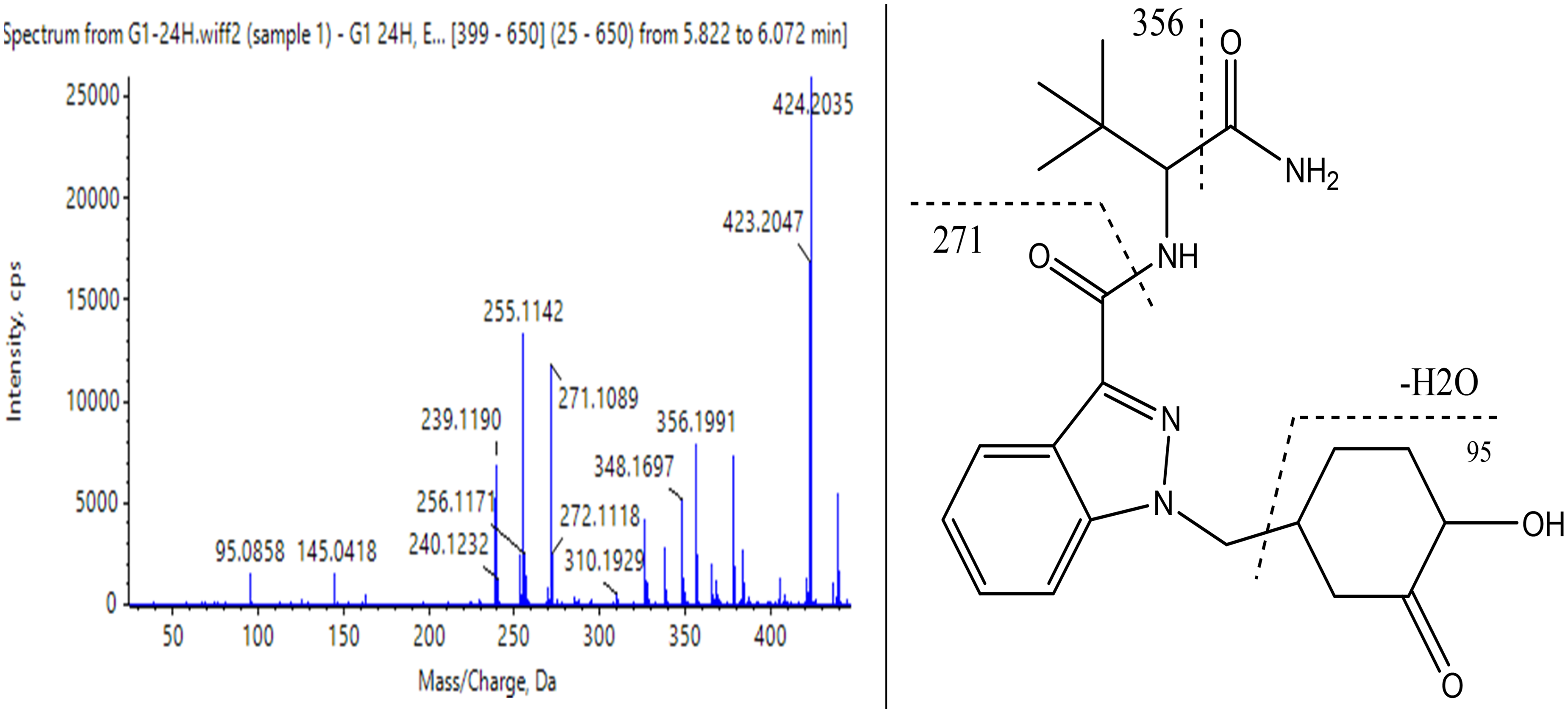
Metabolite product Met 4 Hydroxylation + Ketone formation (cyclohexylmethyl) AB-CHMINACA, proposed fragmentation mechanism and mass spectrum upon ionization (ESI) at 25 MeV.

Metabolite Met 3 Di-Hydroxy AB-CHMINACA, proposed fragmentation mechanism, and mass spectrum upon ionization (ESI) at 25 MeV.
Assumed main metabolites of the study compound, molecular structure, molecular weight of the compounds and their recorded ions, and percentage mass error.

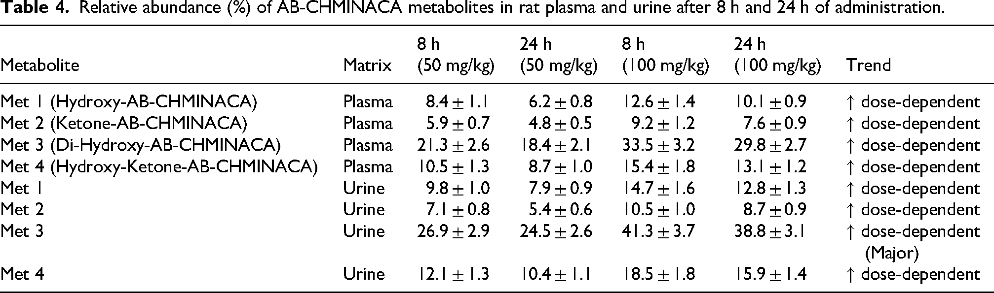
All four metabolites (Met 1–Met 4) were detected in urine samples collected at 8 and 24 h post-administration in both low- and high-dose groups (Table 4). The data revealed a dose-dependent increase in metabolite formation across both blood and urine matrices, with Met 3 consistently exhibiting the highest abundance, followed by Met 4, Met 1, and Met 2. The relative abundance (%) of each metabolite at both time points further confirmed enhanced metabolic activity at higher doses. The proposed metabolic pathways and sequence of metabolite formation are illustrated in Figure 3, highlighting the biotransformation profile of AB-CHMINACA.

Proposed metabolic reactions to the drug AB-CHMINACA in urine samples from rats, and the possible conformation sequence of the metabolites.
Relative abundance (%) of AB-CHMINACA metabolites in rat plasma and urine after 8 h and 24 h of administration.
Effects of AB-CHMINACA at Low, Medium, and High Doses on Experimental Mice Over 24 H Compared With the Control Group.

Comparison between the current study's results and previous studies on the metabolism of some synthetic cannabinoids.

Acute toxicity symptoms
Administration of AB-CHMINACA at doses of 5, 50, and 100 mg/kg body weight produced a clear dose-dependent alteration in behavioral responses among mice (Table 5). At the lowest dose (5 mg/kg), animals exhibited mild hyperactivity shortly after administration, which gradually transitioned to relative lethargy and light sensitivity within 1 h. Complete lethargy was observed by 6 h, followed by full recovery and normal behavior within 24 h.
At 50 mg/kg, the onset of behavioral changes was faster and more pronounced. Mice showed hyperactivity, piloerection, and reduced movement within 15 min, progressing to marked lethargy and partial unresponsiveness between 3 and 6 h. Most mice recovered by 24 h, although mild fatigue persisted.
In contrast, the highest dose (100 mg/kg) elicited severe and sustained toxic effects. Mice demonstrated intense hyperactivity, tremors, and convulsions initially, followed by complete lethargy, loss of appetite, and deep coma in several cases within 6 h. Only four of six animals recovered movement by 24 h, indicating 33% mortality.
Overall, the findings indicate a dose-dependent increase in both the intensity and duration of toxic behavioral effects, with delayed recovery and higher mortality at elevated AB-CHMINACA doses.
Discussion
Method validation
The method validation was performed exclusively for AB-CHMINACA to ensure analytical robustness, reliability, and suitability for forensic and clinical applications. The calibration curve for AB-CHMINACA demonstrated excellent linearity across the range of 0.5–1000 ng/mL, with an r2 > .99. The method exhibited strong precision, with within-day values ranging from 2.3% to 6.5% and between-day precision from 3.1% to 7.2%. Accuracy was maintained between 95% and 105% across all tested concentrations, meeting FDA acceptance criteria.
The method's LOD and LOQ were determined as 0.5 and 1.0 ng/mL, respectively, confirming its high sensitivity for detecting trace levels of AB-CHMINACA. As noted, no separate validation was conducted for metabolites; thus, metabolite data are presented qualitatively to support metabolic interpretation rather than quantitative assessment.
Pharmacokinetics of AB-CHMINACA in rats
This study aimed to characterize the pharmacokinetics of the SC, AB-CHMINACA, in rats. The plasma half-life (t1/2) was found to be 13.8 h, differing from the 7–12 h range reported by Kevin et al. 13 for intraperitoneally administered CUMYL-PICA and 5F-CUMYL-PICA. This discrepancy likely results from differences in administration routes. Since drug delivery methods influence pharmacokinetics, future studies should compare vaporized versus injected administration and their impact on plasma profiles.
The volume of distribution (Vd) was calculated at 228.75 L, reflecting the highly lipophilic nature of SCs. These compounds exhibit rapid distribution into lipid-rich tissues, particularly adipose tissue, leading to an early decline in blood concentrations and prolonged detection windows following chronic use. 14 Previous interpretations attributing this pattern specifically to AB-CHMINACA sequestration have been updated to reflect current evidence. While AB-CHMINACA and related analogs display strong lipophilicity and extensive tissue distribution, direct quantitative data confirming adipose sequestration remain limited. Studies on structurally related SCs, including 5F-AMB, NNEI, and MN-18, support the hypothesis of preferential partitioning into fat due to their physicochemical properties and high logP values. 15 This behavior likely contributes to delayed elimination and variable post-mortem redistribution patterns observed in this case. Additionally, blood protein binding may further influence elimination kinetics, although this parameter was not assessed in the present study.
Plasma clearance (CL) was 11.43 L/h, indicating relatively simple linear kinetics at the doses tested. This elimination rate appears faster than for other SCs. Studies examining various administration routes (intraperitoneal, intravenous, pulmonary) report faster peak concentrations and higher systemic exposure.13,16,17
Drug elimination primarily depends on plasma volume passing through filtration organs, facilitating removal via urine, bile, dialysate, or exhaled gases. If significant drug amounts are sequestered in tissues or bound to plasma proteins, clearance from plasma slows. This relationship underscores the importance of volume of distribution in pharmacokinetics.
Metabolite profiling
Metabolite profiling using LC-Q/TOF-MS confirmed four major Phase I metabolites of AB-CHMINACA, mainly formed through hydroxylation and ketone formation: Met 1 (Hydroxy-AB-CHMINACA), Met 2 (Ketone-AB-CHMINACA), Met 4 (Di-Hydroxy-AB-CHMINACA), and Met 3 (Hydroxy-Ketone-AB-CHMINACA). All four were detected in urine at 8 and 24 h post-administration in both low- and high-dose groups. Met 3 was the most abundant, followed by Met 4, Met 1, and Met 2. Metabolite concentrations increased with dose, demonstrating dose-dependent metabolism. Similar metabolic interactions with comparable functional groups were reported in related SC compounds (Table 6).
Previous human hepatocyte incubation studies predicted these metabolites in vivo. 18 Confirmation using authentic human urine samples would strengthen these findings but was unavailable.
Compared to Erratico et al. 19 who identified M21 (a carboxylic acid metabolite formed via amidase hydrolysis) as the major Phase I metabolite, this study found hydroxylation and ketone formation as dominant pathways. Amidase enzymes selectively cleave the outer amide group, leading to M21 formation. CYP3A4 likely mediates hydroxylation at the methyl-cyclohexyl moiety, consistent with this study's findings, where Met 4 (Di-Hydroxy-AB-CHMINACA) was the major hydroxylated metabolite. These patterns align with metabolic profiles of similar SCs like MDMB-CHMICA. 20
Acute toxicity
AB-CHMINACA caused dose-dependent acute toxicity in rats, including lethargy, uncoordinated movements, excessive salivation, seizures, coma, hypothermia, and mortality at higher doses. These effects correspond with known toxic profiles of SCs, which include cardiovascular events (myocardial infarction, ischemic stroke), acute kidney injury, seizures, psychiatric symptoms (psychosis, paranoia, and suicidal ideation), hyperemesis, tachycardia, agitation, and nausea. 21 The observed toxicity underscores the risks of SC use, especially at high doses, emphasizing the need for caution in clinical and forensic contexts. Understanding the pharmacodynamics and pharmacokinetics of SCs is crucial for managing overdoses and adverse effects.
Conclusion
In conclusion, the validated method presented in this study is robust for detecting and quantifying AB-CHMINACA and its metabolites, offering valuable insights into the pharmacokinetics and toxicity of this SC. The metabolite identification and profiling results are consistent with existing literature, and the pharmacokinetic parameters suggest that AB-CHMINACA behaves as a lipophilic compound with a long detection window in biological samples. The study also highlights the importance of acute toxicity data in understanding the potential risks associated with SCs and the need for continued research to develop more effective detection methods.
Footnotes
List of abbreviations

Ethics approval
Ethical approval was obtained from the Experimental Animals Ethics Committee of Naif Arab University for Security Sciences, in accordance with international standards for the care and use of experimental animals (Approval reference: NAUSS-REC-23-06).
Consent for publication
All authors give consent for publication.
Funding
The authors received no financial support for the research, authorship, and/or publication of this article.
Declaration of conflicting interests
The authors declared no potential conflicts of interest with respect to the research, authorship, and/or publication of this article.