
Abstract
Coal gangue is the main solid waste generated during the mining and processing of coal. Its resource utilization is a pressing issue that needs to be addressed urgently at present. Meanwhile, lead pollution has become one of the major environmental challenges faced by global soil and water bodies. This study utilized the bacterium (Bacillus megaterium) to conduct microbial modification of coal gangue, with the aim of enhancing its application performance in the remediation of heavy metal pollution. By evaluating the tolerance of the strain to different Pb2+ concentrations, the adsorption behavior of the modified coal gangue (CG-W) was systematically investigated, and the adsorption mechanism was revealed by combining techniques such as XRD, XPS and FT-IR. The results show that Bacillus megaterium can still grow normally under the condition of 800 mg/L Pb2+, demonstrating a high tolerance; the modified coal gangue achieved a removal rate of 94% for Pb2+ under the optimal adsorption conditions (initial pH 6, temperature 40°C, and time 60 min) with an initial Pb2+ concentration of 200 mg/L. Under the optimal modification conditions (bacterial concentration of 5.88×10¹²CFU/mL, load temperature of 30 °C, and coal gangue pH of 6.0–8.0), the repair mechanism mainly results from the complexation effect of extracellular polymers secreted by microorganisms on Pb2+, as well as the ion exchange process involving mineral components. This study provides an effective approach for the resource utilization of coal gangue and the treatment of heavy metal pollution.
1. Introduction
Coal gangue increased rapidly with the increase of coal energy consumption as a by-product of coal mining and washing. In China, the cumulative pile stock of coal gangue has exceeded 6 billion tons, which is still growing at the rate of 3.0 to 35 million tons per year.1,2 Coal gangue has now become one of the largest industrial solid wastes in China. The accumulation of coal gangue was not properly utilized, resulting in many problems such as resources waste, environmental pollution and treatment, etc.3,4 The accumulation of coal gangue not only occupied a lot of land resources, but also leaded to serious environmental problems. Therefore, it is imperative to develop the full utilization of coal gangue on a large scale and explore the optimal utilization ways of resource conservation and environmental protection.5,6
Therefore, scholars from various countries have been committed to applying coal gangue to different industries to solve the environmental problems caused by coal gangue accumulation. At present, the comprehensive utilization of coal gangue mainly includes power generation with coal gangue, 7 production of building materials,8–10 preparation of chemical products, 11 underground backfilling,12,13 and use for soil improvement.14–17 The comprehensive utilization rate of coal gangue was 73.1% in China. Among them, about 174 million tons of coal gangue were used for power generation (32%), 65.16 million tons were used for materials of building, chemical products and underground backfilling (12%), and 301 million tons were used for land reclamation (56%).18,19 Due to its physical and chemical properties similar to soil, coal gangue had good application prospects in fertilizer preparation, soil improvement, and remediation, which was one of the effective ways for large-scale consumption and utilization of coal gangue nearby. Lv et al. 20 added CaCO3 and corn stover to coal gangue and calcined it, resulting in a mineral fertilizer with an effective silicon content of up to 23%. Munira et al. 21 found that the composite of thermally decomposed coal gangue and biochar had a certain improvement effect on the leaching characteristics, bioavailability, and bioaccumulation of heavy metals in copper tailings. These studies indicate that coal gangue could alter soil pH value, redox potential, nutrient content, and even the content of different forms of heavy metals. Thus, coal gangue demonstrates significant potential not only in soil improvement but also in the remediation of heavy metal-contaminated environments.
In order to stimulate and enhance the reactivity of coal gangue, physical modification, chemical modification, 22 thermal modification, 23 and microbial modification24,25 could usually be used. Among them, the microbial modification has become a research hotspot for soil remediation of heavy metal due to its advantages of green environmental protection, low cost, and no secondary pollution. Coal gangue contained a large amount of organic matter, which could provide a suitable living environment for microorganisms of nitrogen fixing, phosphorus and potassium solubilizing. These microorganisms had the ability to degrade and dissolve elements such as phosphorus, potassium, and silicon in coal gangue from minerals into available phosphorus, available potassium, and available silicon, which could be directly absorbed and utilized by plants, and provide nutrients for the soil. Zhu et al. 26 isolated and screened a strain of Stenotrophomonas maltophilia, which was used to treat coal gangue to prepare mineral fertilizers. The content of available phosphorus, available potassium, and available silicon in the treated coal gangue reached 278.4 mg/kg, 1305.3 mg/kg, and 522.7 mg/kg, respectively. Meanwhile, the strain had high tolerance to lead and chromium, where the adsorption rates of lead and chromium reached up to 96% and 92%, respectively. Therefore, the use of microbial modified coal gangue to remediate lead containing soil was both theoretically and technically feasible.
Lead pollution has become one of the major environmental challenges faced by global soil and water bodies. The excessive rate of lead pollution in soil has reached 1.5%, which is the fifth largest heavy metal pollutant among the main inorganic pollutants in soil in China. 27 The harm of lead to animals and humans is well known. 28 Previous studies have found that many extracellular polymers produced by bacteria could precipitate and adsorb heavy metal lead.29,30 Among them, Bacillus megaterium has attracted particular attention due to its strong tolerance to heavy metals, rapid reproduction, and ability to secrete extracellular polymeric substances (EPS) that effectively bind Pb2+.29,30 Teng et al. 31 studied the possibility of using bacteria to remove lead toxicity from wastewater, and the results show that the Leclercia adecarboxylata strain can adsorb lead through soluble extracellular polymers produced by metabolism. The strain converted Pb2+ into colloidal lead, reduce its toxicity to fix the lead ions in wastewater. The interactions between different microorganisms and heavy metals mainly include adsorption, mineralization, oxidation-reduction, and other methods.32,33 The nutrients in coal gangue can be dissolved by utilizing the degradation and adsorption capabilities of microorganisms, and the specific surface area and pore volume of coal gangue were increased to enhance its adsorption capacity for heavy metals. 32 Meanwhile, microorganisms could use coal gangue as a carrier and reactor to fix heavy metals. The synergistic effect of microorganisms and coal gangue could adsorb and convert heavy metals into low toxicity products.34,35 However, there is currently limited research on the use of coal gangue as a carrier for Bacillus megaterium to synergistically immobilize Pb2+, and the underlying mechanisms remain poorly understood.
Therefore, this article aimed to repair and improve lead containing soil, in which the coal gangue was used as raw material and modified with screened Bacillus megaterium. The modified coal gangue (CG-W) was added to the solution system, where the strain tolerance to different concentrations of Pb2+ and secretion composition was analyzed. The effect of CG-W on the removal rate of Pb2+ in the solution system under different modification and adsorption conditions was investigated. The adsorption process and mechanism of Pb2+ by using CG-W were explored. The result may provide an effective way to achieve comprehensive utilization of coal gangue with resources and harmlessness.
2. Materials and methods
2.1. Experimental materials
Chemical composition of coal gangue.
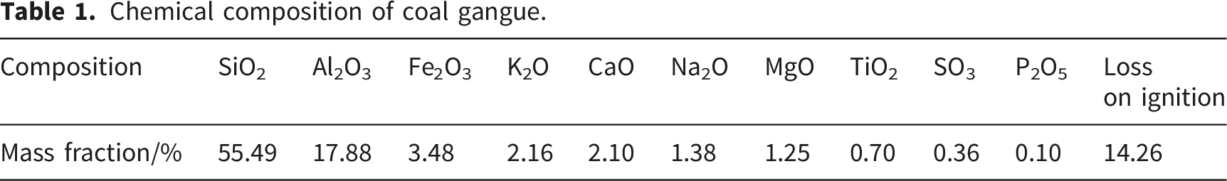
Table 1 shows that the main chemical components of coal gangue were SiO2 and Al2O3, in which a small amount of Fe2O3, KO,2 CaO, Na2O, etc. were existed. Among them, the nutrients content of silicon, potassium and phosphorus for plants were higher.
2.2. Experimental methods
For evaluating the tolerance of Bacillus megaterium to Pb2+, both LB solid and liquid media were prepared. The LB solid medium was prepared with agar to allow for the growth of colonies on solid surfaces, while the LB liquid medium was prepared without the addition of agar for bacterial culture in suspension. The detailed preparation method is as follows: 10 g of peptone, 3 g of beef paste powder, 5 g of sodium chloride, and 15 g of agar (for solid medium only) were dissolved in 1000 mL of distilled water with heating to 50 °C, yielding a final concentration of 3.3% (w/v) solid components. The pH was adjusted to 7.2, and the mixture was sterilized in an autoclave at 121 °C for 20 min. The LB liquid medium was prepared using the same procedure without the addition of agar.
For the resuscitation and preservation of Bacillus megaterium powder, 1 g of the bacterial powder was added to 100 mL of sterilized LB liquid medium under aseptic conditions and shaken for 120 min to obtain a bacterial suspension. The cell concentration of the suspension was determined by measuring the optical density at 600 nm (OD600) using a spectrophotometer and adjusted as required for subsequent experiments. The cell concentration was further calibrated to colony-forming units per milliliter (CFU/mL) based on a pre-established standard curve, as used in subsequent adsorption experiments. The above bacterial suspension of 0.2 mL was placed in LB solid medium for striating isolation and culture. A sterile L-shaped coating rod was applied to test the purity and activation of the strains. According to the characteristics of the strains, the medium was cultured in an incubator at 30 °C. After 2 days of culture, the strains were stored in the refrigerator at 4 °C for 1 week.
Treatment of coal gangue modified by Bacillus megaterium: After 2 days of culture, Bacillus megaterium colonies were scraped from the solid medium using a sterilized inoculation loop and suspended in 5 mL of sterile water. The initial concentration of the bacterial suspension was adjusted by controlling the number of colonies collected, and the concentration was determined by measuring the optical density at 600 nm (OD600) using a spectrophotometer. To remove residual culture medium components, the bacterial suspension was centrifuged at 4000 r/min for 10 min. The supernatant was discarded, and the cell pellet was resuspended in sterile water. This washing procedure was repeated with three times. The bacterial suspension was then mixed with 0.2 g of coal gangue in a 100 mL beaker. The mixture was shaken at 150 r/min for 24 h and then filtered to obtain the resulting microbial modified coal gangue (CG-W).
For Pb2+ adsorption by Bacillus megaterium cells, the strain was first cultured to the logarithmic growth phase, and the cell concentration was adjusted to a consistent optical density (OD600 = 0.2). The bacterial suspension was then mixed with Pb2+ solution at a predetermined concentration and incubated under shaking conditions. After adsorption, the cells were separated by centrifugation, washed with deionized water to remove unbound Pb2+, and the Pb2+ content in the collected cells was determined by atomic absorption spectrometry.
Adsorption of Pb2+ by modified coal gangue with microorganism: A 100 mL aliquot of Pb2+ solution with a concentration of 200 mg/L (containing 20 mg of Pb2+) was transferred into a 500 mL conical flask. A certain amount of CG-W was added and the bottle was sealed with plastic wrap. The bottle was shaken in a constant temperature oscillation box at 150 r/min for 2 h. The mixture was centrifuged after adsorption, and then the concentration of remaining Pb2+ in the supernatant was determined.
2.3. Analytical methods and data processing
The mineral composition of the sample was determined by the German D8 Advance X-ray diffractometer. The chemical composition and content of the sample were determined by X-ray fluorescence spectrometer. The functional groups on the surface of the sample were determined by the German INVENIO R Fourier infrared spectrometer. The apparent morphology and element distribution of the samples were measured by Merlin Compact scanning electron microscope of Zeiss. The element content and chemical morphology of the samples were measured with Thermo Scientific ESCALAB 250Xi X-ray photoelectron spectrometer. Statistical analysis was performed using Origin 2024. All experiments were conducted in triplicate, and data are presented as mean ± standard deviation (SD).
3. Results and discussion
3.1. Adsorption of Pb2+ with Bacillus megaterium
3.1.1. Tolerance of Bacillus megaterium to Pb2+
Following the procedures described in Section 2.2, the tolerance of Bacillus megaterium to Pb2+ was first evaluated. In order to improve the adsorption performance of coal gangue, the microorganisms were used to modify it. Among them, Bacillus megaterium was fast reproduction speed, strong vitality and non-toxicity, which also had the functions of phosphorus dissolution, potassium dissolution and heavy metal degradation. Therefore, the Bacillus megaterium was selected and the tolerance of Bacillus megaterium to Pb2+ was investigated. The lead nitrate was added to LB liquid medium and prepared into the mixed medium with different Pb2+ concentrations. The same bacterial solution of 1 mL was added into the medium with different concentrations, and cultured at 30 °C in the dark. The OD600 value of the bacteria was measured every 24 hours. The result is shown in Figure 1. Tolerance of Bacillus megaterium to Pb2+.
Figure 1 shows that the OD600 value of Bacillus megaterium firstly increased and then decreased with the increase of culture days, which reached the highest value on the 5th day. It indicates that the strain could still grow and multiply in the medium containing Pb2+, and began to decline after the 5th day. The OD600 value of Bacillus megaterium also gradually decreased with the increase of Pb2+ concentration in the medium, which also indicates that the presence of Pb2+ would inhibit the growth and reproduction of Bacillus megaterium. Although the growth of the strain was inhibited at the beginning in the 800 mg/L Pb2+ system with the prolongation of culture time, it could still achieve a higher OD600 value in the later stage. It indicates that the strain had a strong adaptability to Pb2+. Therefore, it is possible to study the adsorption and fixation of Pb2+ in solution by Bacillus megaterium with the strong tolerance.
3.1.2. Adsorption of Pb2+ with Bacillus megaterium
Following the procedures described in Section 2.2, the strain was cultured to the logarithmic growth phase and used for Pb2+ adsorption experiments. While the strain felled heavy metal stress, it would regulate its own metabolism, such as secreting sugars, proteins and other substances to adsorb and chelate heavy metals. Therefore, the effects of different Pb2+ concentrations on the contents of protein, polysaccharide and lead in the extracellular polymer of the strain were determined in order to study the adsorption of Pb2+ by the extracellular polymer of Bacillus megaterium. The results are shown in Figures 2 and 3 respectively. Influence of adsorption time on the component content the extracellular polymer. Influence of adsorption time on lead content in extracellular polymer.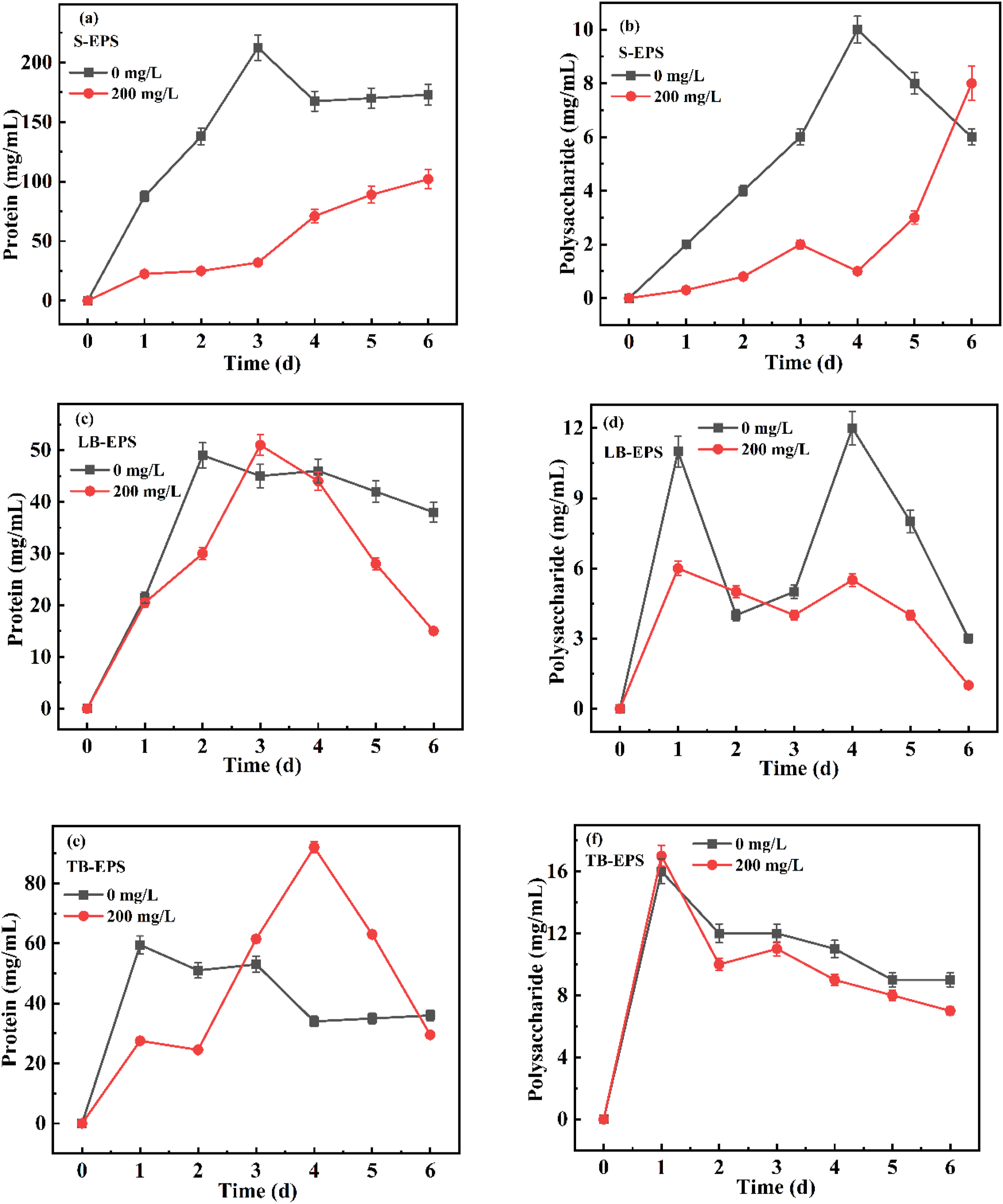

Figure 2 shows that the content of protein in the extracellular polymer was significantly higher than that of polysaccharide. The extracellular polymer was mainly composed of protein and supplemented by polysaccharide. The changes of protein content in S-EPS were observed. It is found that the protein content in S-EPS was significantly reduced after adsorption of Pb2+ compared with that before adsorption. But it was always increasing with the extension of time. Combined with Figure 3, it can be seen that Pb2+ was mainly concentrated in S-EPS, which indicates that proteins in S-EPS can bind to Pb2+. The lead content in the extracellular polymeric substances and intracellular components was quantified, and the results are presented in Figures 3 and 5, respectively. In order to resist the invasion of Pb2+, proteins were secreted inside the cell and transported to the outside of the cell, and buffer areas were formed in the S-EPS layer to prevent the entry of Pb2+. After adsorption, the Pb2+ content and protein content in LB-EPS and TB-EPS showed a trend of first increasing and then decreasing, which was due to the gradual diffusion of Pb2+ in S-EPS to LB-EPS and TB-EPS. Upon sensing the entry of Pb2+, a large number of proteins were mobilized to bind and fix Pb2+.
About the changes of polysaccharide content in extracellular polymers, the increase rate of polysaccharide content in TB-EPS was significantly faster than that in S-EPS and LB-EPS within one day. The polysaccharide content in S-EPS increased with the extension of time from 1 to 6 days, while the polysaccharide content in TB-EPS decreased. It indicates that polysaccharides produced by bacteria were excreted from inside to out. 36 Although the polysaccharide content in S-EPS after adsorption was obviously lower than that before adsorption, it was always increased due to resisting the diffusion of Pb2+ in the environment to cells. A large amount of polysaccharide in S-EPS was bound to Pb2+, which was consistent with the situation in Figure 3. The ever-increasing polysaccharide content was due to the continuous secretion of polysaccharide in the interior of the cell to prevent the entry of Pb2+ and the transport to the outermost layer to prevent the invasion of Pb2+. The change trend of LB-EPS polysaccharide was similar before and after adsorption, but the polysaccharide content was relatively low after adsorption. It is because of that the polysaccharide in LB-EPS was gradually transported to the S-EPS layer under the stress of external Pb2+, which prevented the entry of Pb2+. Furthermore, the Pb2+ concentration in LB-EPS continued to increase at 1 to 3 days, where the polysaccharide in LB-EPS combined with it resulting in a decrease in polysaccharide content.
In order to explore the adsorption of Pb2+ by cells of Bacillus megaterium, the contents of protein, polysaccharide and lead in cytoplasm and cell membrane were determined respectively. The results are shown in Figures 4 and 5. Effect of adsorption time on the component content in bacterial cells. Effect of adsorption time on lead content in cell components.
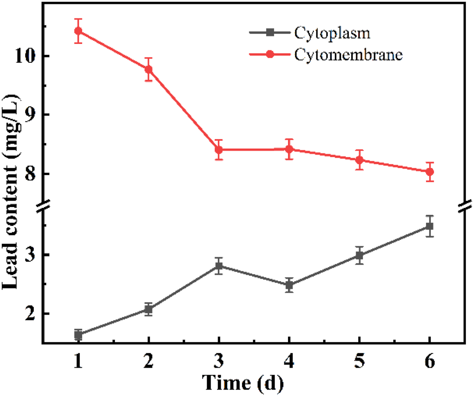
Figure 4 shows that the protein content in cytoplasm and membrane decreased at 0-2 days and increased at 3-6 days after adsorption. Combined with Figure 5, the content of Pb2+ in the cell membrane gradually decreased with the extension of time, while the content of Pb2+ in the cytoplasm gradually increased. This is because that the strain transported proteins outward to bind Pb2+ in the early stage of adsorption in order to prevent the persecution of Pb2+ on cells, which resulted in a decrease in protein content compared with that before adsorption. With the extension of time, the strain used some transporters on the cell surface to transfer Pb2+ into the cell and accumulate in the body. Upon sensing the entry of Pb2+, the cell could synthesize a large number of proteins to polymerize with Pb2+ and seal it in the cell to reduce the toxicity of Pb2+, so the protein content increased in 3 to 6 days compared with before adsorption. With the further extension of the adsorption time, the protein content gradually decreased due to large amount of Pb2+ accumulation in the cell inhibiting the synthesis of proteins. The high concentration of Pb2+ stress would cause damage to the plasma membrane system, resulting in the increase of membrane permeability and intracellular protein loss. For polysaccharide, the contents of cytoplasm and cell membrane did not obviously change at 1 to 2 days after adsorption of Pb2+, while the content of polysaccharide in cell membrane was significantly higher at 3 to 6 days. In addition, the content of polysaccharides in the cytoplasm also increased on day 3, even exceeding the level before adsorption. It indicated that the strain cells began to secrete polysaccharide to prevent the entry of Pb2+ in the middle and late stage. When a large amount of Pb2+ entered the cytoplasm, it combined with Pb2+ to reduce its toxic effect on bacteria. Furthermore, the content of protein and polysaccharide basically changed from the 3rd day. It may be that Pb2+ was mainly concentrated in the outside of the cell and the cell membrane at 0-2 days with less toxicity to the inside of the cell. With the extension of time, more Pb2+ entered the cytoplasm, and the toxic effect became stronger and reached the limit value on the 3rd day. Therefore, the cytoplasm and the cell membrane secrete protein and polysaccharide to reduce the harm of Pb2+ in the cell and reduce the entry of Pb2+ at the beginning of the 3rd day, which was also consistent with the change trend of the lead content in the cytoplasm and the cell membrane on the 3rd day (Figure 5). The result indicates that both protein and polysaccharide had a role in the adsorption process of Pb2+. The content of polysaccharide in both cytoplasm and cell membrane was significantly less than the content of protein, indicating that the protein played a major role in the adsorption process of Pb2+ inside microorganisms.
3.1.3 Adsorption mechanism of Pb2+ by Bacillus megaterium
In order to study the intracellular accumulation of Pb2+ by Bacillus megaterium, TEM was used to observe the morphological changes of the cells before and after adsorption. The result is shown in Figure 6. TEM images of Bacillus megaterium (a) before and (b) after Pb2+ adsorption. After adsorption, electron-dense precipitates (black granular matter) are observed in close association with the cell surface.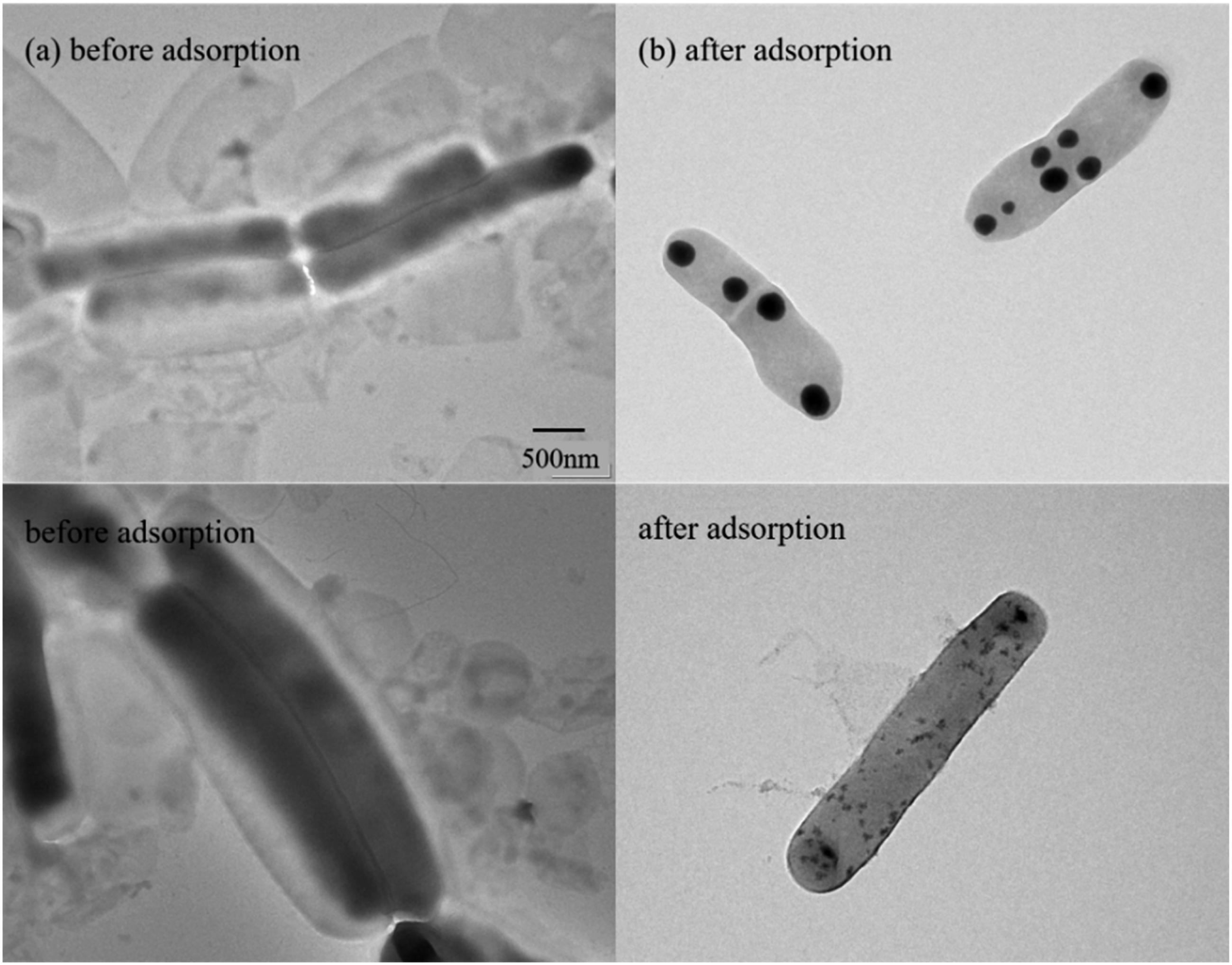
Figure 6 shows that Bacillus megaterium had a complete cell structure before and after adsorption of Pb2+, and the cell morphology did not obviously change. However, after adsorption, electron-dense precipitates were observed on the surface of the cells, appearing as black granular matter closely associated with the cell envelope, and cell shrinkage occurred to a certain extent. This suggests that the strain responded to Pb2+ stress through surface binding and extracellular complexation. It has been reported in the literature that in the environment of high heavy metal concentration, some microorganisms with heavy metal resistance can immobilize heavy metal ions through surface adsorption and extracellular polymer binding, thereby reducing the heavy metal concentration in the environment.37,38 These findings indicate that the immobilization of Pb2+ by the strain involves not only passive adsorption on the cell surface but also complex interactions with extracellular polymeric substances.
In order to explore the adsorption of Pb2+ by microbial functional groups, the infrared spectra before and after adsorption of Pb2+ by Bacillus megaterium were compared. The results are shown in Figure 7. FTIR diagram of Bacillus megaterium before and after the adsorption of Pb2+.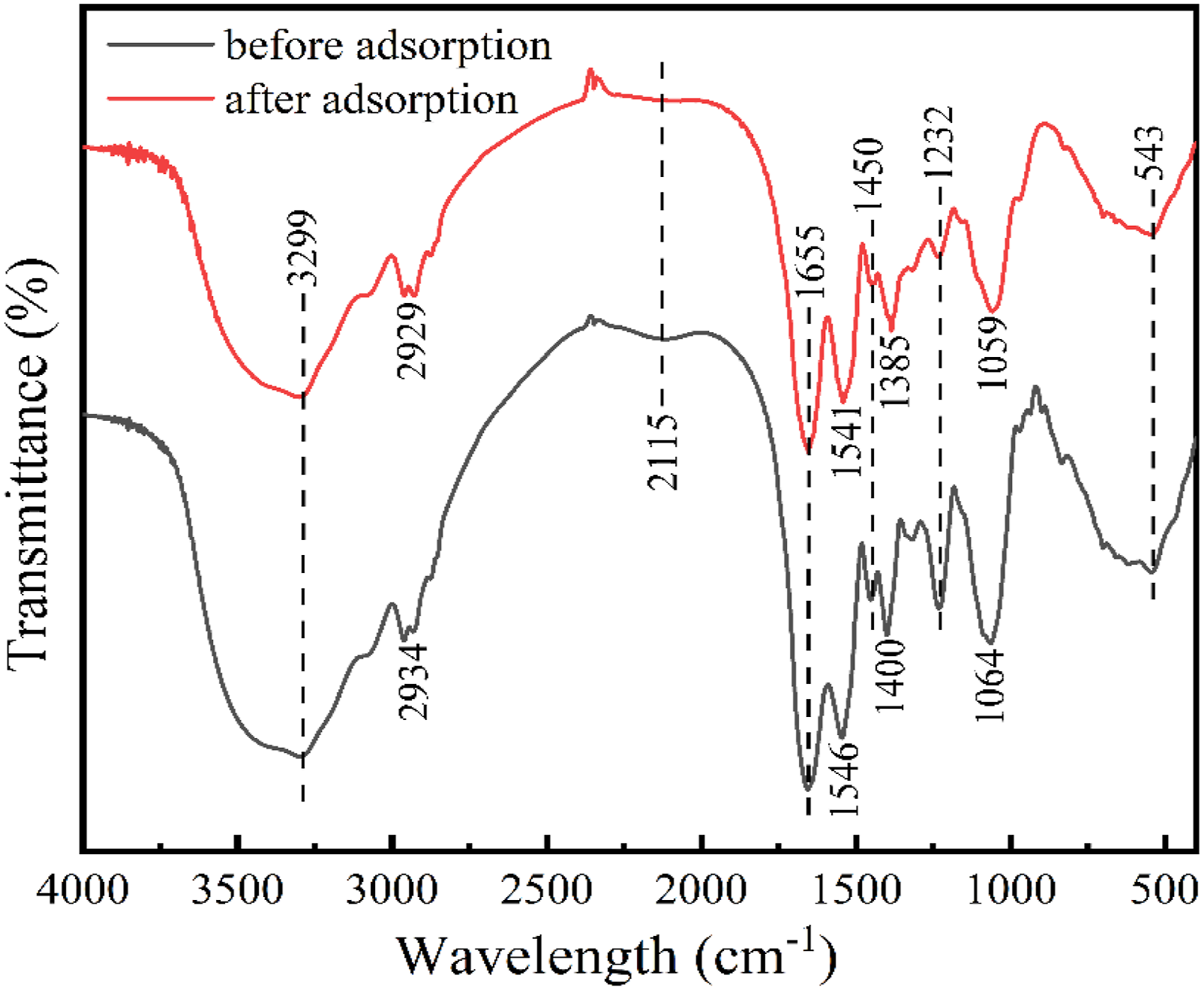
Figure 7 shows the FTIR spectra of Bacillus megaterium before and after Pb2+ adsorption. Certain changes in peak positions and intensities were observed after adsorption. The absorption peak at 3299 cm-1 was the overlapping peak of O-H stretching vibration and N-H antisymmetric stretching vibration, exhibited a slight decrease in intensity, suggesting the involvement of hydroxyl and amino groups in Pb2+ binding. The peak at 2934 cm-1, corresponding to alkyl C-H stretching, shifted to 2929 cm-1 after adsorption, indicating a minor contribution from alkyl groups. The absorption peaks at 1655 cm-1, 1546 cm-1 and 1450 cm-1, corresponding to amide I, II, and III bands, showed minor shifts or intensity reductions, indicating that protein amide groups participated in the adsorption process.29,30,39 The peak at 1400 cm-1 (symmetric C=O stretching of carboxyl groups) shifted to 1385 cm-1 after adsorption, possibly due to interactions between Pb2+ and microbial metabolism. The absorption peaks at 1232 cm-1 and 1064 cm-1, attributed to phosphoryl group and C-O-C in polysaccharide, also showed slight changes after adsorption. These spectral changes, though subtle, were reproducible and consistent with the functional group transformations identified by XPS analysis. According to the above analysis, functional groups such as hydroxyl, amino, carboxyl, and phosphate were present in Bacillus megaterium, confirming that the main cellular components (proteins, polysaccharides, and phospholipids) contributed to Pb2+ adsorption.
The bacterial strain cells and three extracellular products before and after adsorption of heavy metals were scanned using 3D-EEM. The excitation wavelength was set at 200-400 nm and the emission wavelength was set at 250-550 nm. The results are shown in Figures 8 and 9. 3D-EEM images of extracellular polymers with Bacillus megaterium. 3D-EEM diagram of cell components with Bacillus megaterium.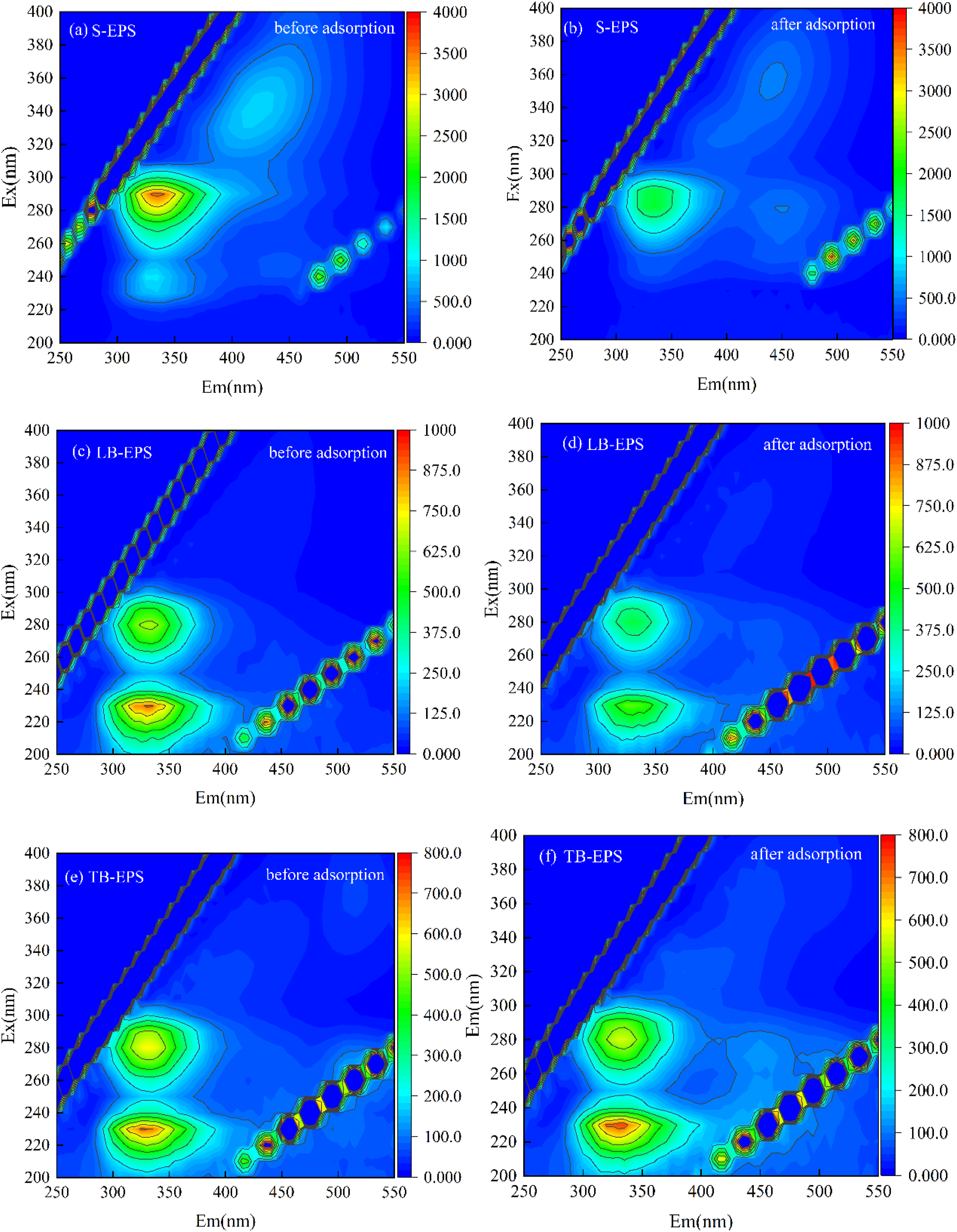
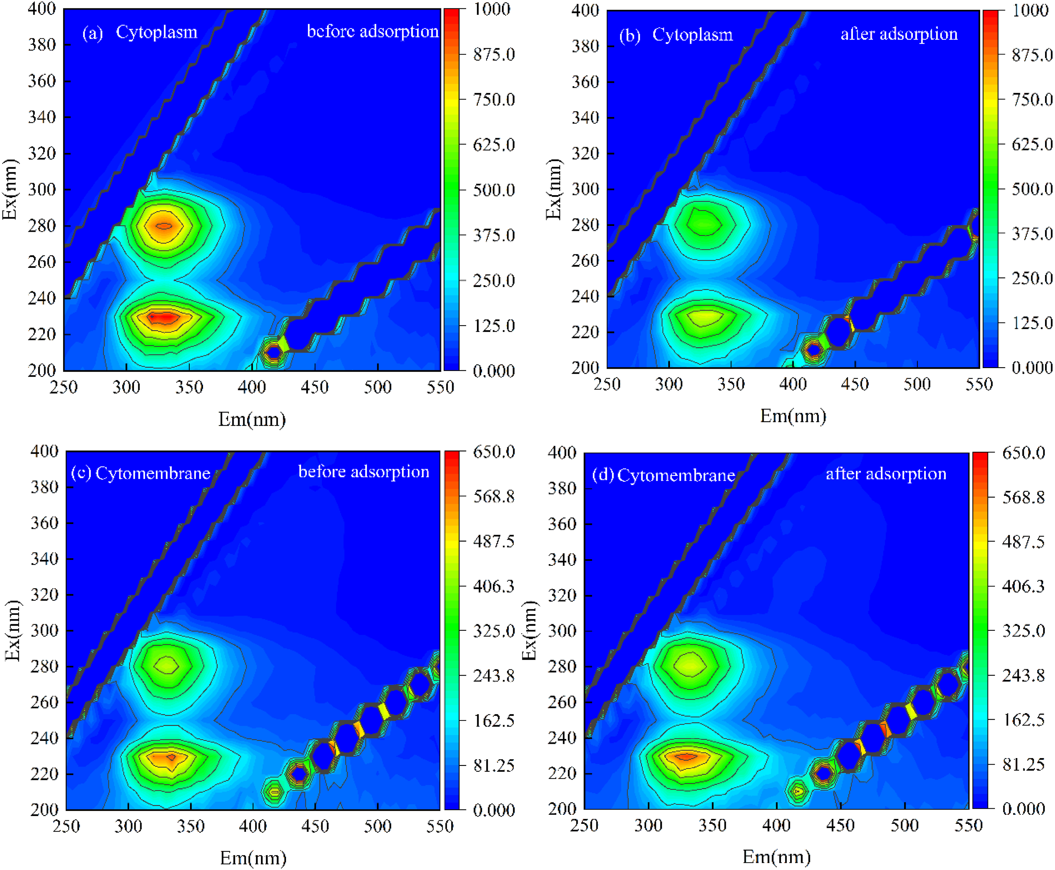
Figure 8 shows the 3D-EEM spectra of three extracellular products before and after adsorption of Pb2+ by Bacillus megaterium. Before adsorption, S-EPS peaks existed at Ex/Em=285/335 nm, Ex/Em=235/330 nm and Ex/Em=340/423 nm, corresponding to tryptophan, tyrosine and humus in proteins, respectively. Compared with the spectra before and after adsorption, it is found that the peaks at these three places were significantly weakened after adsorption of Pb2+, and the peaks at Ex/Em=340/423 nm shifted to Ex/Em=355/442 nm. Furthermore, a new peak appeared at Ex/Em=280/450 nm, which was attributed to the xanthohumic acid. Combined with the high fixation amount of Pb2+ by S-EPS, it can be inferred that these substances adsorbed and fixed a large amount of Pb2+. The humus reacted with Pb2+ to form fulvic acid substances. At the same time, the presence of Pb2+ could also affect the activity of amino acids in proteins, which made the peak intensity of tryptophan and tyrosine weaken. Although LB-EPS and TB-EPS showed similar fluorescence peak positions, the changes were different before and after adsorption. The peak intensity of tryptophan, tyrosine in LB-EPS and tryptophan in TB-EPS decreased after adsorption, but the peak intensity of tyrosine in TB-EPS increased slightly compared with that before adsorption. It indicates that tryptophan and tyrosine played a key role in the adsorption of Pb2+. TB-EPS called part of tyrosine from inside the cell to fix Pb2+ to the outside of the cell, preventing Pb2+ from entering the cell. By comparing the changes of these three extracellular products before and after adsorption, it can be seen that the composition of S-EPS and LB-EPS greatly changed, while the changes in TB-EPS were not obvious. It indicates that the extracellular adsorption mainly relied on S-EPS and LB-EPS to adsorb and fix the Pb2+.
Figure 9 shows the 3D-EEM diagram of cell membrane and cytoplasm before and after adsorption of Pb2+ by Bacillus megaterium. The components of cytoplasm and cytoplasm were similar to those in LB-EPS and TB-EPS, both of which were attributed to tryptophan and tyrosine in proteins. The strength of tryptophan and tyrosine in the cytoplasm decreased significantly after Pb2+ was added, indicating that tryptophan and tyrosine in the cytoplasm played an important role in the fixed adsorption process of Pb2+. After the reaction, they were stored and accumulated in the cell to reduce the concentration of Pb2+ in the environment. The content of each component in the cell membrane changed little, and the peak intensity was slightly enhanced after adsorption of Pb2+. It was to prevent Pb2+ continuing to enter the cell resulting in high content to damage the cell, which could secrete some proteins to inhibit the entry of Pb2+, and adsorb it to the surface of the cell membrane. Comparing the contents of various substances inside and outside the cell of the strain, it can be seen that tryptophan and tyrosine in the protein play a main role in the adsorption process of Pb2+. The humus substances also had obvious contributions outside the cell, which also proves that the adsorption of Pb2+ by the strain was mainly the role of protein consistent with the above analysis results.
3.2. Preparation of microbial modified coal gangue and its application
3.2.1. Influence of preparation parameters
Based on the above understanding of Pb2+ adsorption by the strain, the microbial modified coal gangue (CG-W) was prepared according to the method in Section 2.2, and its adsorption performance was systematically investigated. Under the conditions of 5 mL bacterial solution, 0.2 g coal gangue, Pb2+ solution with a concentration of 200 mg/L for 120 min, the effect of bacterial solution concentration, pH value of coal gangue and load temperature in the preparation process on the removal rate of Pb2+ was investigated. The result is shown in Figure 10. Effect of preparation parameters on the removal rate Pb2+.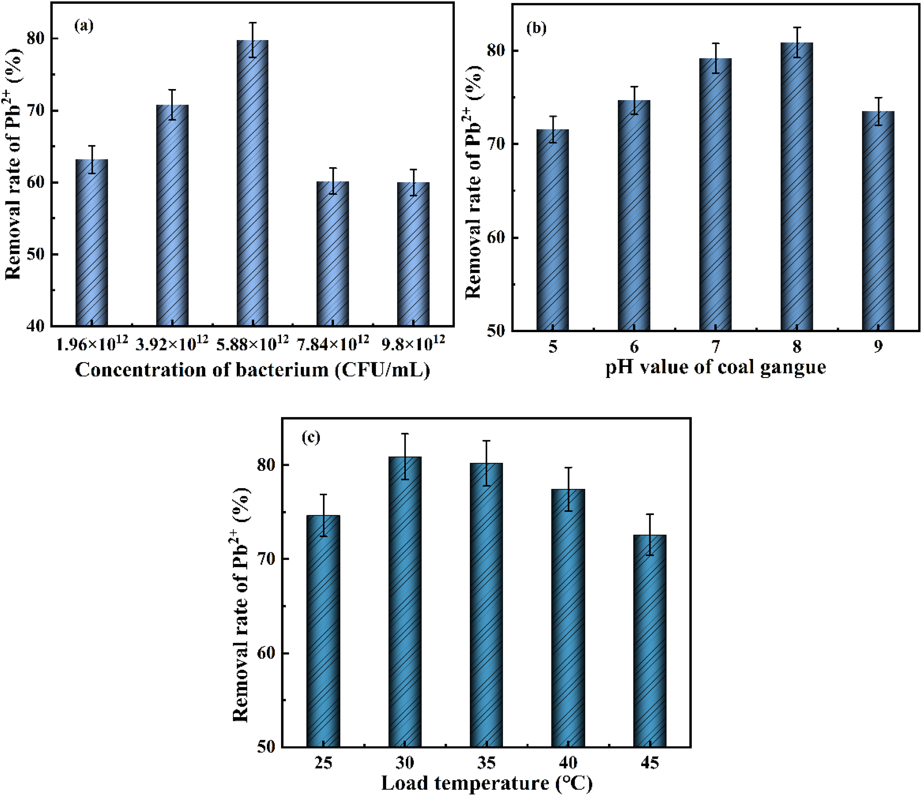
As can be seen from Figure 10(a), the adsorption of Pb2+ by CG-W first increased and then decreased with the increase of bacterial solution concentration. The removal rate of Pb2+ could reach 80.8% with the bacterial solution concentration of 5.88×10¹²CFU/mL. This is because the cell structure of Bacillus megaterium could adsorb a part of Pb2+, and its metabolites could also bind to Pb2+ providing more adsorption sites. The adsorption rate of Pb2+ decreased with later increase of bacteria, because the coal gangue provided limited nutrients, which was not enough to make all bacteria survive. Therefore, a number of Bacillus megaterium died due to lack of nutrients resulting in decrease in the removal rate of Pb2+. Figure 10(b) shows that the removal rate of Pb2+ first increased and then decreased with the pH increase of coal gangue. There was little difference with pH value of 7 and 8, both of which showed good adsorption effect of Pb2+. This is because the optimum pH value existed in the growth and reproduction process of Bacillus megaterium, which ranged from 6.5 to 8.5. Too much acid or alkali was not conducive to the growth of bacteria, resulting in the death of some bacteria and the decrease of removal rate of Pb2+. A large amount of H was attached to the surface of coal gangue at low pH value, which enhanced the competition for the Pb2+ adsorption site and limited the combination of adsorbent and Pb2+. With the increase of pH value, more negatively charged ligands were exposed to the surface of the adsorbent, where positively charged Pb2+ occupied more binding sites resulting in enhanced adsorption of Pb2+. However, the pH value of the solution would increase during the adsorption process with an increase of coal gangue pH value resulting in the precipitating of Pb2+. Since the pH value of coal gangue was between 7.5 and 8.2, the subsequent experiment would not adjust the pH value of coal gangue for the convenience of experimental operation. Figure 10(c) indicates that the load temperature also had a certain influence on the adsorption effect of Pb2+, and the removal rate of Pb2+ was the highest by CG-W with the load temperature of 30 °C. This is because the growth and metabolism ability of microorganisms could be inhibited at too low temperature. The high temperature would lead to the deterioration of important biomacromolecules in microorganisms, resulting in the death of microorganisms and a decrease in the adsorption capacity of adsorbents.40,41
3.2.2 Influence of adsorption parameters with microbial modified coal gangue
Since lead exists in different forms at different solution pH values, the precipitation will occur when the pH value is greater than 6, so the initial pH value of the solution was set below 6. Under the conditions of Pb2+ concentration of 200 mg/L, CG-W (optimal loading condition) dosage of 0.2 g, adsorption temperature of 25 °C and adsorption time of 120 min, the effect of pH value of solution on the removal rate of Pb2+ was investigated, as shown in Figure 11(a). The effect of adsorption temperature, adsorption time and initial Pb2+ concentration were also studied, and the results are shown in Figure 11. Influence of adsorption parameters on the removal rate of Pb2+.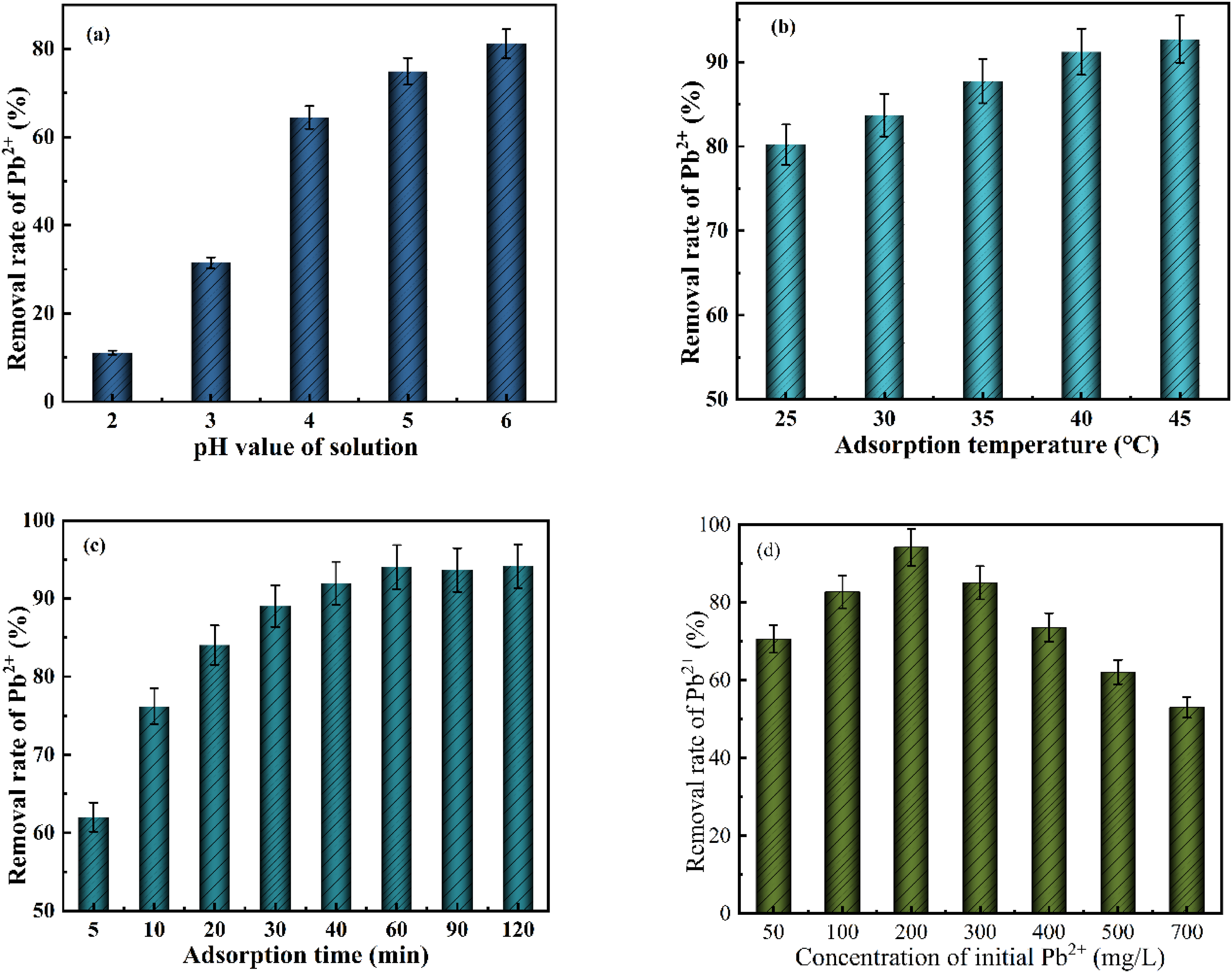
Figure 11(a) shows that the pH value of solution had a significant impact on the adsorption of Pb2+. The removal rate of Pb2+ with CG-W also kept increasing with the increase of pH value of solution. The removal rate of Pb2+ was only 11% at pH value of 2, while the removal rate reached 82% at pH value of 6. On the one hand, the concentration of H in the solution was high with low pH value, where there was a competitive adsorption between H+ and Pb2+. Most of the adsorption sites in CG-W would be occupied by H+, thus reducing the adsorption of Pb2+. On the other hand, Bacillus megaterium was suitable for survival in a neutral environment and difficult to survive in an acidic environment, which affected its normal growth and metabolism. More negatively charged functional groups were exposed on the surface of CG-W with an increase of pH value of solution, providing more adsorption sites.42,43 The influence of Bacillus megaterium on acidic environment was also reduced, and most strains could survive normally. Extracellular polymers secreted by strains and their metabolites also had a passivation effect on Pb2+, which was conducive to the removal of Pb2+. This trend is consistent with previous studies showing that Pb2+ adsorption by microbial-modified materials is typically favored in near-neutral conditions due to reduced competition from H+ and increased availability of binding sites.42,43
Figure 11(b) shows that the removal of Pb2+ by CG-W first increased and then tended to equilibrium with the increase of temperature. The increase of temperature would increase the number of activated groups on the surface of CG-W, which can adsorb more Pb2+ free in solution. At the same time, the increase of temperature could not only effectively improve the activity and movement rate of Pb2+, but also better overcome the diffusion resistance. Therefore, more Pb2+ could contact with CG-W particles, which was conducive to the adsorption reaction. Similar temperature-dependent adsorption behavior has been reported for other biosorbents, where higher temperatures enhance the mobility and diffusion of Pb2+ ions. 40
Figure 11(c) shows that the removal rate of Pb2+ by CG-W gradually increased with the extension of adsorption time within 0-60 min, and the growth rate gradually slowed down. the removal rate of Pb2+ basically leveled off, and the adsorption reached equilibrium with adsorption time of 60-120 min. On the one hand, the surface of CG-W had rich functional groups, which could provide sufficient adsorption sites for free Pb2+ in the environment. The concentration of Pb2+ in the initial solution was high, and the solute transport power was large. The Pb2+ soon occupied the adsorption site while the adsorption began. As the adsorption continued, the number of adsorption sites decreased and the external Pb2+ concentration decreased. Therefore, the driving force was small and the adsorption amount no longer increased with the extension of the time. On the other hand, Bacillus megaterium would secrete a large number of extracellular polymers and metabolites during the growth process, which could effectively bind Pb2+. However, the microorganisms were gradually eroded by Pb2+, and the growth and metabolism of the bacteria were slow with the progress of adsorption. The processing capacity of heavy metal ions is close to saturation. Figure 11(d) shows that the removal rate of Pb2+ by CG-W showed a trend of first increasing and then decreasing with the increase of initial concentration. When the concentration of Pb2+ increased from 50 mg/L to 200 mg/L, the removal rate of Pb2+ increased significantly. This is because the initial concentration of Pb2+ was high, which could generate enough driving force for mass transfer and promote the adsorption of Pb2+ by CG-W. The removal rate reached 94.02% at 200 mg/L of Pb2+ concentration. With the further increase of Pb2+ concentration, the removal rate gradually decreased. It is because that the amount of CG-W and the number of adsorption sites were fixed. With the continuous increase of Pb2+ concentration, the adsorption of Pb2+ by CG-W has reached the maximum saturation state, and the adsorption sites were all occupied. The amount of remaining Pb2+ in the solution increased with the increase of the initial concentration, and the removal rate showed a trend of gradual decrease. This saturation phenomenon is commonly observed in adsorption processes with limited active sites, and the maximum removal rate obtained in this study (94.02%) is comparable to or higher than those reported for other coal gangue-based adsorbents.26,32
The removal performance of Pb2+ by CG-W was compared with a recently reported efficient adsorbent, coal gangue-modified by roasting with KOH (CG-R), where a adsorption capacity of 60-268 mg/g reached. 44 In this study, CG-W achieved a removal rate of 94% at an initial Pb2+ concentration of 200 mg/L (adsorption capacity of 94 mg/g). Although the capacity of CG-W was lower than the maximum adsorption capacity of CG-R, CG-W was prepared via a simple microbial modification process without high-temperature pyrolysis or chemical activation, which significantly reduces energy consumption and environmental impact.
3.2.3. Surface properties analysis
To investigate the changes in physical structure induced by microbial modification, the adsorption-desorption isotherms were measured for raw coal gangue (GC) and microbial modified coal gangue (CG-W). The isotherms are presented in Figure 12, and the calculated BET parameters are summarized in Table 2. Adsorption-desorption isotherms of GC (a) and CG-W (b). BET surface properties of GC and CG-W.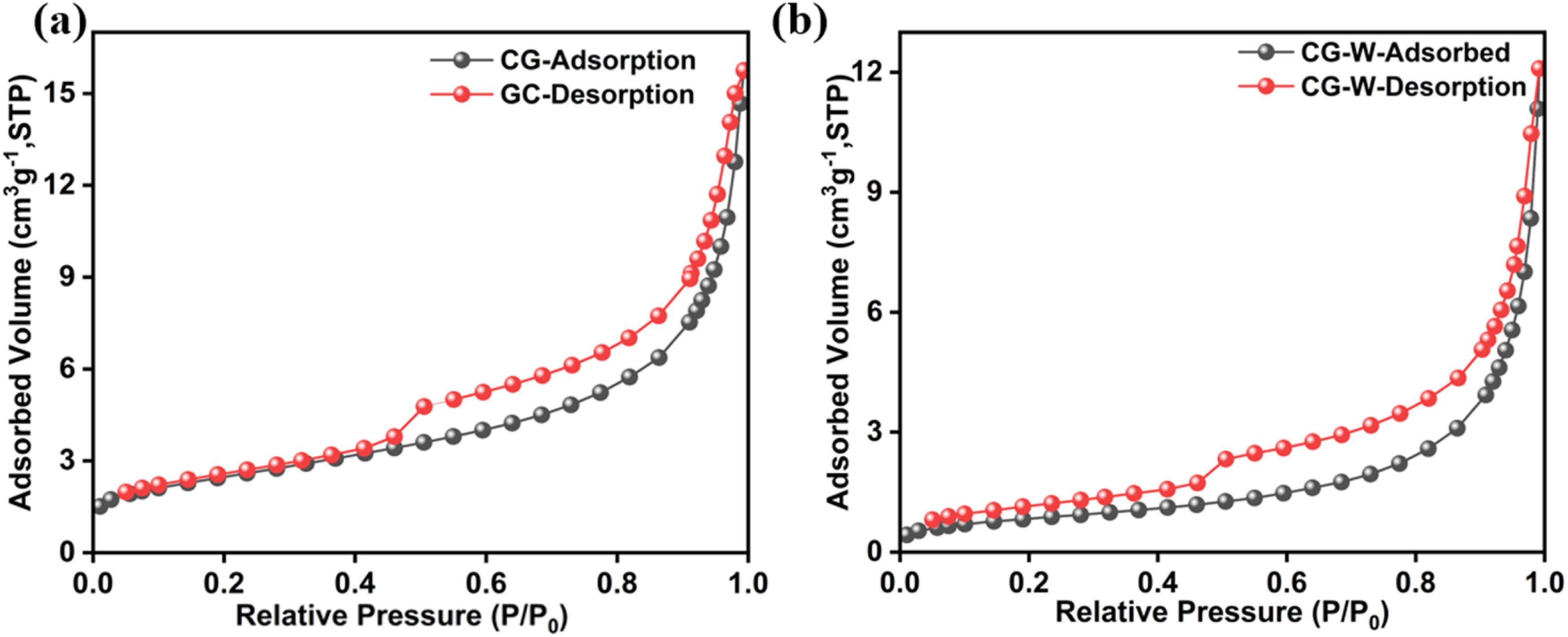
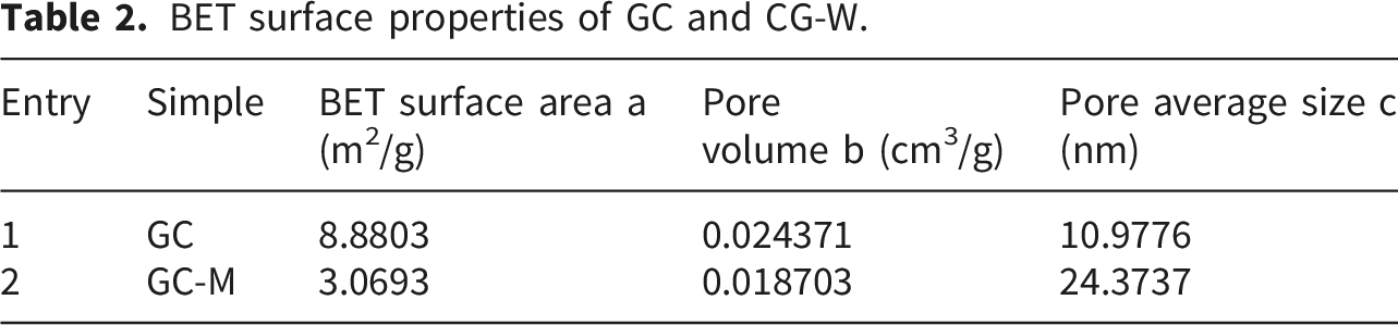
After microbial treatment, the BET surface area decreased from 8.88 m2/g to 3.07 m2/g, and the pore volume decreased from 0.0244 cm3/g to 0.0187 cm3/g. However, the average pore size increased significantly from 10.98 nm to 24.37 nm. The reductions in surface area and pore volume were likely due to the attachment of bacterial cells and extracellular polymeric substances (EPS) onto the coal gangue surface and within pore channels, partially blocking the pores. The increase in average pore size may result from the dissolution of fine mineral particles by organic acids produced by Bacillus megaterium, leading to the merger of adjacent small pores. Despite the decreased surface area, CG-W exhibited substantially higher removal of Pb2+ compared to raw coal gangue. This indicates that the improved adsorption performance was not governed by physical surface area but rather by chemical complexation. The functional groups introduced by the microorganism (e.g., hydroxyl, carboxyl, amino, and phosphoryl groups) may play an important role in binding Pb2+.
3.3. Adsorption mechanism of Pb2+ by CG-W
To elucidate the underlying mechanisms of Pb2+ adsorption by CG-W, the samples before and after adsorption were characterized by FT-IR, XRD, and XPS, and the results are discussed below. The FT-IR spectra of raw coal gangue, CG-W and samples adsorbed Pb2+ are shown in Figure 13. FT-IR diagram of CG, CG-W and CG-W adsorbed Pb2+.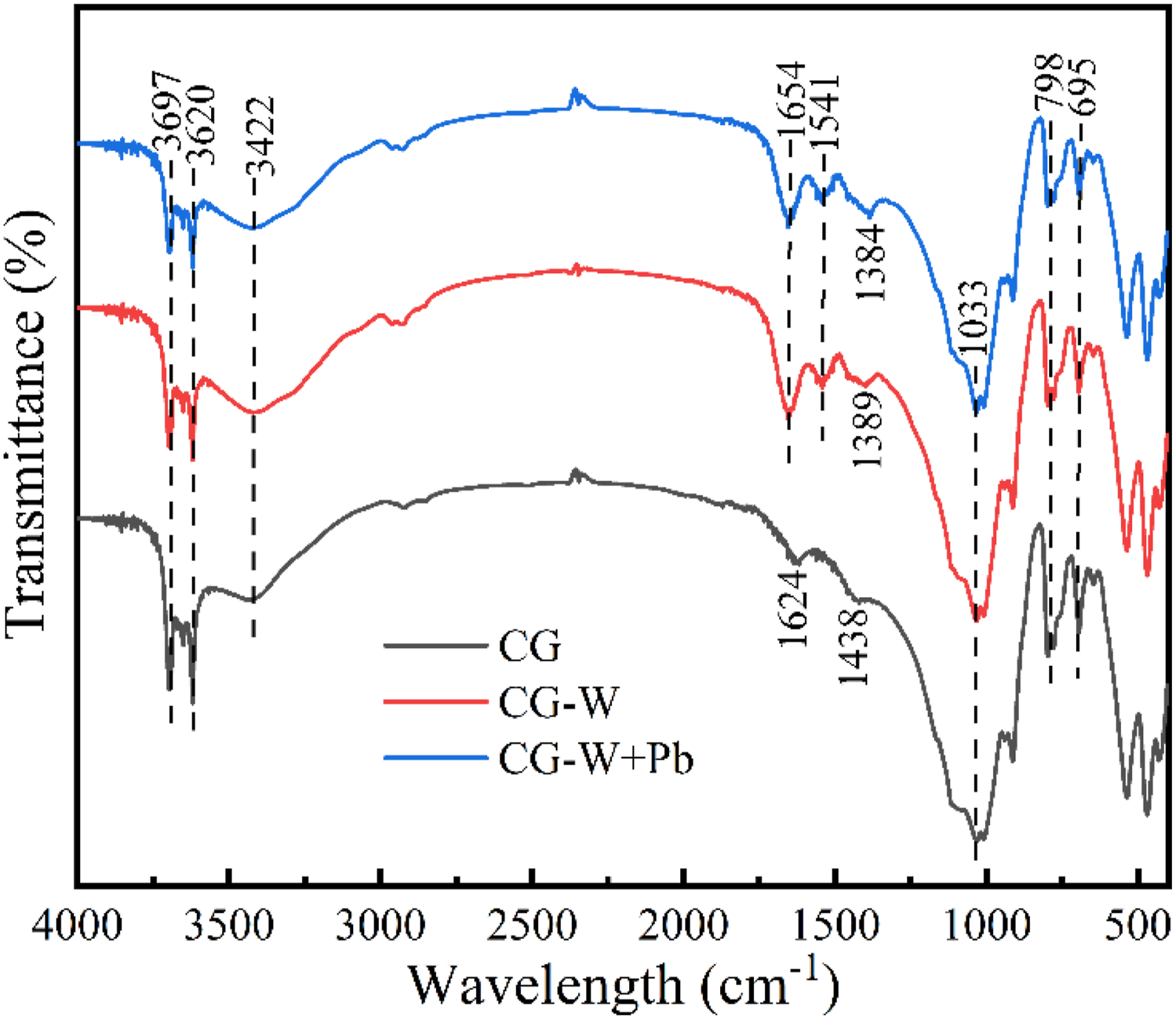
Figure 13 shows the FTIR spectra of raw coal gangue (CG), microbial modified coal gangue (CG-W), and CG-W after Pb2+ adsorption. The raw coal gangue exhibited characteristic absorption bands: -OH stretching vibrations at 3697 cm-1 and 3620 cm-1, a broad peak near 3422 cm-1 attributed to -OH in structural water, and an H-O-H bending vibration at 1624 cm-1 from physically adsorbed water. The peak at 1438 cm-1 corresponds to O-C-O bonds, while the absorption peaks at 1033 cm-1, 798 cm-1 and 695 cm-1 are attributed to Si-O-Si antisymmetric stretching, Si–O symmetric stretching, and Si-O bending vibration, respectively.
45
Compared with raw CG, CG-W showed weakened characteristic peaks at 3697 cm-1, 3620 cm-1, 1033 cm-1, 798 cm-1 and 695 cm-1, likely due to the reduction of the proportion of coal gangue after bacterial loading and partial dissolution of minerals by microbial metabolites. New absorption peaks appeared at 1654 cm-1, 1541 cm-1 and 1389 cm-1 in CG-W, attributed to microbial amide I (C=O), amide II (C–N, N–H), and lipid chain (C–H) vibrations, confirming successful bacterial loading. After Pb2+ adsorption, certain peaks showed minor changes: the peaks at 3697 cm-1, 3620 cm-1, 798 cm-1 and 695 cm-1 decreased in intensity, suggesting the involvement of -OH groups and silicate minerals; the peaks at 1654 cm-1 and 1033 cm-1 were weakened, and the peak at 1389 cm-1 shifted to 1384 cm-1(Figures 14 and 15). These subtle spectral changes, while not dramatic, were reproducible and consistent with the XRD (Figure 14) and XPS (Figures 15 and 16) results, which more clearly revealed the formation of lead-containing phases and ion exchange mechanisms. XRD patterns of CG-W before and after adsorption of Pb2+. XPS diagram: (a) Full spectrum of XPS for CG-W before and after adsorption of Pb2+; (b) High-precision spectra of Pb4f for CG-W after adsorption of Pb2+. High-precision spectra of XPS for main elements of CG-W.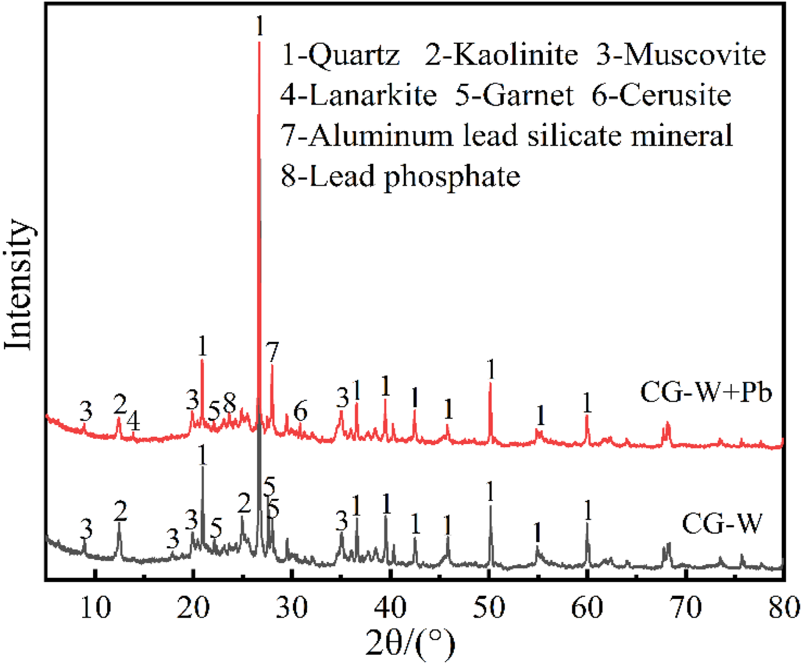

The XRD patterns of CG-W samples before and after adsorption of Pb2+ were compared, and the results are shown in Figure 13.
As can be seen from Figure 14, the diffraction peak intensity of kaolinite (Al[Si2O](2OH5)), muscovite (KAl2[AlSiO](OH)310), sodium garnet (NaAlSi3O8) and some partially amorphous silica (SiO2) in CG-W was weakened after adsorption of Pb2+. And the chrysolite (Pb2[SO4]O), white lead (PbCO3), lead phosphate (Pb3(PO4)2) and lead silicoaluminate minerals (Pb[AlSi3O8]2) were existed. This is because that the Pb2+ would be complexed with the hydroxyl group on kaolinite, where the structure of kaolinite could change. The Pb2+ could also have ion exchange with K and Na in muscovite and sodium garnet, resulting in the reduction of the diffraction peak strength of kaolinite, muscovite and sodium garnet, and the formation of lead silicaluminate minerals. In addition, Pb2+ could also combine with SO42-、CO32- and PO43- in CG-W to produce lanarkite, cerussite and lead phosphate precipitation, which was also consistent with the above infrared spectral analysis results.
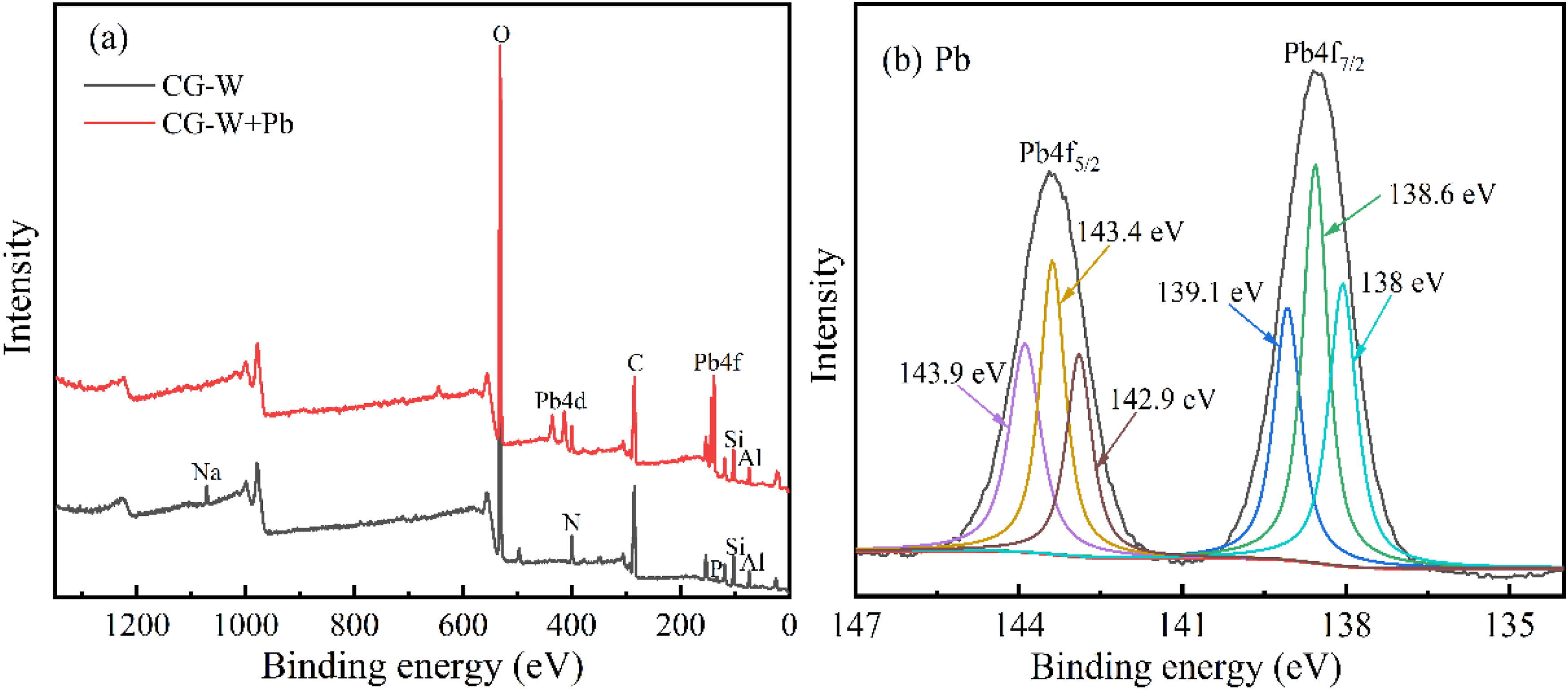
In order to further explore the adsorption effect of CG-W on Pb2+, XPS analysis was performed on the CG-W samples before and after adsorption of Pb2+. The results are shown in Figure 15.
Figure 15(a) shows that new energy peaks of Pb4d and Pb4f appeared in 409-421 eV and 134-150 eV segments for CG-W after adsorption of Pb2+, respectively. Figure 15(b) shows the corresponding energy peaks of Pb4f and Pb4f at 143.4 eV and 138.5 eV, respectively. Sub-peak fitting was carried out on these two peaks, and then the multiple peaks appeared in the fitting results. It indicates that CG-W successfully adsorbed Pb2+, and there were multiple binding ways for CG-W to adsorb Pb2+. The energy peak of Na at 1072 eV disappeared after adsorption of Pb2+, suggesting that ion exchange occurred between Pb2+ and Na+ in CG-W during the adsorption process.32,33 In addition, the energy peak intensities of Si, Al, C, N, P and S all changed in Figure 15 indicating that these elements may react with Pb2+ during the adsorption process. In order to explore the role of these elements in the adsorption process, the peak sub-fitting was performed respectively, and the results are shown in Figure 16
For Si and Al, the absorption peaks at 101.8 eV in Si and 74 eV in Al decreased in intensity after adsorption of Pb2+. The absorption peaks at these two positions belonged to silicaluminate minerals, indicating that the silicaluminate minerals in CG-W reacted with Pb2+ or generate lead silicaluminate through ion exchange during the adsorption process. It is also consistent with the analysis results of FT-IR and XRD. It can be seen from the peaks of C element that it had energy peaks at the positions of 284.5 eV, 286 eV, 287.8 eV and 293 eV, which belonged to the absorption peaks of C-C, C-O, C=O and O-C=O, respectively. The peak intensity of C-O, C=O and O-C=O was weakened after adsorption of Pb2+. On one hand, the carbonates in CG-W reacted with Pb2+ to produce cerussite. On the other hand, the proteins in microorganisms and organic acids produced in the metabolic process could combine with Pb2+. Compared with the spectra of N before and after adsorption, two peaks were fitted at 399.3eV and 399.8eV, belonging to -NH2- and -NH-, respectively. After adsorption, the binding energy of these two peaks shifted to 399.5eV and 400.1 eV respectively. The binding energy increased and the peak intensity of -NH- decreased. This is due to the complex reaction between -NH2- and -NH- and Pb2+. N contained lone pair electrons, and Pb2+ shared electrons with N to increase the electron cloud density, which further proves that the proteins in microorganisms can promote the adsorption of Pb2+. For P, the absorption peaks of P-O and P=O at 132.9eV and 133.7eV decreased significantly after adsorption of Pb2+. Combined with XRD analysis, the phosphates in CG-W reacted with Pb2+ to produce lead phosphate. The peak position of S was shifted after adsorption of Pb2+, and the strength was also significantly increased. The result indicates that S played a significant role in the adsorption process, where the lanarkite was formed and attached to the surface of CG-W combined with the analysis of XRD.
The FT-IR analysis reveals that the functional groups involved in Pb2+ adsorption mainly include –OH, Si–O–Si, Si–O, C=O, C–N, N–H, and C–H, originating from both coal gangue minerals and microbial components. Furthermore, XRD and XPS results indicate that Pb2+ can replace Na+ in sodium feldspar (NaAlSi3O8) through ion exchange, and may also substitute Ca2+ and Si4+ in silicate minerals, forming lead silicate and aluminosilicate phases. In addition, Pb2+ combines with SO42-, CO32-, and PO43- present in the coal gangue to form precipitates such as Pb2[SO4]O, PbCO3, and Pb3(PO4)2. These combined mechanisms contribute to the efficient immobilization of Pb2+ by CG-W. The formation of insoluble lead-containing mineral phases and ion-exchange products suggests that Pb2+ was immobilized in a chemically stable form.
4. Conclusion
The tolerance of Bacillus megaterium to Pb2+ and the adsorption performance of CG-W were investigated. Bacillus megaterium had a high tolerance for Pb2+ up to 800 mg/L and could enrich Pb2+ into and beyond the cell. The concentration of Pb2+ inside the cell was higher, mainly by secreting substances such as proteins and polysaccharides to bind to Pb2+. Extracellular fixation of Pb2+ was mainly through the secretion of extracellular polymers including proteins, polysaccharides and humus substances. Quantitative analysis revealed that the lead fixed by Bacillus megaterium was mainly concentrated in the soluble extracellular polymeric substances (S-EPS), indicating the dominant role of extracellular adsorption. The suitable modified conditions were bacterial concentration of 5.88×10¹²CFU/mL, coal gangue initial pH value of 6.0-8.0, and the load temperature of 30 °C. The optimal adsorption conditions were the solution pH 6, adsorption temperature 40 °C, adsorption time 60 min and initial Pb2+ concentration of 200 mg/L. Under these conditions, the removal rate of Pb2+ reached 94% with CG-W. CG-W could adsorb Pb2+ by ion exchange with Na+ in NaAlSi3O88 to produce Pb[AlSi3O8]2. The SO42-, CO32- and PO43- in the CG-W could bind Pb2+ to precipitate Pb2[SO4]O, PbCO3, and Pb3(PO4)2), as well as binding with proteins and polysaccharides secreted by Bacillus megaterium. This study only conducted the adsorption performance and mechanism experiments in a single Pb2+ system, without considering the interference effects of competitive coexisting ions such as Ca2+, Mg2+, Zn2+, as well as the stability and desorption analysis of Pb2+, which will be focused on in the subsequent research.
Footnotes
Ethical considerations
This manuscript is original and has not been published elsewhere, nor is it under consideration by another journal. All authors have made substantial contributions to the research and approved the final version of the manuscript.
Author contributions
Wang Li: Investigation, Writing-Original Draft. Chenxi Shao: Data Curation, Writing -Review & Editing. Xiaobo Zhu: Funding Acquisition, Supervision. Xueyu Bai: Investigation. Shenxu Bao: Resources.
Funding
The authors disclosed receipt of the following financial support for the research, authorship, and/or publication of this article: The research is financially supported by National Natural Science Foundation of China (52574314), Natural Science Foundation of Henan Province (262300421343) and Hubei Key Laboratory of Mineral Resources Processing and Environment (Wuhan University of Technology) (ZHJJ202403).
Declaration of conflicting interests
The authors declared no potential conflicts of interest with respect to the research, authorship, and/or publication of this article.
Data Availability Statement
The data that support the findings of this study are available from the corresponding author upon reasonable request.