






Abstract
Objective
A large number of substrates after harvesting the fruiting bodies of Cordyceps militaris have been discarded or only used as feed and fertilizer. In this study, we aimed to investigate the potential utilization value of residual substrates under industrial cultivation of C. militaris.
Methods
The various bioactive substances in C. militaris fruiting body and substrate were measured and compared by colorimetry, HPLC, burning method, Kjeldahl legal nitrogen method, and GC-MS et al. The fruiting body was taken as a reference to better reflect the internal quality of substrate. Finally, a response surface experiment was conducted to investigate the effects of drying temperature, humidity, and time on the retention rate of C. militaris polysaccharide.
Results
In the substrate, the content of polysaccharide was 4.68%, the cordycepic acid content was 7.95%, which were significantly higher than the content in the fruiting body. The content of cordycepin was as high as 1.13%, which was more than 4 times of the fruiting body. The crude fat content was also higher than that in fruiting body. In addition, 17 amino acids were detected in the substrate, including 7 essential amino acids. Moreover, several volatile components with drug properties were detected in the substrate. Finally, the optimum drying parameters of the substrates are as follows: drying temperature, 59°C; humidity, 16%; time, 24 h.
Conclusions
This study highlighted the available value of substrate is no less than the fruiting body, which is an ideal food supplement and pharmaceutical agent, which can be served as a good source of healthy substances potentially bioavailable for humans.
1. Introduction
Cordyceps militaris is an entomopathogenic fungus and a traditional Chinese medicinal mushroom, which containing some prominent bioactive compounds, such as polysaccharide, cordycepic acid, adenosine, and cordycepin. 1 It can be used in medical care, functional food development and so on.2,3 C. militaris polysaccharides have a wide variety of biological activities, such as lipid-lowering, anti-atherosclerosis, antitumor, hypoglycemia and anti-inflammatory activities, 4 it is an important indicator to measure the quality of C. militaris. 5 Cordycepic acid is the main active substance of C. militaris, which has certain benefits in regulating blood lipids and removing free radicals in the body. 6 In addition, it is often used in some food additives. 7 Adenosine and Cordycepin were the main bioactive compounds that correlated with a wide range of functions in C. militaris. 8 Adenosine as the basic structural unit of nucleic acid, it plays a role in improving blood circulation, metabolism, and physiological regulation in human. 9 Cordycepin has antibacterial, anti-inflammatory, antiviral, anti-tumor, immuno-stimulant and other effects. 10 Aroma is another essential feature, which is critical element influencing consumers acceptability. 11
Since C. militaris could be cultivated artificially, industrial cultivation has become an effective means to solve the serious shortage of wild C. militaris resources. Wheat has become one of the main raw materials for artificial culture of C. militaris due to its advantages of wide source, low price, and reliable quality. 12 Related researchers cultivated C. militaris mother species through separation, breeding, rejuvenation and other processes, propagated and transferred to the prepared inactivated wheat solid medium. Added with various essential nutrients, and finally successfully cultivated C. militaris, which has similar effective components and pharmacological effects to the natural varieties. 13 At present, for C. militaris, the focus of research is on its fruiting body, while its substrate is mostly treated as waste.14–16
In this study, the bioactive substances in C. militaris substrate were measured and compared to fruiting body, including polysaccharide, cordycepic acid, cordycepin, adenosine, amino acid, protein, ash, crude fat, and volatile components. Furthermore, although many investigators have reported the correlation of fermentation conditions with polysaccharide production,17,18 there are still few researches conducted the effects of drying condition on polysaccharide production. The drying condition can not only significantly affect the moisture and apparent properties, but also significantly affect its polysaccharide content and other biological activity. 19 When the drying temperature was too high, the Maillard reaction occurred, resulting in gelatinization of starch, destruction of polysaccharide structure, thus the polysaccharide content was decreased. 20 The factors affecting Maillard reaction are including temperature, reaction time, water activity, pH value, reactant concentration, metal ions, etc. 21 Therefore, this study selected three important factors: drying time, temperature and water activity, with polysaccharide content as response value, the single factor test and response surface method were used to obtain the optimal process parameters of substrate drying.
2. Materials and methods
2.1. Materials
The C. militaris strain CM1 was collected, identified and stored by the Institute of Fungus Resources, Guizhou University (GZUIFR), Guiyang, Guizhou, China. The strain is approved by the research institute and used for subsequent experiments. Wheat (food grade) was purchased from the market (Guiyang, China). Methanol (chromatography pure) was purchased from ThermoFisher Technologies (Shanghai, China). Glucose, mannitol, sodium periodate, hydrochloric acid, sulfosalicylic acid, concentrated sulfuric acid, and phenol were purchased from Sinopharm Chemical Reagent Co. LTD (Shanghai, China), all reagents used were of analytical grade.
Potato dextrose broth medium (PDB): potato 200 g, glucose 20 g, water 1 L. PDA solid medium was added with 18 g agar.
Nutrient solution formula: granulated sugar 2%, KH2PO4 0.1%, MgSO4 0.5%, prepared with tap water.
Wheat fermentation medium: 300 g wheat was loaded into a 28 × 28 cm plastic box, the ratio of nutrient solution to wheat was 1.5:1. Then autoclaved at 121°C for 30 min, and cooled to room temperature.
2.2. Activation and cultivation of C. militaris strain
The strain was grown on PDA plates at 25°C for 14 d. Three pieces of the plate culture (0.5 cm2) were cut using a sterilized cutter and transferred to the sterilized 100 mL PDB in a 300 mL cylindrical glass bottle, then incubated at 25°C on a rotary shaker incubator at 160 rpm for 3∼5 days. 22
Vaccination and fermentation stages: the prepared sterilized wheat solid mediums were uniformly inoculated with 5 mL liquid strains and fermented for 5 days at 25°C. Mycelium growth period: C. militaris was incubated in the dark at 20°C and 60%∼70% relative humidity until mycelia overgrew the substrate (about 14 days). Then, light and temperature difference were given to enter the fruiting body growth period. The solid culture medium full of hyphae was maintained at 22°C with 80% relative humidity, and exposed to the light intensity of 1000 Lx for 14 h. They were maintained in the dark at 17°C for 10 h, taking advantage of the temperature difference between day and night to obtain the primordial fruiting bodies (about 7 days). 23 At the end of fruiting body growth period, the samples were harvested when the fruiting body length reached 3∼5 cm. The samples were harvested along the root of the the fruiting bodies and divided into two parts as much as possible: wheat substrates and fruiting bodies. Samples were subsequently dried to constant weight and grounded to determine the contents of bioactive compounds.
2.3. Determination of polysaccharide content
To each group of fruiting body and substrate powder (0.1 g), 2 mL of NaOH (1 mol/L) was added and mixed evenly. The mixture was then heated in a water bath at 70°C for 70 min. After the water bath, filtered through 0.45 µm microporous membrane and make up the volume to 10 mL with deionized water. The color reaction was carried out using the phenol-sulfuric acid method, with deionized water serving as the blank control. The absorbance value was measured at 490 nm. Using the mass concentration of the glucose standard solution as the abscissa (X) and the absorbance at 490 nm as the ordinate (Y), a standard curve was plotted to obtain the regression equation. The content of polysaccharides was calculated based on the regression equation. 24
2.4. Determination of cordycepic acid content
To each group of fruiting body and substrate powder (0.5 g), extracted with 30% ethanol solution twice, centrifuged at 4000 rpm for 15 min, combined with two times of supernatant, and then filtered through 0.45 µm microporous membrane, respectively. The cordycepic acid content was determined by sodium periodate colorimetric method, and the standard curve and sample treatment methods were referred to the literature. 25 With the mass concentration of mannitol standard solution (X) as the abscissa and OD412 nm (Y) as the ordinate, the cordyceps acid standard curve was drawn.
2.5. Determination of adenosine and cordycepin by HPLC
Known amounts of adenosine and cordycepin (Chromatography grade, Sigma-Aldrich, St. Louis, MO, USA) were dissolved in mobile phase solution to give various concentrations for calibration. The 0.25 g sample was dissolved in a 50 mL volumetric bottle with 30 mL water, extracted ultrasonic at 30°C for 3 h, then fixed to 50 mL and shaken well. 1 mL sample solution was taken and centrifuged at 5 000 rpm for 10 min. The supernatant filtered through 0.45 µm filter membrane, the filtrate was analyzed by HPLC analyzer (Shimadzu, Japan). The determination method referred to Kaushik et al. 26
The chromatographic conditions were determined after corresponding improvement: used a SB-C18 analytical HPLC column 4.6 mm × 250 mm column and a UV–vis detector. The mobile phase was a mixture of water and methanol, the adenosine was (16:84, v/v) and cordycepin was (20:80, v/v). The injection volume was 10 µL, the column temperature was 30°C, the flow rate was set at 1 mL/min, and the eluent was monitored at 254 nm, the injection method was automatic. The amount of adenosine and cordycepin analyzed by HPLC was regarded as being the actual values in the samples. Adenosine and cordycepin concentrations were determined in three duplicates.
2.6. Determination of ash, protein and crude fat contents
The ash content was determined by burning method, the protein was determined by Kjeldahl legal nitrogen method.27,28 The crude fat was determined by soxhlet extraction method, 0.25 g samples were accurately weighed, wrapped in neutral filter paper and placed in soxhlet extracter. The samples were extracted by reflux with petroleum ether at 60°C for 5 h. The solvent was dried to remove, then weighed the crude fat mass. Crude fat content (mg/g) = (A-B) × 103/m, A is the mass of extract bottle and crude fat (g); B is the empty bottle mass (g), m is the sample mass (g).
2.7. Determination of free amino acid content
The content of various amino acids was compared by determining the free amino acid content in the fruiting body and substrates of the control and experimental group. The free amino acid content of the sample was determined according to Ha et al.
29
The samples were incubated by sulfosalicylic acid (the control group was replaced with the same volume of ultra-pure water, and other experimental procedures were handled in the same way), and determined by an automatic amino acid analyzer (Hitachi High-Tech Science Corporation, Japan). Proline was determined at 440 nm, and the other 16 amino acids were determined at 570 nm. Amino acids were identified by comparing retention times and areas to an authentic standard mixture. Calculate the amino acid content (mg) in 100g of sample (represented by A) according to the following formula.


2.8. Determination of volatile components
The volatile components of the samples were analyzed by SPME-GC-MS. Added 40 mL double steaming water to 5 g dried sample powder, then sealed and soaked for 5 h. The supernatant was placed in a 20 mL headspace bottle and equilibrated in 50°C water for 20 min. The solid phase extraction head had been aged at 50°C for 30 min in advance, and then inserted into the headspace of the sample bottle through the polytetrafluoron spacer. After extraction and adsorption at 90°C for 30 min, the extraction head was extracted and quickly inserted into the GC-MS injector in pre-running state. Subsequently, the SPME fiber was desorbed at 250°C for 5 min in the GC–MS injector. Each sample was analyzed in triplicate. The volatile compounds were analyzed via GC–MS (TSQ8000, Thermo Fisher Technology Co., LTD, Shanghai, China) equipped with a fused-silica capillary column (30 m × 0.25 mm × 0.25 µm, HP-5MS, Restek, USA). 30
GC–MS analysis: the initial temperature was 60°C, retained for 5 min; increased to 180°C at a rate of 10°C/min and retained 5 min, finally increased to 250°C at 10°C/min, and held for 20 min. High-purity helium (purity > 99.999%) was used as carrier gas (flow rate: 1 mL/min). The MS electron energy was 70 eV. The temperatures of the ion source and the quadrupole mass detector were 230°C and 150°C. In full scan mode, the mass scanning range was 45–400 atomic mass units. 7
2.9. Single factor experiment for drying conditions
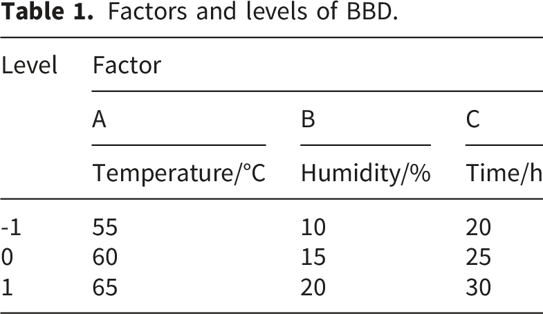
In order to obtain the most favorable drying conditions for polysaccharide retention in C. militaris substrate, the drying temperature, humidity, and time was designed as single factor variables respectively, and other factors remained unchanged. The drying equipment used is STIK precision blow drying oven (IBAO-150H, Stikgroupllc Co., LTD, Shanghai, China). Spread the substrate evenly on a dry tray (45 × 30 × 2 cm), with a loading capacity of 300 g per tray and the thickness of substrate is 1 cm. Using the fixed value mode (its convection mode is ‘Forced’, heating power is 1800w) to set temperature is 45, 50, 55, 60, 65, 70 and 75°C, the humidity is 5%, 10%, 15%, 20%, 25% and 30%, and the time is 10, 15, 20, 25, 30 and 35 h. Three parallel experiments were set in each group with the content of polysaccharide as index, providing reference for subsequent Box-Behnken design (BBD) experiment parameter setting. The 100 g substrate was weighed for single factor experiment, respectively. The processed sample was weighed to calculate the water content (initial moisture content was 87.53%), and ground into powder with liquid nitrogen to determine the polysaccharide content. Finally, according to the water content to convert the polysaccharide content.
2.10. Response surface method experimental design
Factors and levels of BBD.
2.11. Statistical analysis
SPSS 25.0 and Excel 2019 were utilized as the software to analyze the data. The volatile data were retrieved in the NIST mass spectrometry library of Xcalibur. Combined with the manual chromatographic analysis and identification, the peak area of each volatile component was determined by the normalization method. Response surface plot was drawn by Design Expert 13.0 software. All experiments were repeated and measured in triplicate. The results are presented as mean ± standard deviation, the statistical significance of the difference between the means of individual groups was assessed using a one-way analysis of variance (ANOVA) with F-test and performed Tukey posthoc tests, p < 0.05 was considered significant.
3. Results and discussion
3.1. Polysaccharide and cordycepic acid contents determination
Contents of polysaccharide, cordycepic acid, adenosine, cordycepin, ash, protein and crude fat.
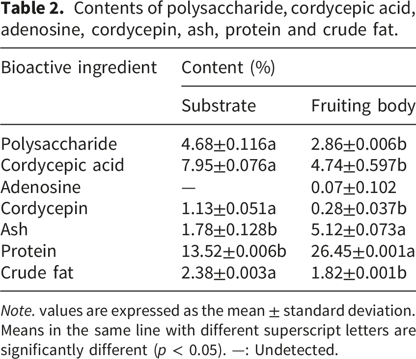
Note. values are expressed as the mean ± standard deviation. Means in the same line with different superscript letters are significantly different (p < 0.05). —: Undetected.
3.2. Results of adenosine and cordycepin content determination
The HPLC chromatogram of the samples are shown in Figure 1, adenosine and cordycepin concentrations are presented in Table 2. There were differences in adenosine and cordycepin contents between the fruiting body and the substrate. The content of adenosine was 0.07% in the fruiting body and not detected in the substrate. The cordycepin concentration was 0.28% in the fruiting body and 1.13% in the substrate. The adenosine concentration was lower than the concentration of cordycepin. The content of cordycepin in the substrate was significantly higher than that of fruiting body (p < 0.05), the difference can be more than 4 times. The result contrasts with the previous studies. Karol et al.
35
found that the cordycepin content in the culture substrates of four strains of C. militaris used in the experiment was significantly lower than that in fruiting body. This markedly different outcome may be attributed to the variations in fungal strains, culture substrate, culture periods and extraction methods. According to previous studies, different strains exhibit differences in morphology, growth conditions and metabolites. These differences may lead to their distinct preferences in the utilization of substances during the cultivation process, and there are also significant differences in their ability to produce active substances. In the same culture substrate, the cordycepin content in the fruiting body and substrate of sample 1 was 85.83 mg/100 g d.w. and 0.81 mg/100 g d.w., respectively. In contrast, the content in sample 3 eached 377.82 mg/100 g d.w. and 31.06 mg/100 g d.w.
35
The type of substrate also has different effects on the production of cordycepin. Kang et al.
36
found that regardless of the strain type, the C. militaris produced the highest amount of cordycepin on the silkworm pupa medium, followed by the brown rice medium and potato dextrose broth resulted in the lowest. And adding different minerals, nitrogen sources, growth factors, etc. to the culture medium can all contribute to the production of cordycepin. Li et al.
37
utilized extensive targeted metabolomics techniques to identify metabolites in C. militaris cultured for different durations, discovering that 61 metabolites, including cordycepin and 2′-deoxyadenosine, exhibited a sustained upregulation trend over time during the experimental period. It indicates that the accumulation of substances is closely related to the duration of cultivation. Cordycepin is an alkaline substance. Different extraction methods may cause varying degrees of loss or damage to it, resulting in differences in its content. Je˛drejko et al.
38
found 556 mg/100 g d.w. of cordycepin in the dried whole fruiting body. However, the content of cordycepin ranged from 354.8 mg/100 g d.w. to 616.6 mg/100 g d.w. in the methanol extract and water extract. These results might be due to the individual heterogeneity of the sample in this study or the limitations of the testing technique. Whether the results are coincidental requires further in-depth exploration with more samples. HPLC chromatograms of adenosine and cordycepin. (A) Adenosine standard. (B) Cordycepin standard. (C) Fruiting body. (D) Substrate.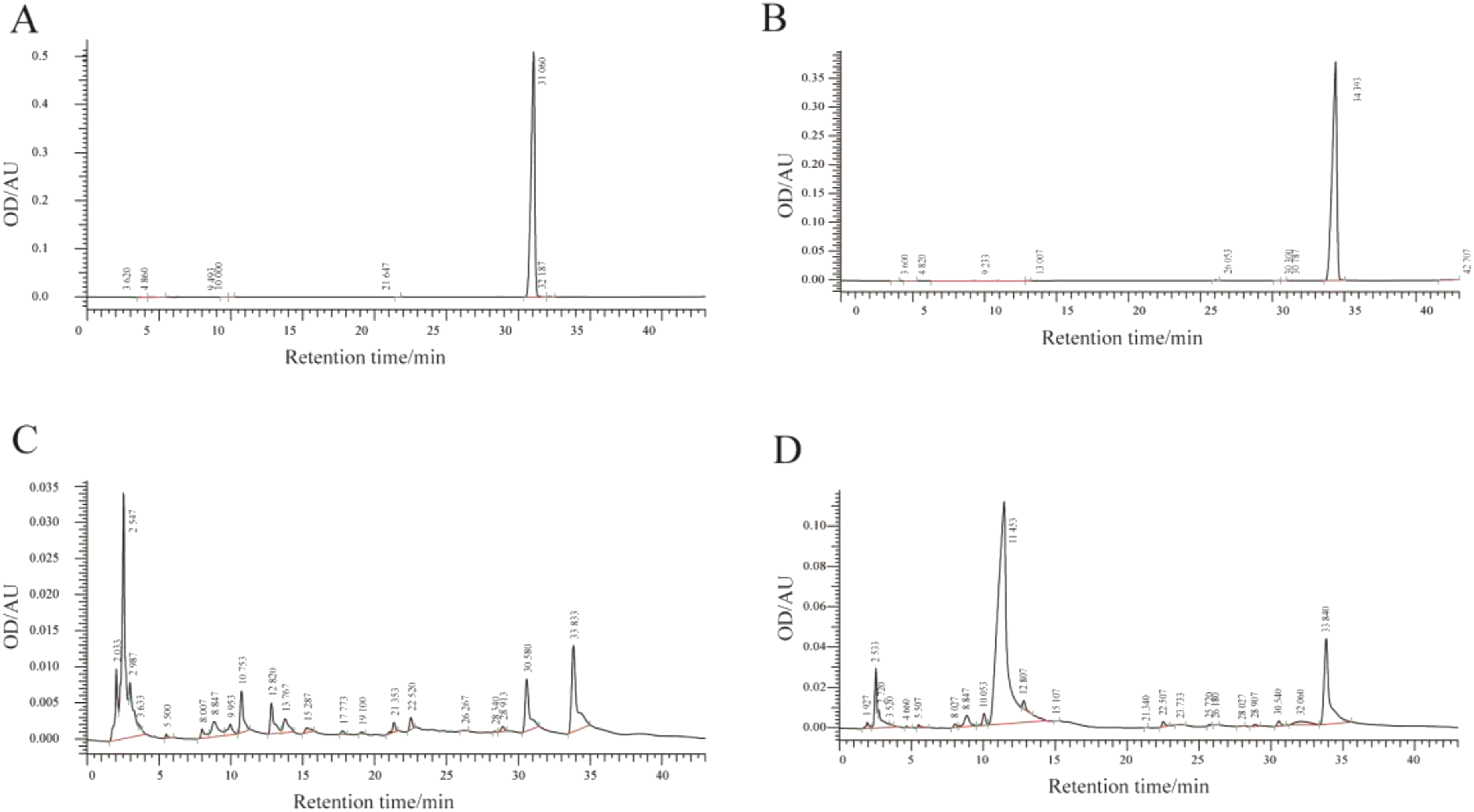
Combined with the existing literature, the initiation of the synthesis pathway of cordycepin can be explained as glucose or adenosine, thus the adenosine may be converted into cordycepin, which may also be explained in part why adenosine don’t detect in the substrate in this study.39,40 In addition, substrates were spread throughout mycelia, and mycelia were rich in cordycepin, this also explain the high cordycepin content in the substrate. 41 As for the absence of adenosine in the substrate, in addition to the synthetic cordycepin just mentioned, it may also be due to the unstable decomposition of adenosine, resulting in its concentration was too low to be detected. 42 Nevertheless, the numerous results mentioned above suggest that post-cultivation substrates, in some cases, should not be considered waste for disposal but rather valuable materials for secondary reuse, serving as an alternative source for obtaining bioactive substances. For example, the substrate can be used to extract cordycepin, brewing and developing functional health food, processing into health wine and so on.43–45
3.3. Determination of ash, protein and crude fat content
The ash, protein, and crude fat contents of samples are presented in Table 2. Contents of ash and protein in the substrate were significantly lower than that in the fruiting body (p < 0.05). The protein content in the fruiting body is approximately 1.96 times that of the substrate, and the ash content even reaches 2.88 times. This may be due to the nutrients contained in the wheat substrate (such as carbohydrates, proteins, etc.) being decomposed and absorbed by the mycelium during the cultivation of C. militaris, and then converted into the nutritional components of the fruiting body. It reflects the accumulation of substances during the formation of the fruiting body. This might also be related to the detection method used in this study, as the Kjeldahl legal nitrogen method measures the total organic nitrogen content, rather than just the protein nitrogen. For the samples treated by the burning method, the nutrients and bioactive ingredient in the substrate and fruiting body are volatilized during the combustion process. The main components in the ash are inorganic salts. The ash content of fruiting bodies was significantly higher than that of substrate, which might be related to the accumulation of minerals in the fruiting bodies. Secondly, the samples used in the experiment were the dried fruiting bodies and substrates. Whether there was any loss of substances during the sample pre-treatment process that led to this result needs to be further explored. However, the content of crude fat was rich in the substrate, which was significantly higher than that of fruiting body (p < 0.05). This may be due to the fermentation of C. militaris, the starch in wheat was consumed and more fat be synthesized. 46
3.4. Free amino acid content detection
The chromatograms of the standard substance, control and experimental group are shown in Figure 2, amino acid compositions are presented in Table 3. All the 17 amino acids achieved good separation effect, both the substrate and fruiting body contained 7 essential amino acids, which were lysine, phenylalanine, threonine, isoleucine, valine, leucine, and histidine, respectively. In the control group, the content of individual amino acids in the substrate and fruiting body of C. militaris ranged from 0.01 to 6.58 mg/g and from 0.07 to 31.67 mg/g, respectively. The total content of amino acid (dry weight) was lower in the substrate (50.48 mg/g) than in the fruiting body (183.01 mg/g). The total content of amino acids in the substrate was significantly lower than that in the fruiting body, and the ratio of essential amino acids to total content of amino acids was 41.26%. The most abundant amino acids of C. militaris were glutamic acid, aspartic acid and glycine in the substrate, and glutamic acid, aspartic acid and tyrosine in the fruiting body. The content of glutamic acid and aspartic acid in substrate were significantly lower than that in fruiting body. Both the substrate and fruiting body contain four types of umami amino acids (aspartic acid, glutamic acid, glycine and alanine). The total content of these umami amino acids in the substrate is 19.23 mg/g, while the total content in the fruiting body reaches 73.26 mg/g, which is much higher than that in the substrate. These umami amino acids may be one of the sources of unique umami flavor of the fruiting body. Chromatograms of free amino acids in the control and experimental groups. (A), (B), (C) represent the free amino acids of the standard substance, fruiting body, and the substrate in the control group, respectively. (D), (E), (F) represent the free amino acids of the standard substance, fruiting body, and the substrate in the experimental group, respectively. (The amino acid names of each peak are abbreviated, which full name are shown in Table 3.) Amino acid composition and content in substrate and fruiting body (dry matter content, mg/g). Note. *indicates essential amino acid. #indicates umami amino acid. Values are expressed as the mean ± standard deviation. Capital letters indicate significant differences in amino acid contents of the substrate and fruiting body among the same treatment groups. Lowercase letters indicate significant differences in amino acid content of fruiting body among different treatment groups. ♦♦ and ♦ indicate significant differences in amino acid content of substrate among different treatment groups. All significant differences are with p < 0.05.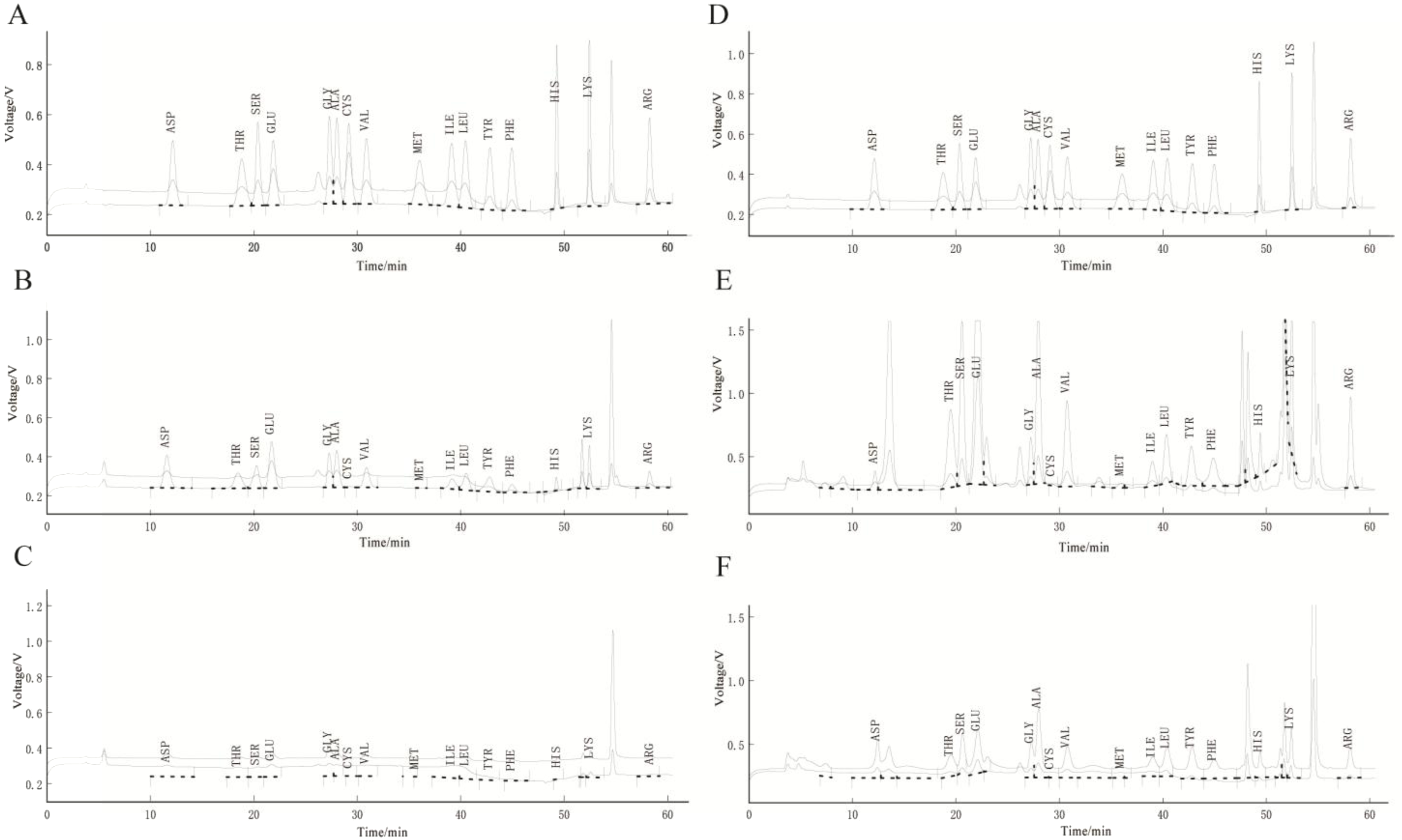
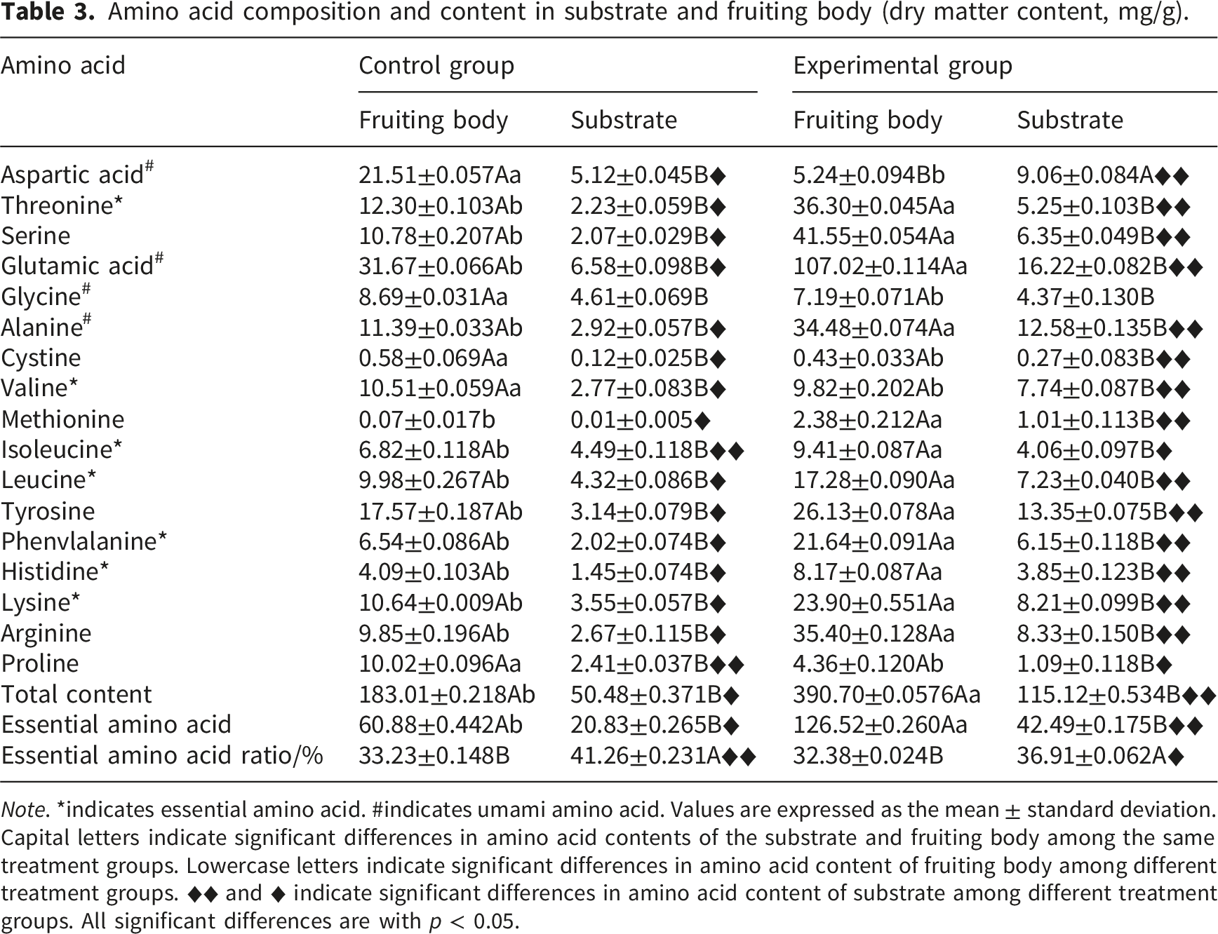
In the experimental group, the content range of individual amino acids in the substrate and the fruiting body was from 1.01 to 16.22 mg/g and from 0.43 to 107.02 mg/g, respectively. It shows a significant increase compared to the control group. However, the most abundant of amino acids in the substrate change to glutamic acid, alanine and tyrosine, while the amino acids that are most abundant in the fruiting body change to glutamic acid, serine and threonine. The total content of umami amino acids in the substrate and fruiting body reached 42.23 mg/g and 153.93 mg/g, respectively. In the acidic experimental environment, the substrate did not produce as much free amino acids as the fruiting body did. However, compared with the control group, the total content of amino acid content was approximately 2.28 times of that (the fruiting body was about 2.13 times). As an important participant in flavor substances, free amino acids can be used as nutritional supplements and feedstock for aquaculture, which are widely used in food, medicine, and animal husbandry industries.47,48
It is worth noting that the proportion of essential amino acids in the substrate was higher than that in the fruiting body, regardless of the experimental group or control group. From this perspective, the substrate is also rich in nutritional value. The high-content components in the wheat substrate after C. militaris cultivation can be developed and utilized in a targeted manner to provide high-quality raw materials for food and health products.
Comparison of experimental data revealed that the content of some amino acids (such as glycine and proline) in the experimental group was lower than that in the cntrol group, which probably because that their structures were damaged and lost during the hydrolysis process. Alternatively, these free amino acids may be involved in oxidation reactions or combine with metal ions to form insoluble or non-free forms, resulting in a decrease in their content during detection.
3.5. The volatile compounds of substrate and fruiting body by GC-MS
Relative content of volatile components in substrate and fruiting body.
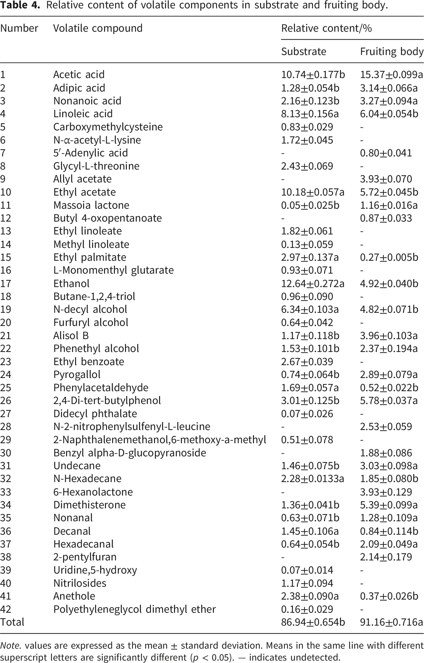
Note. values are expressed as the mean ± standard deviation. Means in the same line with different superscript letters are significantly different (p < 0.05). — indicates undetected.
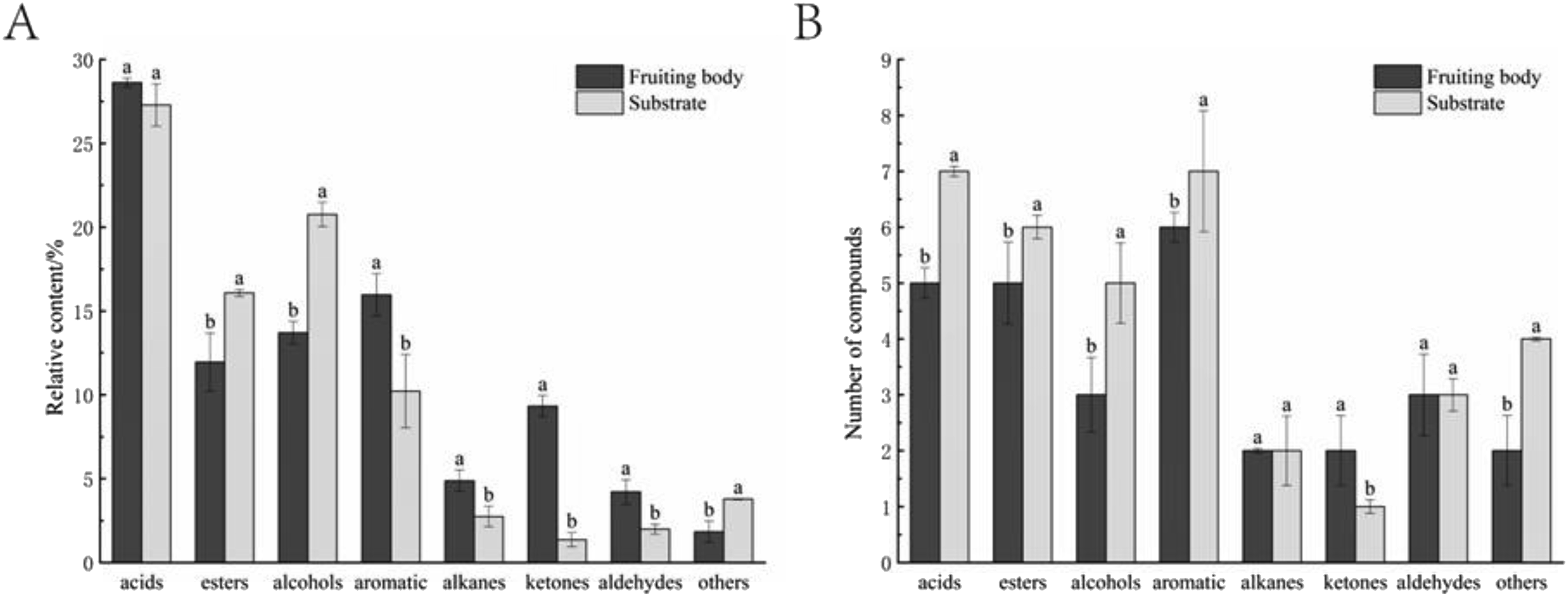
The relative content (A) and number (B) of various compounds in the substrate and fruiting body.
The esters and alcohols are the main volatile components in the substrate, which types and contents are higher than the fruiting body. The pharmacologically active components of alismol B and amygdalin were detected in the substrate. Alisol B is a novel and potentially therapeutic compound for orthopedic diseases, with functions such as treating hepatitis and lowering blood sugar. 49 Amygdalin can specifically inhibit the increase of blood glucose caused by arurea and has anticoagulation effect. 50 In addition, other volatile components were also detected, such as linoleic acid, which is an essential fatty acid in human and animal nutrition. 51 Linoleic acid also can be used in medicine to treat hyperlipidemia and arteriosclerosis,52,53 thus the substrate of C. militaris is a better source of essential fatty acids and has potential medicinal value.
3.6. Single factor experiment for drying conditions
The polysaccharide content increased with the increase of temperature within the range from 45°C to 60°C, indicating that the polymer movement was faster and the polysaccharide dissolution rate was higher. The content of polysaccharide decreased when the drying temperature exceeded 60°C, indicating that the structure of polysaccharide was destroyed and the content of polysaccharide decreased when the temperature was too high. When the drying temperature was 60°C, the polysaccharide content was the highest (9.90%) (Figure 4(A)). Effects of drying temperature (A), humidity (B), and time (C) on polysaccharide contents.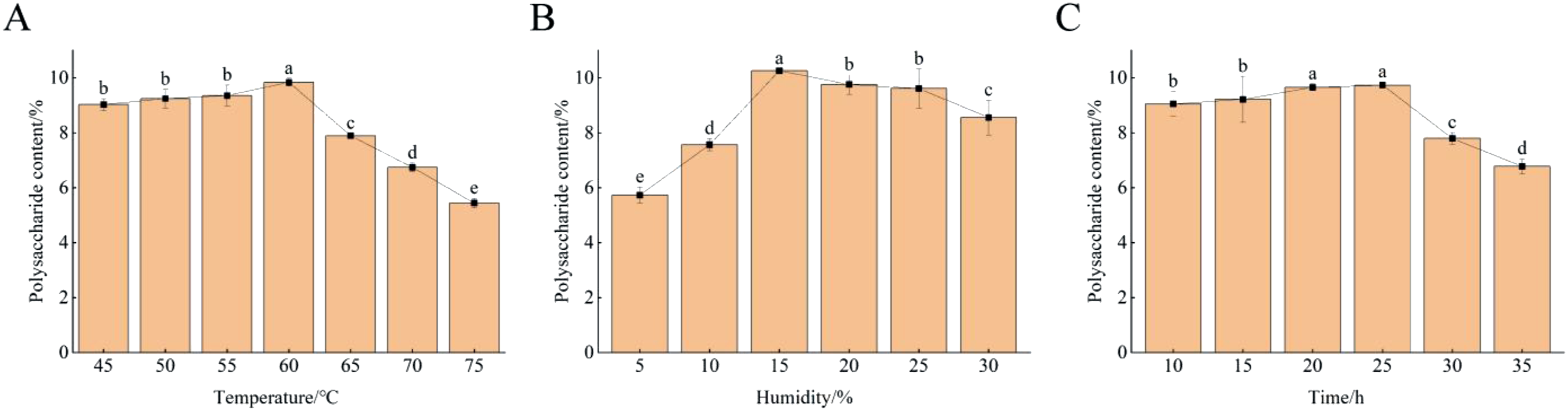
When the drying humidity was 5∼15%, the polysaccharide content increased with the increase of humidity, and decreased when the humidity was more than 15%. The highest polysaccharide content was 10.20% when the humidity was 15% (Figure 4(B)).
The polysaccharide content was significantly increased when drying time was 10∼20 h, and almost unchanged when drying time was 20∼25 h, indicating that polysaccharide could be further separated with the increase of drying time, but this was limited. The time was inversely proportional to the polysaccharide content when the drying time between 25 and 35 h, which might be because the polysaccharide degradation occurred under the action of high temperature for a long time. When drying time was 25 h, the polysaccharide content was the highest (9.86%) (Figure 4(C)).
3.7. Response surface test
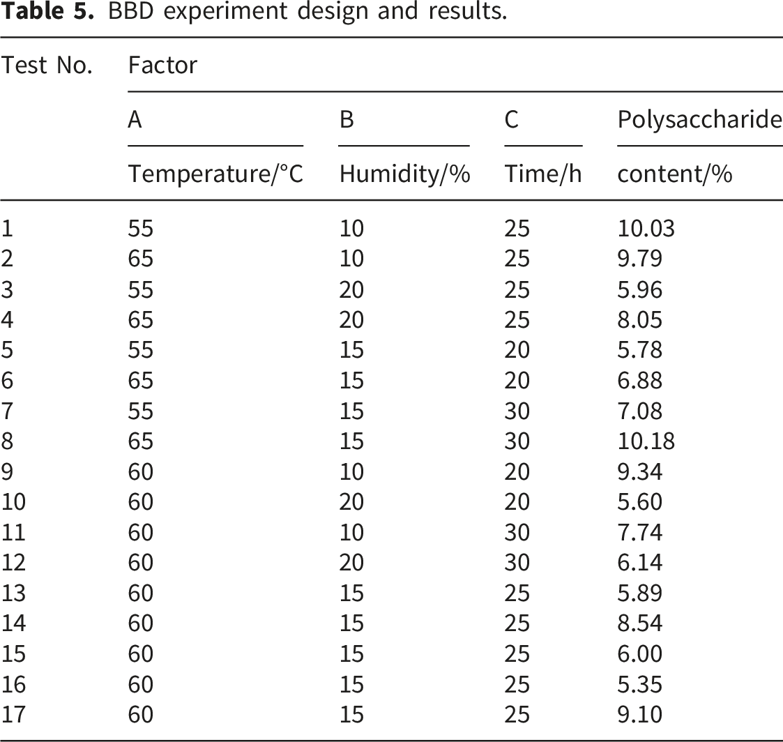
BBD experiment design and results.
Response surface regression model analysis of variance.
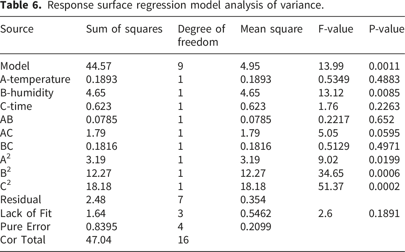
According to the response surface shape fitted by the pairwise interaction of experimental factors (Figure 5). The steeper the response surface curve is, the more significant its influence is, and the smoother the curve is, the less its influence is. The openings of all figures are downward, indicating that drying temperature, humidity, and time all affect the content of polysaccharide in the substrate among the experimental parameters. The influence of each factor on polysaccharide content was B (humidity)>A (temperature)>C (time). Response surface diagram of influence of humidity and temperature (A), temperature and time (B), humidity and time (C) on substrate polysaccharide content.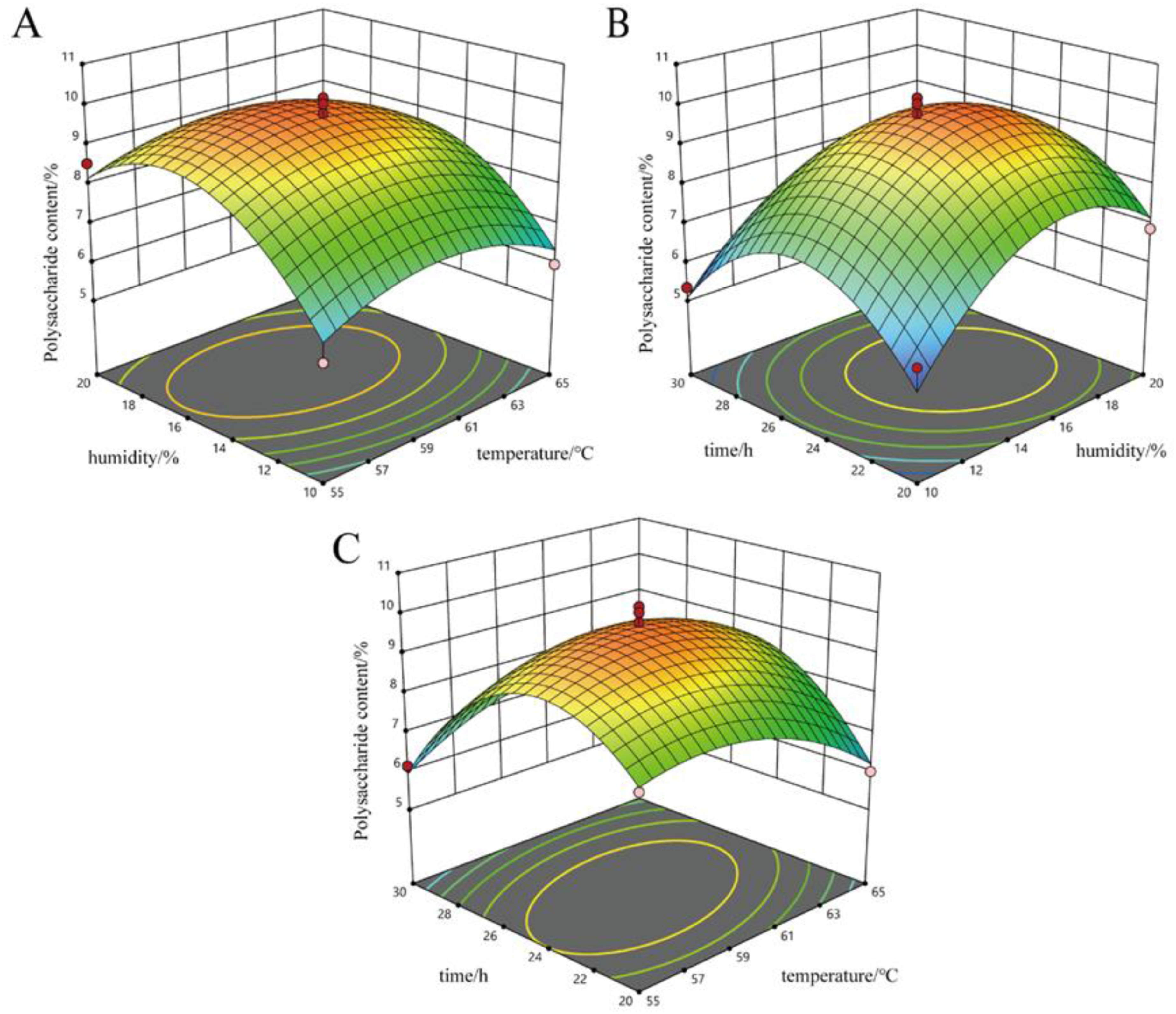
According to the regression model, the optimal drying conditions of substrate are as follows: the drying temperature was 59.268°C, the humidity was 16.177%, the time was 24.485 h. Under these conditions, the polysaccharide content was 9.803%. Combined with the actual situation, the parameters were adjusted appropriately. The adjusted drying conditions are as follows: the drying temperature was 59°C, the humidity was 16%, the time was 24 h. Under this condition, the actual polysaccharide content was 9.779%. It’s close to the theoretical predicted value, indicating that the model is true and reliable.
4. Conclusion
This study found that the culture substrates after harvesting C. militaris fruiting bodies have similar nutrient compositions to the fruiting bodies. Contents of bioactive components in the substrate were still high, even higher than the fruiting body. These beneficial components can be extracted and separated from the substrate to prepare antioxidants and immunomodulators, so as to fully realize the efficient resource utilization of C. militaris substrate in food processing, medical care, and other aspects. It is suggested that targeted development and utilization of high content of bioactive ingredients in C. militaris wheat substrate should be carried out, providing high-quality raw materials for food and health products, and reducing substrate pollution caused by cultivation of C. militaris. Finally, the results showed that the best drying conditions of substrate: temperature is 59°C, humidity is 16%, and time is 24 h. Too high temperature, humidity, and time were not conducive to the retention and quality of substrate.
Supplemental material
Supplemental material - Exploration of the potential utilization value in Cordyceps militaris substrate after industrial cultivation in terms of nutritional and pharmaceutical applications
Supplemental material for Exploration of the potential utilization value in Cordyceps militaris substrate after industrial cultivation in terms of nutritional and pharmaceutical applications by YouCui Yang, Lu Qiao, Min Yao, YeMing Zhou, JiaoJiao Qu, Hui Dai, Gui Gao, YaLi Guo, Xiao Zou in Science Progress
Footnotes
Acknowledgements
We are grateful to members of the Institute of Fungus Resources, Guizhou University, For their help with factory and laboratory cultivation. All authors declare that no artificial intelligence (AI) tools were used to generate or modify any scientific data in the text, images, or other content of the paper. And authors are fully responsible for ensuring the accuracy, originality and integrity of all submitted material.

Consent for publication
All authors agree to publish this article in the Science Progress.
Author Contributions
Y.C.Y. was responsible for article writing and data collation. L.Q. was responsible for experimental and data processing. M.Y. was responsible for data collection. H.D, G.G. and Y.L.G. was responsible for acquiring funding and managing projects. J.J.Q. was responsible for proofreading the manuscript and approving the final version. Y.M.Z was responsible for conceiving and designing the experiment. X.Z. was responsible for revising the article, approving the final version and acquiring funding. All authors have read and agreed to the published version of the manuscript.
Funding
The authors disclosed receipt of the following financial support for the research, authorship, and/or publication of this article: This research were funded by Guizhou Tabacco Company Qianxinan Branch (Zhong Yan Qian Ke [2023] No 6) and Guizhou Guiwang Biotechnology Limited Company (Zun Shi Ke He CXZX [2025] No 3).
Declaration of conflicting interests
The authors declared no potential conflicts of interest with respect to the research, authorship, and/or publication of this article.
Data Availability Statement
All data generated or analyzed during this study is included in this published article and its supplementary information files.
Supplemental material
Supplemental material for this article is available online.
References
Supplementary Material
Please find the following supplemental material available below.
For Open Access articles published under a Creative Commons License, all supplemental material carries the same license as the article it is associated with.
For non-Open Access articles published, all supplemental material carries a non-exclusive license, and permission requests for re-use of supplemental material or any part of supplemental material shall be sent directly to the copyright owner as specified in the copyright notice associated with the article.
