






Abstract
A 70-year-old woman presented with progressive skin lesions on the face, limbs and trunk in the absence of systemic illness. Three months earlier, she had been prescribed six months prophylactic nitrofurantoin for recurrent urinary tract infections, treated with nitrofurantoin and trimethoprim. Positive immunology and histological inflammatory changes in a skin biopsy were consistent with a diagnosis of sub-acute cutaneous lupus erythematosus. Following treatment with topical steroids, the skin lesions regressed, but alopecia followed and required hydroxychloroquine. One year later, there are no new skin lesions and no evidence of systemic lupus erythematosus. Nitrofurantoin is associated with many side effects and hypersensitivity reactions. Possible drug-induced lupus reactions due to nitrofurantoin include pneumonitis, blood disorders and hepatotoxicity. This is the only published case of isolated sub-acute cutaneous lupus following maintenance nitrofurantoin.
Keywords
Case presentation
A 70-year-old woman presented to her general practitioner (GP) with a six-week history of a slightly itchy purple rash ‘creeping’ over her face and neck (Figure 1). The rash was preceded by a scaly ‘butterfly rash’ over her cheeks and forehead (Figure 2). Lesions then appeared on her arms and back (Figure 3). Alopecia followed.
Serpiginous rash. ‘Butterfly’ rash. Annular lesions.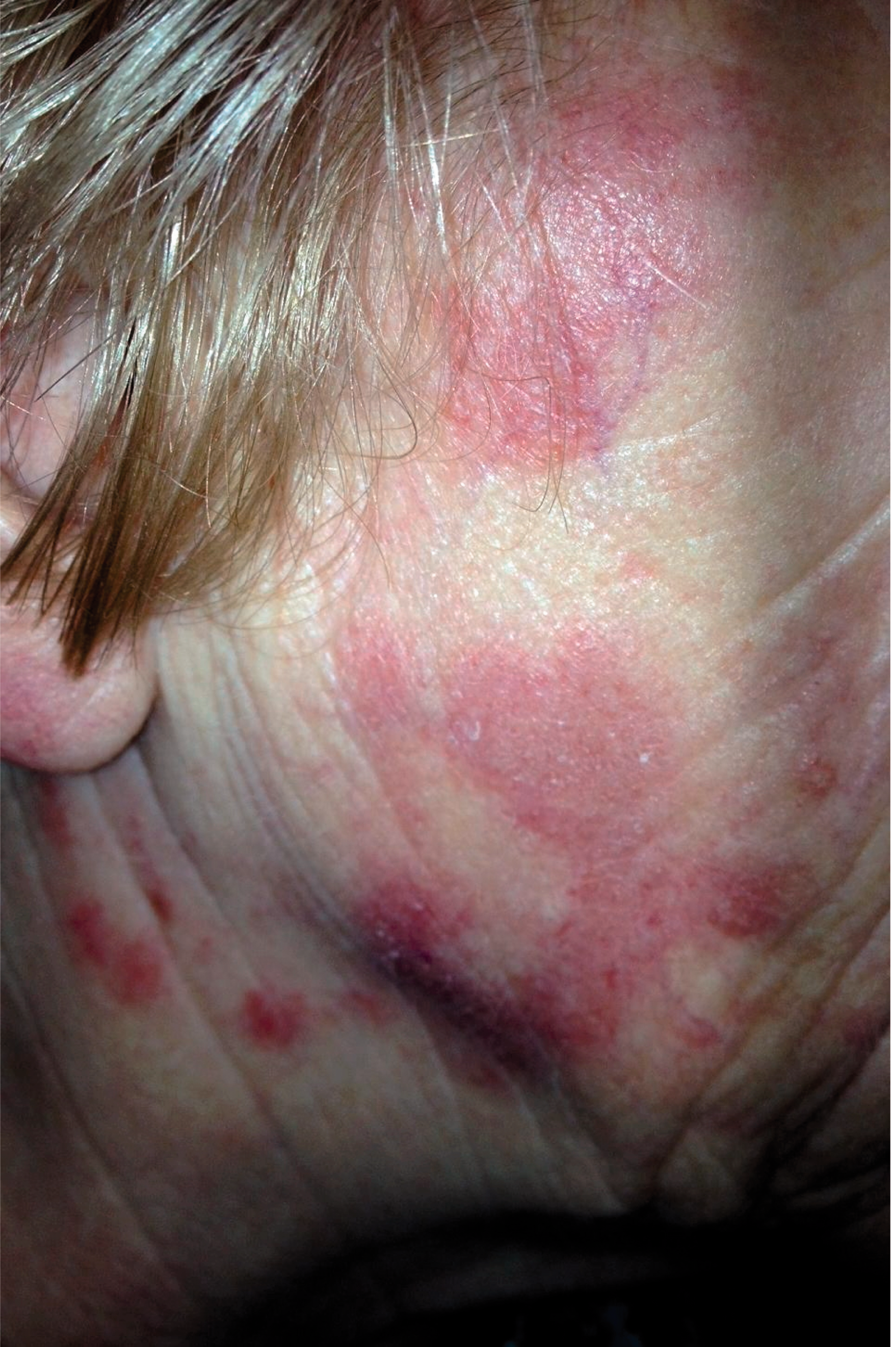
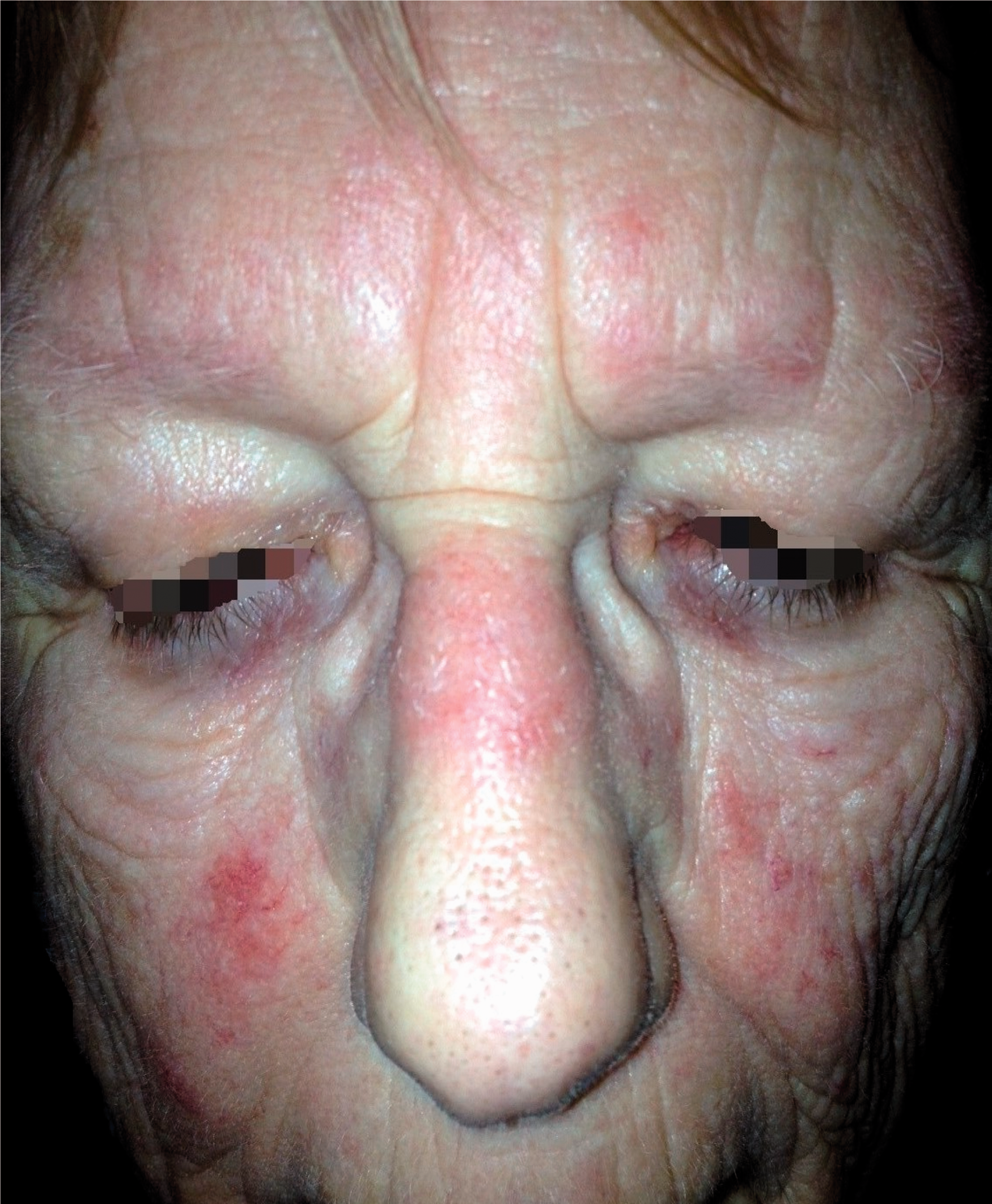
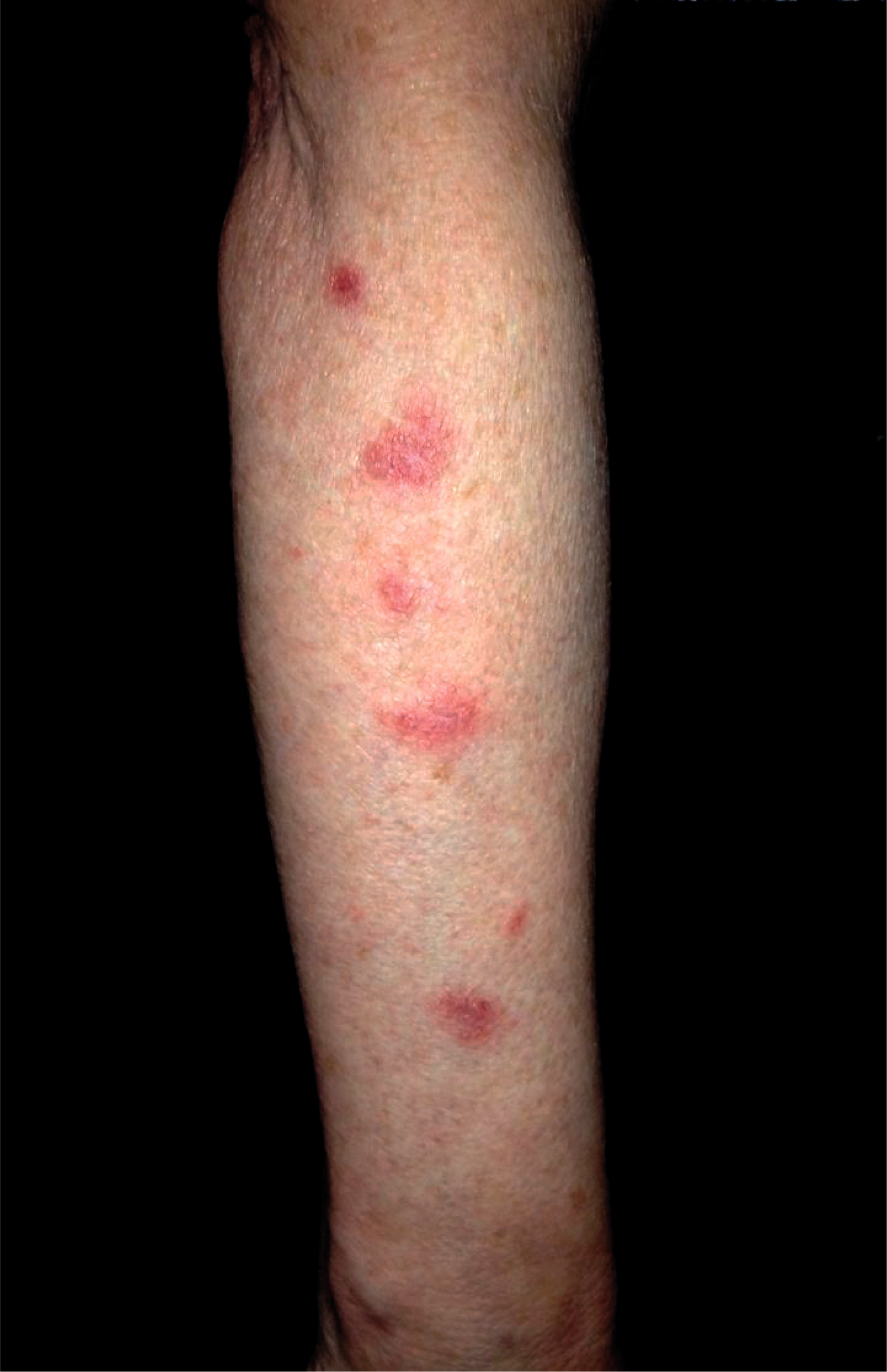
Three months prior to her presentation, she had been prescribed a six-month maintenance dose of nitrofurantoin for prophylaxis against recurrent urinary tract infections (UTIs). In the preceding five years, there had been nine UTIs treated with six courses of trimethoprim and three courses of nitrofurantoin.
She was otherwise well with no fever, dyspnoea or neurological symptoms. There was no other significant medical history, particularly no known hypersensitivity to nitrofurantoin and no renal impairment. There was no family history of systemic lupus erythematosus (SLE) or connective tissue disease.
Other than the nitrofurantoin and trimethoprim, she had not received any other prescribed medication and specifically, none associated with drug-induced lupus (DIL) in the preceding 12 months. Physical examination was unremarkable. The GP prescribed a mild topical steroid and referred the patient to dermatology with the clinical suggestion that she had sub-acute cutaneous lupus.
Differential diagnosis
Sub-acute cutaneous lupus erythematosus Tinea corporis Lyme disease (erythema migrans) Erythema annulare Lichen planus Drug reaction
Investigations
The patient’s full blood count was normal. There was no eosinophilia or leucopenia. Her erythrocyte sedimentation rate (ESR) was 19. There was no evidence of hepatotoxicity or renal impairment. Her auto-antibodies showed weak positive speckled anti-nuclear factor (ANF) with positive anti-Ro and anti-La antibodies and an elevated rheumatoid factor (RF). Other immunological investigations included IgM 1.29, IgA 3.82 g/L and IgG 9.1 g/L.
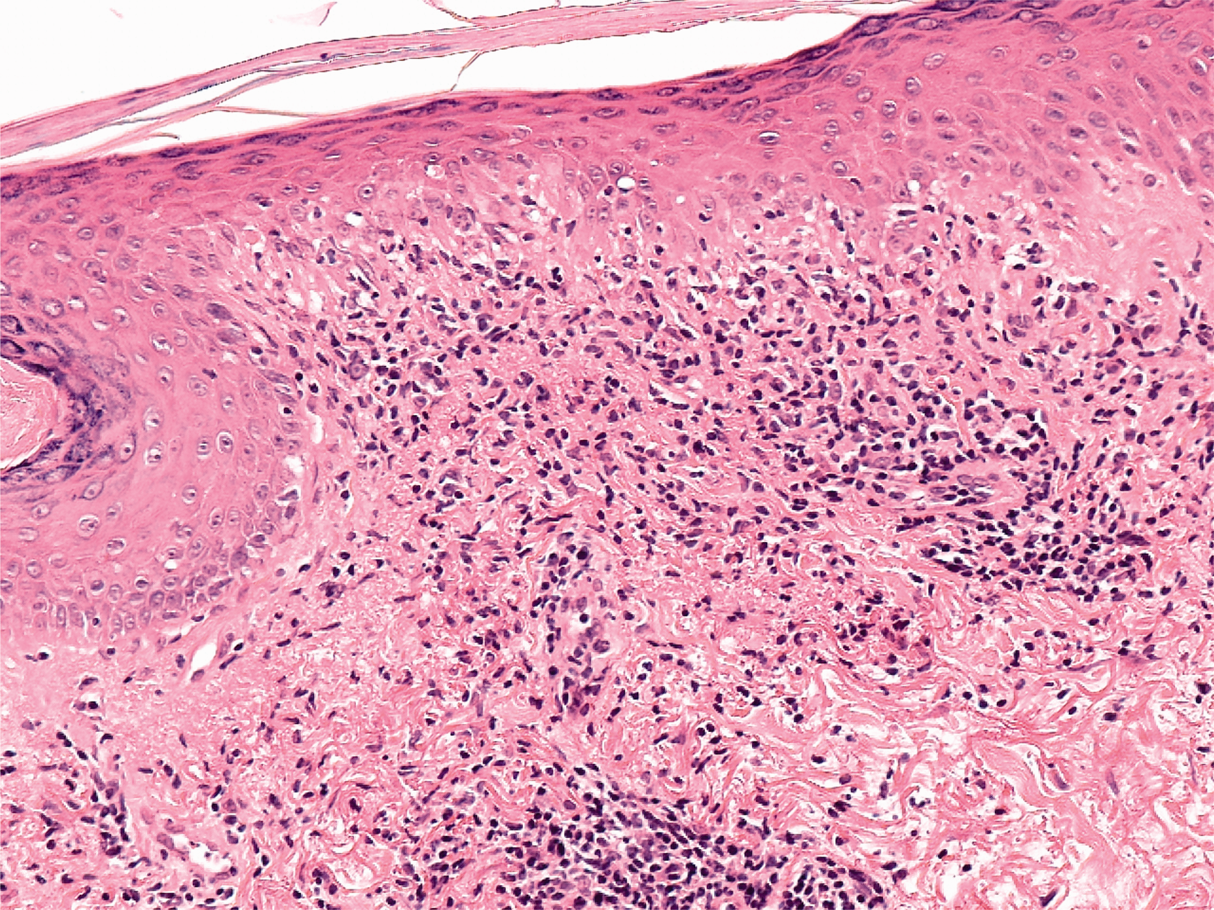
A 4-mm punch biopsy, 4-mm deep of skin from the right arm showed inflammation in the superficial and mid-dermis (Figure 4). High-power magnification confirmed a lichenoid reaction pattern with basal cell damage and vacuolar change. The inflammatory cells comprise predominantly lymphocytes (Figure 5).
Low-power magnification (×40) showing inflammation in the superficial and mid dermis. High-power magnification (×200) showing lichenoid reaction pattern.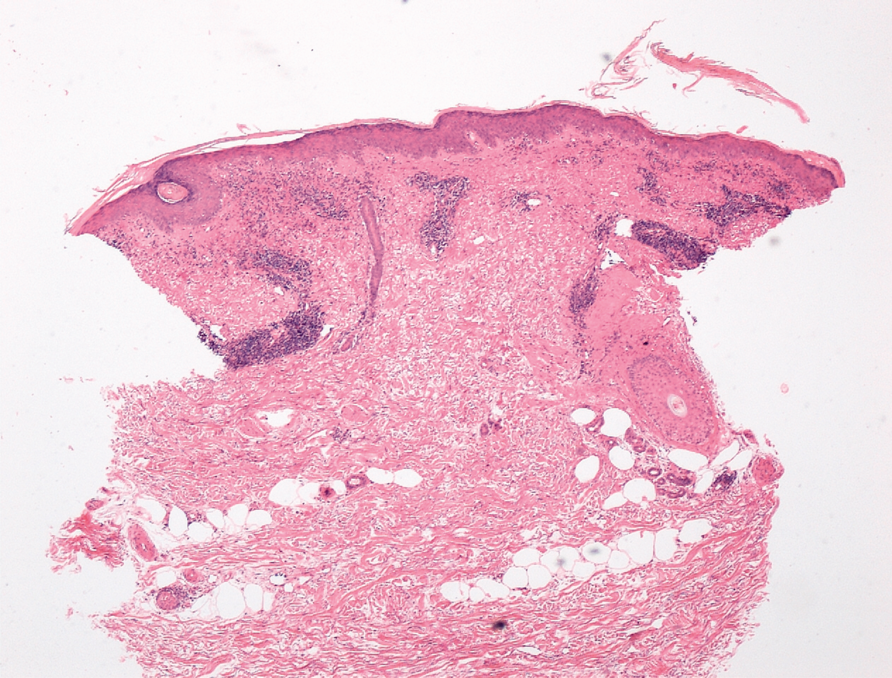
Diagnosis
The overall features were compatible with the ‘clinical suggestion’ of sub-acute cutaneous lupus erythematosus, which was supported by immunology and pathology. The question was raised whether the link to nitrofurantoin was coincidental rather than causative.
Outcome and follow-up
Over the course of the following three months, her condition was monitored. Her ESR rose to 48 and her ANF remained weak positive with positive anti-Ro and anti-La antibodies. Additionally, anti-Sci-70 and anti-Jo-1 were negative. The anti-nuclear antibody analysis conclusion was that her condition was ‘consistent with lupus erythematosus’.
The patient’s rash responded well to potent topical corticosteroid preparations betamethasone (as valerate) 0.1% cream and fluocinolone acetonide 0.025% ointment, both twice daily and emollients. She was able to go on holiday to Spain and did not experience problems with solar exposure, probably because she adhered to advice regarding sun block.
Alopecia appeared six months later. It was associated with scaling, but no obvious scarring. Mycology was negative and there was no response to terbinafine. In order to prevent further hair loss, she was commenced on hydroxychloroquine 200 mg once daily.
At one-year follow-up, her skin rash has cleared, the alopecia has been arrested and she is tolerating hydroxychloroquine. There are no clinical manifestations of SLE. Haematology, liver and renal function, chest x-ray, spirometry and electrocardiogram are normal. The patient will continue to be screened for systemic involvement.
Discussion
Case study
To our knowledge, this is the first published report of isolated sub-acute cutaneous lupus associated with intermittent and then prolonged nitrofurantoin therapy. The risk of adverse reactions to nitrofurantoin is reported to increase with age and with being female, as in this case. 1 The accuracy of the diagnosis is supported by a positive ANF, which is a sensitive screening tool for SLE. However, ANF is non-specific and may be present in the absence of SLE. Similarly, skin biopsy may assist is the diagnosis, but is also non-specific. 2
Whether the link to nitrofurantoin was coincidental rather than causative is debated, but the evidence presented suggests that nitrofurantoin caused a DIL syndrome, after previous sensitisation within the preceding five years. Additionally, rather than the condition disappearing, when the nitrofurantoin was discontinued, this case demonstrates a latent effect three months after six months maintenance therapy was withdrawn. The reaction usually requires weeks or months of treatment of the offending drug 3 and importantly, it may take up to two years for clinical and serological remission. 4
Nitrofurantoin and DIL
Nitrofurantoin is effective and inexpensive. Increasing concerns about cost minimisation, bacterial resistance to antibiotics, methicillin-resistant Staphylococcus aureus and Clostridium difficile infections have resulted in nitrofurantoin being used more frequently as first-line therapy for and prophylaxis against UTIs. The British National Formulary already warns prescribers of a ‘possible association’ between nitrofurantoin and a lupus-like syndrome. 5
There are over 40 medications known to cause DIL: hydralazine, procainamide and isoniazid being the top three. Nitrofurantoin, but not trimethoprim, is included as a causal agent. The risk associated with nitrofurantoin is considered to be ‘very low’. 6
Pulmonary reactions to nitrofurantoin were first reported in 1962 by Israel and Ireland. 7 In 1975, Selroos and Edgren 8 also described chronic pulmonary reactions, which developed into full-blown lupus-like syndromes in three patients, all of whom had been prescribed maintenance nitrofurantoin therapy for UTIs. These were considered type III immunological reactions following the re-administration of nitrofurantoin in previously sensitised patients. It follows that pulmonary function should be monitored in patients receiving long-term maintenance therapy with nitrofurantoin. 9
Other toxic and allergic adverse effects include dose-related polyneuropathy more common in people with renal impairment, hepatotoxicity and haemolytic anaemia, aplastic anaemia and angranulocytosis (BNF ref). SIRS (systemic inflammatory response syndrome) and DRESS syndrome (drug rash, eosinophilia and systemic symptoms), a delayed type IV hypersensitivity reaction, have also been reported in connection with nitrofurantoin.10,11
Progression to systemic lupus erythematosus
Diagnostic criteria for SLE.
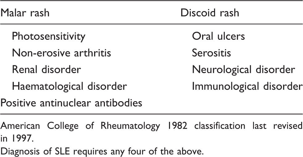
American College of Rheumatology 1982 classification last revised in 1997.
Diagnosis of SLE requires any four of the above.
Conclusion
UTIs are common and nitrofurantoin is frequently prescribed for treatment and prophylaxis. However, cost minimisation and anti-microbial resistance need to be reconciled with nitrofurantoin’s multiple common and rare, but serious adverse effects including SLE. This case report suggests that the relationship between nitrofurantoin and isolated sub-acute cutaneous lupus is causal. It is vital that early DIL is recognised in the course of the disease and that full-blown SLE is avoided.
Learning points
Nitrofurantoin is associated with multiple common and rare, but serious adverse effects including SLE. Isolated sub-acute cutaneous lupus is now reported to be associated with nitrofurantoin. Adverse reactions follow the re-administration of nitrofurantoin in previously sensitised patients. There can be a latent period between treatment and the onset of symptoms. Prescribers should consider the safety of prescribing maintenance nitrofurantoin for recurrent UTIs, particularly if there is renal impairment. Monitoring for systemic side effects, pulmonary function, urinalysis, haematological abnormalities, hepatotoxicity and renal impairment are recommended for patients on long-term therapy.
Footnotes
Funding
This research received no specific grant from any funding agency in the public, commercial, or not-for-profit sectors.
Declaration of conflicting interests
None declared.