
Abstract
This study shows that using a combination of physical and chemical approaches, wool fiber can be systematically broken down to its constituent components. From this it was possible to investigate the antibacterial nature of wool and its two major subcomponents, the cuticle scales and cortical cells. Wool and its constituent components were subjected to two methods of antibacterial testing, with excess and limited amounts of liquid. From this it was shown that the presence of excess water has a significant impact on the antibacterial properties of the wool fiber and its components. Furthermore, the study shows that on treatment with thioglycolic acid, which cleaves disulfide bonds, the fibers exhibit antibacterial activity.
Wool fibers are composite materials with a highly complex physical and chemical composition that has evolved over millions of years to protect sheep from the extremes of heat, cold and rain. With the ability to change the shape of the fiber in response to external stimuli, wool fiber is considered an intrinsically smart material. Wool is classed as a keratin fiber and has two distinct components, the cuticle scales and cortical cells. The physical structure of the wool fiber and of its components are shown in Figure 1. Both have a varying role in the function of the wool fiber and, unsurprisingly, have a drastically different chemical and physical make up that has been described in the literature.1–4 Furthermore, wool has a complex and diverse chemical and morphological structure, resulting in very diverse areas of functionality and properties. When the wool fiber gets wet, water is transported to the cortex via the cell membrane complex, from the hydrophobic cuticle scales. This causes the core of the fiber to swell, in turn causing the cuticle scales to open. This mechanical response from the wool fiber helps protect the animal from environmental challenges.
4
Schematic diagram of wool fiber and its components. Reproduced from CSPIRO, with sowelcome@123me changes to spelling and grammar, under the following license: http://www.scienceimage.csiro.au/pages/about/ CC BY 3.0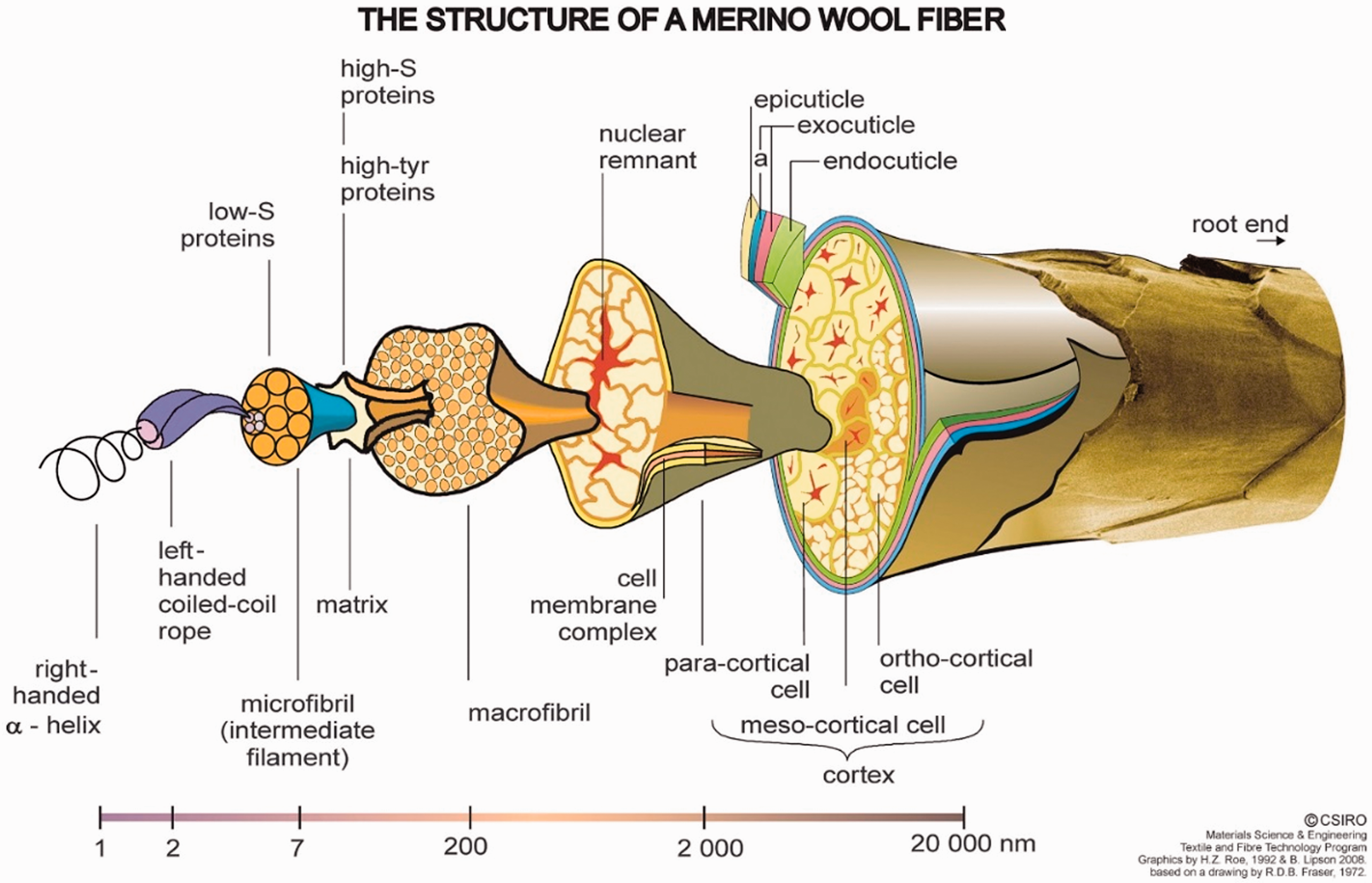
It is also well-known that keratin fibers are particularly inert toward many chemicals and enzymes, which has been ascribed to two main factors5,6: the complex histological structure consisting mainly of cuticle cells, cell membranes and cortical cells, each of which tend to be complementary in inertness toward chemicals; and the high degree of cross-linkage of the cuticle and cortical cells by disulfide bonds originating from cystine. 7 Wool fibers have a truly heterogeneous structure, containing more than 170 different proteins. Each type of protein structure is located in a specific region of the fiber; hydrolyzed wool reveals 18 amino acids. One of the attributes that has been associated with wool is its ability to reduce/resist the onset of odor build up, especially in sport clothing, and therefore it has been associated with having antibacterial properties. However, there is little evidence to support this, with no dedicated research focused on the wool fiber itself; associations alluding to the antibacterial properties are purely anecdotal. 8 Wool fibers and fabrics have nevertheless been used as substrates for antibacterial tests, and the rigorous nature of scientific work has meant that a blank/control sample has been used.9–13 However, it is difficult to make comparisons between the tests made due to the different nature of the wool samples, difference in pre-treatments, types of wool and test methods used to quantify the antibacterial action. Moreover, there is even evidence to show that wool acts as a good host for microbe propagation, 9 which is what common sense would suggest, as wool is a protein-based fiber and therefore a nutrient source for bacteria. In addition to this, it has been shown that a wool fabric under the dynamic shake flask test (SFT) method offers no antibacterial properties. 14 This generates conflicting evidence, as odor is considered to be an indicator of bacterial activity, and wool is reported to be an odor reducing material, 8 but there is clear evidence to the contrary. Moreover, there are several standard methods that can be utilized when determining antibacterial activity; the two most popular for textile applications are the JIS L 1902 Challenge Test Method and ASTM E2149 - 13 a Shake Flask Method. The main difference between the two standards is that the SFT is conducted in an excess aqueous solution, whereas the challenge test is conducted under a limited amount of liquid. In summary of the literature it could be suggested that there are three possible explanations for wool’s supposed antibacterial properties. First, the fiber itself has an antibacterial effect; the epicuticle, lipid monolayer and the cortex have been tentatively suggested as antibacterial. 15 Second, the intrinsic property of wool in having a hydroscopic core and a hydrophobic surface means that the microclimate surrounding the surface of the fiber is hostile to bacterial growth. The third explanation, and one with sound scientific studies supporting it, is that wool components bond to odorous fumes, giving the impression of a lack of bacterial growth,15,16 but not actually preventing or inhibiting bacterial growth. The literature has shown that there is no definite answer to the question of whether wool has antibacterial properties; the intention of this work is to provide significant evidence of wool’s relationship with bacteria. Antibacterial activity is defined in this article as the destruction or inhibiting the growth of bacteria. Therefore, this research concentrated on whether wool fibers are antibacterial and, if so, what mechanisms are utilized by wool. In order for this to be achieved, the wool fiber was reverse engineered into its two major components, the cortical cells, the inner core of the fiber and the cuticles scales that surrounds the cortex. Critical importance was put upon the separation of the major components without damaging their physical and chemical structure.
Experimental procedure and materials
Materials
The merino wool was sourced in sliver form from Schoeller GmbH & CoKG, Hard, Austria; the slivers had been scoured and carded. The fiber diameter, determined through light microscopy, was 18 +/– 3 µm. The wool fiber was washed to remove any impurities that may have migrated to the wool from the processing. The wool was then air-dried and stored in a desiccator, prior to all experimental work.
Preparation of cuticle scales and cortical cells
The two major components of the wool fiber were isolated from each other through a two-stage treatment with formic acid, where 1 g of merino wool fibers were refluxed (100.7℃) for 17 min in 250 ml of 98% conc. formic acid (Carl Roth GmbH + CoKG, Austria). The fibers were then transferred to fresh formic acid at room temperature, 25℃, where upon they were agitated in a laboratory shaker for 16 h. The bulk material, fiber mass, was then separated from the solution, whereupon the resultant solution containing the cuticle scales was filtered using a vacuum pump and a polytetrafluoroethylene (PTFE) membrane filter, pore size 0.45 µm. The cuticle scales where then neutralized and washed before being air-dried. The cuticle stripped fibers, the resulting bulk material from above experiment, underwent ultrasonic degradation in fresh formic acid, at 30℃ for 20 min. This broke the cortex down to the cortical cells. The cortical cells were then neutralized and rinsed.
Preparation of reduced wool
Untreated wool was reduced with thioglycolic acid (TGA) (>98% concentration, Sigma-Aldrich, Austria). A total of 1 g of wool was submersed in a 1 l solution, comprising 0.2 M TGA and 0.1 M Na2HPO4, adjusted to pH 10, with NaOH. The wool was submersed for 16 h in standard laboratory conditions. At the termination of the experiment the fibers were filtered, oven dried at 105℃ and stored in a desiccator.
Examination of bactericidal properties
Two textile industry standard methods of testing of antimicrobial activity were utilized in this study, the JIS L 1902 Challenge Test Method and the ASTM E2149-10 Standard Test Method for Determining the Antimicrobial Activity of Immobilized Antimicrobial Agents under Dynamic Contact Conditions (dynamic SFT). The major differences between the two tests are as follows: Challenge Tests are performed under bacterial growth conditions in a limited amount of medium at 37° C, whereas the SFTs are performed under non-growing conditions in a high amount of buffer at 25° C. The antibacterial activity of both methods was displayed as the reduction of bacteria according to equation (1)
Challenge Test
Inhibition of bacterial growth was tested by a quantitative Challenge Test according to JIS L 1902 (JIS, 1998), with Staphylococcus aureus (ATCC 6538) as the test organism. Briefly, 0.4 g of wool specimen was inoculated with 0.2 ml of bacteria (approximately 1.0 × 10⁵ cells per ml) in a medium containing 5 g of beef extract, 10 g of peptone, and 5 g of sodium chloride per 1000 ml of distilled water. Test tubes were incubated at 37℃ for 18 h. The number of bacteria was determined after washing out the bacteria with physiological saline solution. The solution was then serially diluted, plated on nutrient agar and incubated at 37℃. Bacteria was determined immediately after inoculation, T = 0, and after incubation for 18 h, T = 18. Tests were performed in duplicate.
Dynamic shake flask test
The dynamic antibacterial testing was conducted under the ASTM E2149-01 standard. Briefly, the specimens were submersed in a 50 ml suspension of Staphylococcus aureus ATCC 6538 (approximately 3.0 × 10⁵) in 0.3 mM KH2P04 at a pH of 7.2. The solution and specimen were then sealed in a flask and shaken at 250 rpm at 25℃, and after 24 h aliquots of the solution were removed from the flasks and mounted on an agar nutrient dish. The number of viable cells was determined by incubation at 37℃ for 24 h. Tests were performed in duplicate. The calculation for SFT is as follows

Analytical procedures
Micrographs of the wool fiber and its components were acquired using Field Emitting Gun Scanning Electron Microscopy (FEGSEM) (Leo 1530 Gemini). The samples were sputter coated with a layer of platinum, between 3 and 5 nm in thickness, prior to examination by FEGSEM. Samples were viewed by FEGSEM under a vacuum, 2.0 × 10-05 Torr.
Attenuated total reflectance Fourier transform infrared spectra (ATR-FTIR) were recorded using a Vector 22 Spectrometer (Bruker Analytik, Vienna, Austria) attached with a diamond crystal ATR (PIKE Technologies) unit. Spectra were recorded in the wavenumber range of 400–4000 cm–1, with a scanning repetition of 64 scans and at a resolution of 4 cm–1.
Results and discussion
The utmost importance was placed on the purity of the samples; therefore, additional analytical methods were used to ensure that the wool treatments had in fact produced the correct results. Figure 2(a)–(d) show the systematic breakdown of the wool fiber into its constituent components. Figure 2(b) shows a fiber that has been subject to the reflux treatment only. From this, we can see the importance of the laboratory shaking process after the reflux treatment. Figure 2(c) shows the isolated cuticle cells that were filtered from the supinate after the wool fibers underwent the laboratory shaking process.
(a) An example of an “as received” wool fiber. (b) Wool fiber that has been exposed to the reflux treatment only, showing a cuticle scale partially removed from the fiber core. (c) A collection of isolated cuticles. d) Isolated cortical cells.
Figure 2(d) shows the resultant material after the ultrasonic treatment. Where individual cortical cells have been released from the cortex, from the image magnification we can assess that they are in fact meso-cortical cells. 4
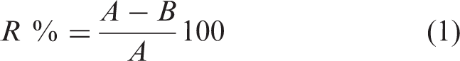
In conjunction with the microscopy, mass loss was also utilized to monitor the processes, as the mass % of the individual components of a merino wool fiber has been well documented.4,17,18 A critical mass loss of 10% by weight is given for the cuticle scales in merino wool. Therefore, optimization of the reflux process to achieve the desired mass loss was required. Figure 3 shows the mass loss against reflux time, where a plateau region can be seen from 60 to 100 min, where the mass loss was 10% +/– 2.
Mass loss against reflux duration.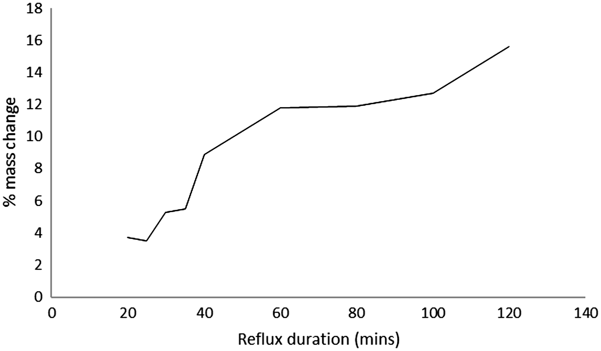
Moreover, the final piece of supporting evidence was the ATR-FTIR data. The traces shown in Figure 4 showed a slight shift at the amide 1 peak 1680 cm–1, which was in accordance with the literature.
18
However, it should be noted that the evidence of FTIR data for distinguishing between a whole fiber and that that has had its cuticle scales removed is not concrete. The cuticle scales have a thickness of 0.3–0.5 µm and the depth of penetration from the PIKE ATR unit ranges from 0.5 to 5 µm. The cuticles scales overlap, and therefore there are regions that are of greater thickness than that of the penetration depth. However, there are regions that are not of significant thickness.
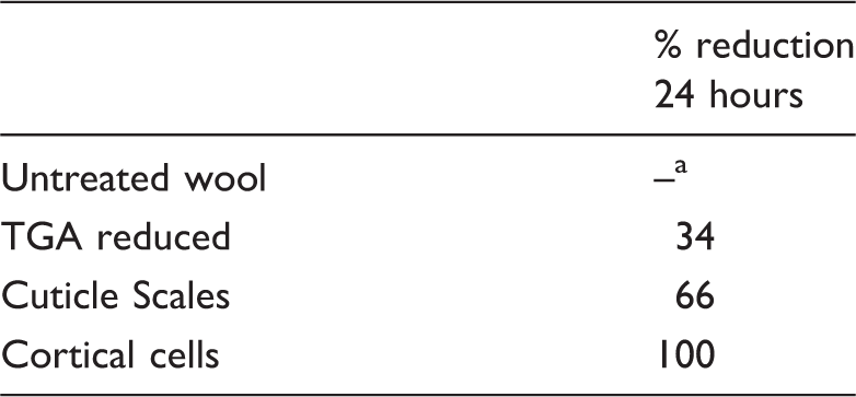
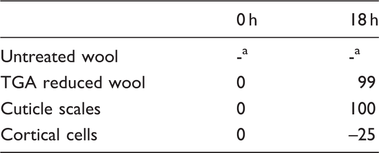
Attenuated total reflectance Fourier transform infrared spectra data, showing untreated and cuticle removed wool. Shake flask test results after 24 h No reduction was seen, but slight growth. TGA: thioglycolic acid. Challenge test results at 0 h and 18 h, values given in percentage reduction. Indicates no reading was possible (significant growth). TGA: thioglycolic acid.
A combination of the above three analytical methods confirmed, without doubt, that a wool fiber had been stripped of its cuticle scales, with minimal damage to the cortical cells, and separated with no cross-contamination.
Dynamic shake flask test
The untreated wool, as described in the literature, showed no antibacterial properties; in fact, it showed that wool is a suitable host for bacterial growth. As wool is a proteinous fiber, this was also expected.
However, as shown in Table 1, all subsequent tested samples showed significant reductions in the CFU after 24 h. The reduced wool, 33% reduction, showed that the cleavage of the disulfide could in fact impart antibacterial properties to the fiber. More interesting, however, is that both the cuticle scales and the cortical cells showed large reductions, 66% and 100% respectively.
Challenge Test
The epicuticle is defined by its unique reaction upon submersion in chlorine water, where it forms bubbles or sacs, known as Allwörden bubbles. These bubbles are formed due to the nature of the membrane being semi-permeable and a rise in pressure caused by the oxidation of the disulfide bonds. 19 The hydrophobic nature of the epicuticle layer made it difficult for the fiber to come into proper contact with the bacterial inoculum solution, and therefore no reading could be obtained for the “Untreated Wool” sample, as shown in Table 2. The epicuticle, which surrounds the cuticle scales, 19 is removed by the processing treatments of thioglycolic reduction and formic acid reflux.
Hence the possibility to get a sufficient contact with the probe for TGA reduced wool and the cuticle scales samples, enabling a bacterial count value. The Challenge Test method showed a clear distinction between the samples exhibiting antibacterial behavior and those that did not. The cortical cells exhibited clear bacterial growth, further proving the fact that the wool fiber is in fact a suitable substrate for bacteria. More importantly, this evidence backs up the shake flask test results that showed large bacterial growth after 24 h.
For the TGA reduced wool and the cuticle scales we can see that there is considerable reduction in bacteria, between t = 0 and t = 18. Both samples have reduced the bacterial count to ≥ 99% of the starting value.
Conclusions
It has been demonstrated that the wool fiber was successfully separated into its constituent components without significant chemical or physical damage, with the exception of the loss of the epicuticle in the TGA reduced wool and the cuticle scale specimens. From this the wool fiber and its components were subjected to bacteria culture, in order to determine its bacterial reduction properties. Using two different standard methods, with excess and limited presence of an aqueous host, a greater understanding of wool’s properties was achieved. The evidence shows that water plays a key role in the wool fiber component’s relationship with bacteria.
Footnotes
Declaration of conflicting interests
The authors declared no potential conflicts of interest with respect to the research, authorship, and/or publication of this article.
Funding
The authors disclosed receipt of the following financial support for the research, authorship, and/or publication of this article: The work was partly supported by the Bergrettung Tirol (Austria) and the Comet K-Project “Sports Textiles” (grant number 820494), funded by the Austrian Research Promotion Company (FFG), Standortagentur Tirol (Austria), and region Vorarlberg (Austria).