
Abstract
In this work, ultraviolet (UV) sensible metallocene isotactic polypropylene (miPP) filaments were produced with different drawing ratios and various concentrations of photochromic pigment. The effects of pigment concentration and drawing ratio on the chromatic properties and the structural modification for the miPP filaments were studied extensively by differential scanning calorimetry (DSC) and small- and wide-angle X-ray scattering (SAXS/WAXS) measurements. The change in melting temperature and the polymeric structures, such as lamellar thickness, long period and degree of crystallinity of miPP with the addition of a UV sensible pigment into miPP and the drawing process, were evaluated. The results show that the pigment concentration and the drawing ratio influence the inner structure of miPP filaments. Finally, our investigation shows that SAXS and also WAXS are appropriate to determine the lamellar thickness and the degree of crystallinity established by the DSC approach. This work attempts to correlate the results of lamellar thickness, the degree of crystallinity and the higher-order structure of the polymer acquired by DSC as well as X-ray diffraction (XRD) techniques in order to develop an appropriate approach to find the influence of pigment concentration and drawing ratio on miPP filaments.
Keywords
Polypropylene (PP) is a highly consumed and commonly used fiber material with low manufacturing cost and good thermo/mechanical performance. 1 The modification and functionalization of polymer materials are essential to introduce new functionalities onto existing polymer structures, which are useful in various technical textiles applications, including sensors, ultraviolet (UV) stability, flame retardants, etc. UV sensible PP filaments should have enough mechanical and chromatic properties for practical use and need to be produced differently. The most common technique is mass coloring with photochromic colorants. Organic photochromic colorants are widely available, and the major compounds are anilines, disulfoxides, hydrazones, semi-carbozones, stilbene derivatives, succinic anhydride, camphor derivatives, o-nitrobenzyl derivatives and spiro compounds. 2 These colorants are normally excited by the UV range from 200 to 400 nm, with the main range of 350–400 nm, and very few are excited in the visible region. This is because the UV indicators used are compounds showing so-called “positive photochromism,” that is, the absorption wavelength maxima of its colorless form are shorter than that of its colored form. The advantage of the mass coloration technology compared to others, such as screen printing, bath dyeing, etc., is in the higher durability of UV sensible textiles, mainly lightfastness. 3
The mechanical properties of semi-crystalline polymer mainly depend on the degree of crystallinity, lamellar thickness and orientation of the polymer chains.4–6 Many factors influence the degree of crystallinity, lamellar thickness and polymer chain orientation. The drawing process and spinning additives applied to the polymer materials also influence them.
On the other hand, Kristofic et al. 7 reported that the drawing ratio has a slight influenced on the dyeability of PP fibers with disperse dyes. Overall, crystallinity is an important factor that considerably affects the overall properties of polymers.8–11 While there have been many studies in this area, numerous unresolved questions about the basic science remain. Flory and Yoon 12 and Peterlin13,14 provided the initial data on morphological changes in semi-crystalline polymers after the application of stress. Spirooxazine-based photochromic pigments were mixed into metallocene isotactic polypropylene (miPP) filaments through mass coloration technology.15–17 The photochromic pigment can be converted from colorless to purple under UV exposure. Likewise, it fades back to a colorless state under the visible spectrum. The chromatic and mechanical properties of UV sensible PP filaments mainly depend on the drawing ratio and the concentration of the photochromic pigment, which predominantly modify the higher-order structures of the polymer.
The main objective of this work is to study the relationship between the drawing ratio, pigment concentration and the structure of miPP filaments. Differential scanning calorimetry (DSC), small-angle X-ray scattering (SAXS) and wide-angle X-ray scattering (WAXS) measurements were performed on the UV sensible miPP filaments to clarify their higher-order structures. The results on the thermal properties and the higher-order structures of UV sensible miPP filaments obtained by DSC and X-ray scattering measurements were compared to depict the change in structure with increasing drawing ratio and pigment concentration.
Materials and methods
Materials
Metallocene catalyst isotactic PP (Metocene HM 562R) with a melting flow rate (MFR) of 26.6 g · min−1 was procured from Lyondell Basell, Italy. The photochromic pigment (Matsui Photopia Purple – MPP) 5-chloro-1,3,3-trimethylspiro[indoline-2,3′-(3H) naphtho(2,1-b) (1,4)-oxazine] (CAS number: 27333-50-2) was purchased from Matsui Shikiso Chemical Co., Ltd, Japan. The photochemical reaction of MPP is shown in Figure S1.
Preparation of colored miPP filaments
The production of colored and colorless miPP filaments involves two steps: the production of 100% concentration colored miPP tape and colored miPP filaments. The detailed production of photochromic miPP filaments is described in our earlier research.18,19 For comparison, the colorless filaments were produced separately without mixing the photochromic pigments. After the production of both colored/colorless miPP filaments, they were subjected to drawing with different drawing ratios (DR-1; DR-2; DR-2.5; DR-3; DR-3.5, DR-4 and DR-5) at 220°C in a laboratory-scale melt spinning unit (MTI Corporation, USA), with the extruder diameter of 16 mm, L/D ratio of 30 and 13 orifices with 0.5 mm diameter. Due to production difficulty observed with the pigment concentration (2.5%) for DR-3, we produced the filaments with DR-2.8. The scheme of the drawing process is shown in Figure S2.
Measurements of photochromic response
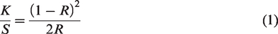
The kinetic properties of UV sensible miPP filaments were measured by a spectrophotometer, a Photochrom-3 (Figure S3),20–22 an instrument with de:0° geometry and aperture diameter of 27 mm. The spectral range of the spectrophotometer is 400–700 nm, with a bandpass of 10 nm. Five measurements were done for statistical data treatments, and average values are plotted the figures in the third section. The reflectance values can be utilized to find the Kubelka–Munk functions (K/S values), which can be computed via Equation (1)
Differential scanning calorimetry
The thermal properties of the produced photochromic miPP filaments were analyzed by Mettler Toledo DSC 3+ apparatus. The measurement was carried based on the ASTM standard D3418-08. Both heating scans (from 25°C to 200°C) and cooling scans (from 200°C to 25°C) were carried out with the heating and cooling rates of 10°C · min−1 under a nitrogen atmosphere (50 mL · min−1). From the DSC measurements, various thermal properties, such as the peak temperature of melting (Tm), melting enthalpy (ΔHm) and melting entropy (ΔSm), were obtained.
From the DSC characterization measurements, the degree of crystallinity Xc (%) of the produced filaments can be calculated by using Equation (2), where ΔH°m is 209 J · g−1 (see Kong and Hay
23
)
The lamellae thickness can be determined from both DSC as well as SAXS techniques. From the DSC, the average lamellar thickness (thickness of the folded-chain crystal) can be determined by associating the lamellar thickness to the melting temperature of miPP filaments based upon the Thomson and Gibbs equation24
SAXS and WAXS
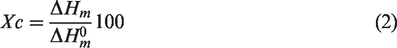
SAXS measurements were used to analyze the higher-order structures of miPP filaments conducted at room temperature at the BL-6A of the Photon Factory in the High Energy Accelerator Research Organization (Tsukuba, Ibaraki, Japan). The beamline was composed of a vertical focusing mirror and a curved monochromator for horizontal focusing. A triangular germanium (111) crystal with an asymmetric angle of 8.0 degrees was used as the monochromator crystal for optimum beam focusing. 27 A PILATUS3-1M (DECTRIS Ltd, Baden, Switzerland) was used as a two-dimensional (2D) detector. The wavelength of the X-ray (λ) was 0.15 nm and the exposure time for SAXS measurements was 10 s. 27 The WAXS measurement was conducted immediately after the SAXS measurement at room temperature. The bundle of the miPP filaments fixed on the sample holder was subjected to the measurements. The filament bundle was made to align filaments in an identical direction, and its size was approximately 1 mm. As schematically illustrated in Figure 1, the fiber bundle was set at the sample position of the 2D SAXS setup.
Schematic illustration for the two-dimensional small-angle X-ray scattering setup for the fiber bundle.
Collagen taken from a chicken tendon was used as the standard sample to calibrate the magnitude of the scattering vector q. The q is defined by q = (4π/λ) sin (θ/2), where θ is the scattering angle. An imaging plate, a BAS-IP MS 2025 (Fuji Photo Film Co., Ltd; size: 200 mm × 250 mm; actual pixel size: 100 μm × 100 μm), was used as the 2D detector for the WAXS measurement. A BAS2500 (Fuji Photo Film Co., Ltd) was used for the development of exposed images on the imaging plate. Polyethylene was used as the standard sample to calibrate the magnitude of the q for WAXS. The 2D WAXS patterns were further converted to one-dimensional (1D) profiles along the fiber axis direction by conducting a sector average. To examine the lamellar thickness (l) and the long period (D), the correlation function γ(r) was calculated from the 1D SAXS profile through Equation (6) (inverse Fourier transform method). 28
Results and discussion
Photochromic response depending upon the pigment concentration and filament drawing ratio
The color of UV sensible miPP filaments turns reversibly from colorless to purple by irradiation with UV light (Figure S4). The detail of the preparation and the coloring behaviors of the filaments are described in a previous paper.
18
The (K/S)
λ
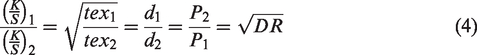
spectral values defined from Kubelka–Munk's theory were obtained by using spectral reflectance of the miPP filament samples to evaluate their color strength. Here, the dependences of their color strength upon the supply amount of pigment concentration and filament drawing ratio were estimated with the maximum signal value of (K/S)max at 570 nm. The maximum values of (K/S)max plotted against the drawing ratio (DR) of miPP filament samples with a systematic change in the pigment concentration are shown in Figure 2(a). The (K/S)max increases naturally with an increase in the pigment concentration of the miPP filaments, as shown in the Figure 2(b). The data in Figure 2(a) are rearranged into a relationship between photochromic pigment concentration (c) and (K/S)max and the obtained figure is shown in Figure 2(b) to confirm. The straight dotted lines depicted in Figure 2(b) were drawn on the basis of the data obtained and within the limits of them, as a kind of empirical and experimental line. It is found that the relationships between the c and (K/S)max values show mostly linear dependency for each of the DR samples. The results indicate that the pigment particles are dispersed almost homogeneously in the filaments, and the visible coloring increases with the increasing supplied amount of pigment within the range. Theoretically, the trajectory of optical rays in filaments is not impacted by the fineness of filaments. When the colors of two filaments, in which the amount and concentration of colorants are the same, are compared, the color of the finer filament (i.e., higher drawn filament) is lighter than that of the thicker one. The primary reflectance is maximized on the boundary of the air/filament. The amount of light reflected at the surface of the filament for a finer filament was higher than that of a thicker one because the surface area of the finer one is larger than that of the thicker one. Then, the less light reaches the colorant molecules in the filament. This effect decreases optical color density, that is, (K/S)max (Figure 2(a)). The reduced (K/S)max is the consequence of the reduced path length of light in the filaments with respect to their fineness. When the colorant concentrations in the filaments are the same, the ratio of K/S values for filaments ((K/S)1 and (K/S)2) is equal to the ratio of their diameters (d1 or d2) and the square root of fineness (strongly correlated with fineness, and represented by DR), as shown in Equation (4)29
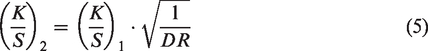
DR–(K/S)max relationships of ultraviolet sensible metallocene isotactic polypropylene filaments (a) and photochromic pigment concentration. (b) c–(K/S) max relationships.
The obtained plots shown in Figure 2(a) were fitted with solid line curves according to Equation (5), where the K/S values for the filament with DR-1 were used as the (K/S)1. The change in (K/S)max mostly fits Equation (5), as shown in Figure 2(a). This indicates that the optical density of the sample colors correlates with the filament diameter and the drawing ratio.
Evolution of the fiber microstructure with drawing ratios/pigment concentration by DSC
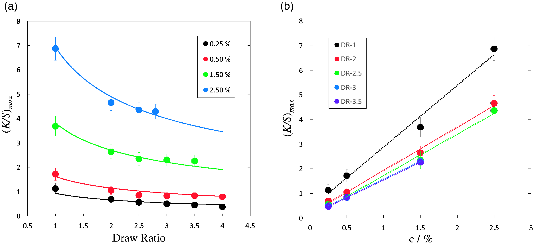
DSC was used to analyze the thermal behavior of UV sensitive miPP filaments. The results are shown in Figure 3. The UV sensible miPP filament is a semi-crystalline polymer with typical polymorphic behavior. It forms a higher-order structure at higher temperature, particularly close to the melting temperature when the relaxation of the polymer chain in the amorphous phase is prevented mechanically, keeping fibers under tension.30,31 Therefore, it significantly changes the various thermal properties, which influence their mechanical properties. Quantities of structural characterization of iPP (isotactic polypropylene) were studied first by Samuels, 32 and it was concluded that iPP fibers provide a higher melting temperature and enthalpy of fusion. The orientation of the crystalline phase will not have been affected by the melting temperature of the filaments. Therefore, the thickness of the lamella was analyzed using both DSC and SAXS methods. The results are compared and discussed in the upcoming section. The melting temperature (Tm) of the undrawn filament shows 143.86°C, whereas the DR-4 shows 151.02°C. Figures 3(a) and (b) show the results of DSC measurements of miPP/UV sensible miPP filaments with increasing the drawn ratios and photochromic pigment concentration, which were made in order to estimate the melting temperature (Figures 3(c) and (d)), the degree of crystallinity and lamellar thickness (Figures 3(e) and (f)). Areas of melting peaks on thermograms related to the degree of crystallinity were gradually increased with drawing ratios. The melting temperature of the polymer is known to change with the crystallization temperature as, generally, it is dependent on the crystal lamellar thickness. From the DSC results, Tm and ΔHm were found to increase with an increase in the drawing ratio. This is due to the lamella thickening and increase of the crystallinity. In the case of DR-4, considering that transition from the amorphous component to the crystalline component takes place in the miPP filament, the fraction of the amorphous region decreases and boosts the differences in thickness between the crystal and amorphous layers.
Normalized differential scanning calorimetry curves for metallocene isotactic polypropylene filaments with different draw ratios without the addition of pigment (a), as well as the different concentrations of photochromic pigment (Matsui Photopia Purple – MPP) (b). Melting temperature versus drawing ratio (c). Pigment concentration (d). Lamellar thickness versus drawing ratio for MPP = 0% (e). Pigment concentration for DR-2 (f).
Consequently, it appears that the transmission capacity of infrared light declines over four times drawing (DR-4) to miPP filaments. In this work, the anticipated lamellar thickness was determined from the DSC results by utilizing Equation (3). The outcomes are shown in Figures 3(e) and (f) as a function of the drawing ratio along with pigment concentration. The results showed that the shape of the curve is different, but the lamellar thickness, I, increases with an increase in the drawing ratio and pigment concentration. The drawing ratio in particular has a direct impact on the lamellar thickness, which is boosted 13% at DR-4 as compared to DR-1. The outcomes were fitted with non-linear regression; they show strongly non-linear relationships (Figure 3(e)). Throughout the DSC measurement, there was an uncertainty error due to the variability of DSC measurement and additional computations.
Next, SAXS and WAXS measurements of the UV sensitive miPP filaments were made in order to obtain the information from other measurement analyses and clarify the sample structures, including the lamellar thickness.
Evolution of the fiber microstructure with the influence of drawing ratios/pigment concentration by SAXS/WAXS
Figure 4(a) shows the 2D SAXS patterns for the initial miPP filament (DR-1) without the addition of UV sensible pigment, and Figures 4(b)–(g) show those for the drawn filaments with different draw ratios, as indicated by the numerical values in the specimen code names after “DR,” while Figures 5(a)–(d) show the 2D SAXS patterns for the same specimens with the addition of UV sensible pigment (2.5%). Overall, the 2D SAXS patterns show similar features with parallel layer peaks and are parallel to the equatorial direction. However, some of those indicate an “arc” (Figures 4(b)–(e), Figures 5(b)–(d)) or a “streak” (Figures 4(f) and (g)) and the extents of the intensity accumulation differ depending on the individual specimens. The appearance of the two spots indicates the existence of the preferentially oriented lamellar stacks, parallel to the fiber axis, of the crystalline lamellae sandwiching the amorphous layer. While the “arc” peaks suggest orientation distribution of the lamellar stacks, the parallel “streak” peaks indicate the existence of the crystalline lamellae with finite width, which are repeating one-dimensionally as the lamellar stacks. Another notable feature in the 2D SAXS patterns for all of the specimens is the existence of high-intensity streaks near the beam-stopper, which are prolonged in the equatorial direction. Although some sharp streaks should be ascribed to the total reflection of the X-ray beam by the surface of the miPP fibers, other gentle streaks (see, e.g., Figures 4(d) and 5(b)) suggest the existence of “Shish” according to the SAXS/SANS (small-angle neutron scattering) study of the formation of the “Shish-Kebab” in polyethylene. 33 Then, the above-mentioned lamellar stacks should be considered as a “Kebab,” with the “Shish-Kebab” structure in a fiber filament, which has been schematically illustrated in Figure 6. The formation of the “Shish-Kebab” in iPP has been reported in the literature.34–37
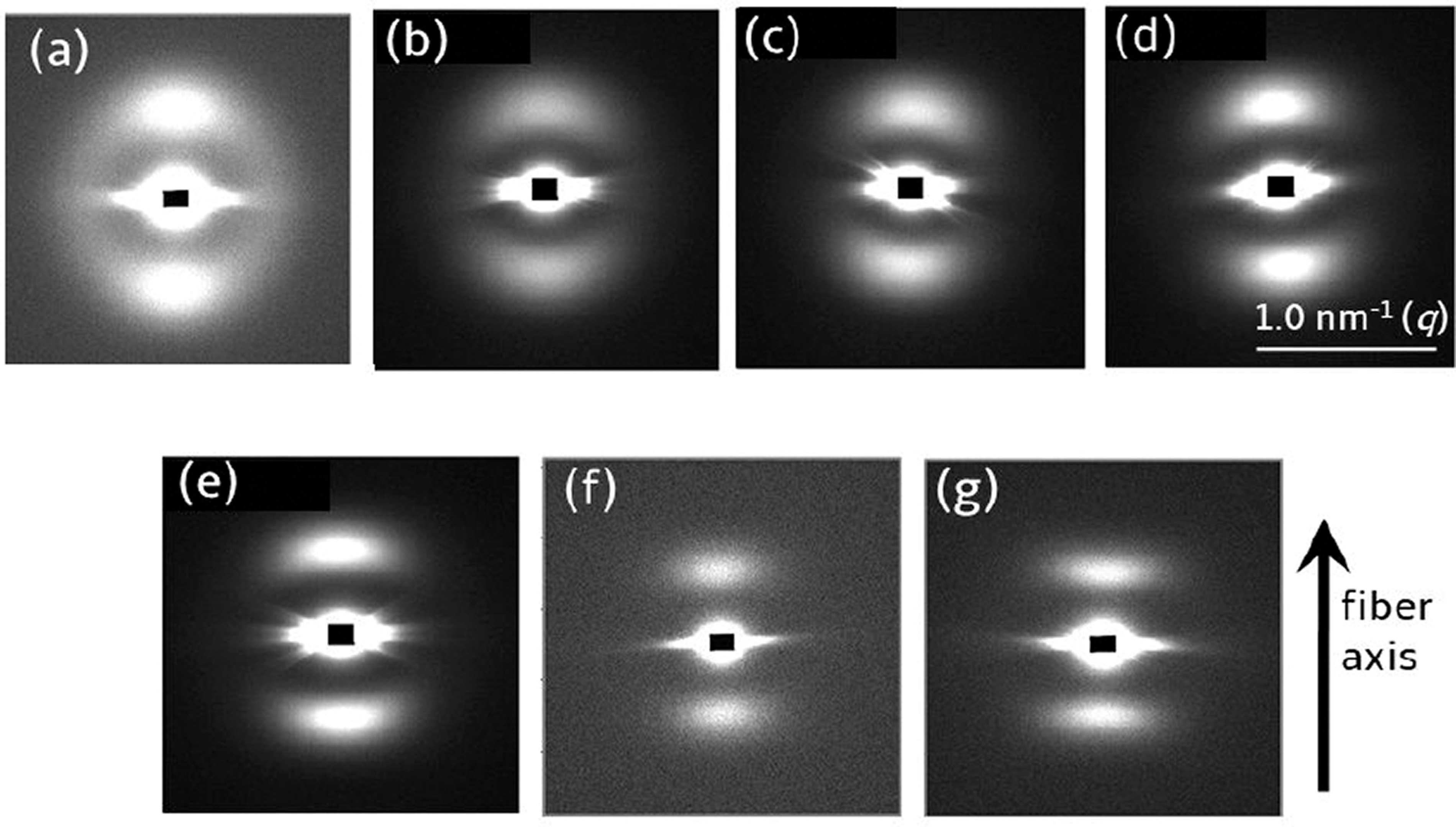
Two-dimensional small-angle X-ray scattering patterns of the metallocene isotactic polypropylene filaments (without photochromic pigments): (a) DR-1; (b) DR-2; (c) DR-2.5; (d) DR-3; (e) DR-3.5; (f) DR-4; (g) DR-5.
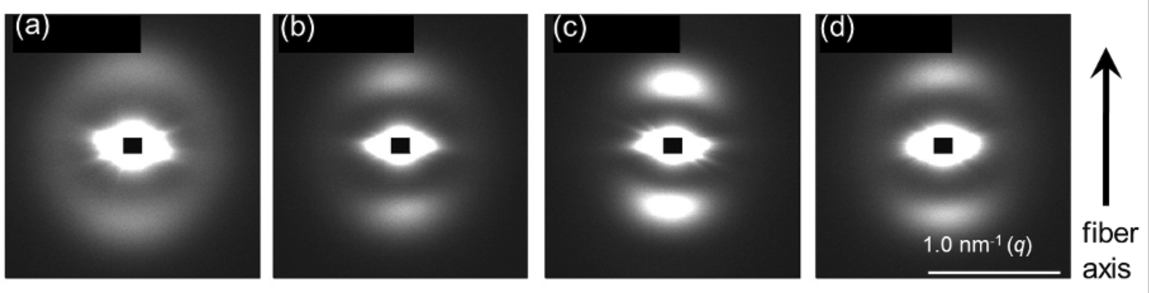
Two-dimensional small-angle X-ray scattering patterns of the ultraviolet sensible metallocene isotactic polypropylene filaments (2.5% Matsui Photopia Purple): (a) DR-1; (b) DR-2; (c) DR-2.5; (d) DR-3.
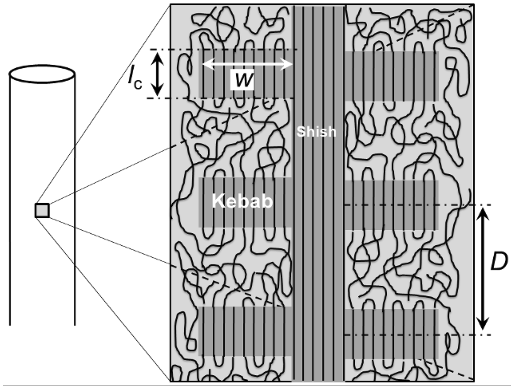
Schematic representation of lamella thickness, long period and “Shish-Kebab” in the polymer structure.
It is noteworthy that almost isotropic donut-shaped ring peaks overlapped in the two-spot pattern in Figures 4(a) and 5(a), while no such peak was discernible in the others. This result suggests that the lamellar stacks (“Kebab” without “Shish”) with random orientation co-exist with the preferentially oriented “Shish-Kebab” parallel to the fiber axis in the initial miPP filament. Further, these un-oriented lamellar stacks were subjected to be oriented parallel to the fiber axis upon the mechanical drawing, which may be transformed into the “Shish-Kebab” structure. It seems that the intensity of the two spots was more accumulated with increasing the draw ratio for both cases of the filaments with and without the addition of the UV sensible pigment. Furthermore, the position of the two-spot peak shifted to the lower q direction, suggesting that the long period of the lamellar staking is increased as a function of the draw ratio. This is reasonable because of the elongation effect acting on the amorphous layer being expanded accompanying the stretching of the molecular chains. We conducted a quantitative evaluation of the long period (D) as a repeat distance of the crystalline lamellae and their thickness (lc), according to the Strobl method.
38
Firstly, a sector average of the scattering intensity was conducted to obtain a 1D scattering profile in the fiber axis direction. The air-scattering intensity was subtracted by considering the transmission of the X-ray beam for each specimen. The background intensity due to the fluctuation of density was further subtracted. Then, the correlation function, γ(r), was calculated from the thus-obtained 1D profile, I(q), according to the following equation (inverse Fourier transform method)
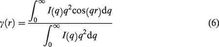
Figure 7 shows γ(r) for all of the specimens. According to Strobl, 38 based on the two-phase model with crystalline and amorphous phases, the thickness of the thinner phase can be evaluated from the plot of γ(r) in Figure 7 at the crossing point of the horizontal line, which passes through the minimum point of γ(r), and the approximated line of γ(r) in the small r range. Since the degree of crystallinity evaluated by DSC was less than 0.50, the obtained value of the thickness of the thinner phase can be considered as the lamellar thickness (lc).
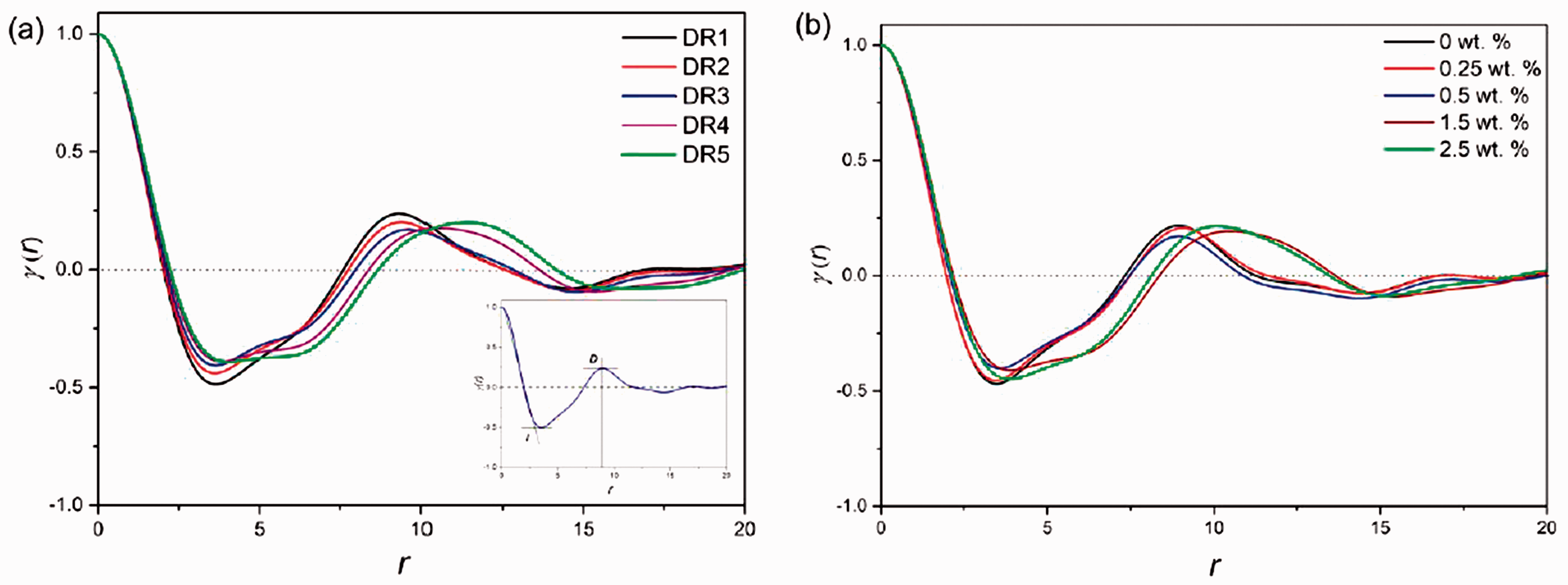
(a) Small-angle X-ray scattering correlation functions for the metallocene isotactic polypropylene filaments without pigment for various draw ratios. (b) Those for various pigment concentrations (DR-2.5).
The intensity accumulation of the pair of peaks in the direction parallel to the fiber axis is correlated to the orientation of the lamellar stacks (namely, the orientation of the Shish-Kebab). Thus, this feature can be used to evaluate the lamellar orientation factor (f), according to the following equation from the value of 〈cos
2
ϕ〉, where ϕ denotes the orientation angle with respect to the fiber axis. Note that the 1D cylindrical symmetry was assumed


Before discussing these structural parameters, other important parameters such as the degree of crystallinity and the crystallite orientation factor should be evaluated. For this purpose, WAXS measurements were conducted. The results are shown in Figure 8. As shown in Figure 8(e), four Debye–Scherrer ring peaks were clearly observed for the undrawn filament with pigment. The appearance of the Debye–Scherrer ring indicates almost no preferential orientation of crystallite. In contrast, other drawn filaments exhibited oriented WAXS patterns with three reflections in the equatorial direction and two arcs as the first-order layer reflections. To evaluate the degree of crystallinity (dc), the 2D WAXS patterns were circularly averaged and the 1D WAXS profiles were obtained. Further, the computational peak decomposition of the 1D WAXS profile was conducted. 40 From areas of the decomposed reflection peaks and that of the decomposed amorphous halo peak, dc was evaluated by taking a ratio of the summed peak areas of the crystalline reflection and the total area of all peaks, including the amorphous halo. As for the evaluation of the crystallite orientation factor, the intensity distribution of the (110) reflection peak (the innermost peak appearing on the equator) as a function of the azimuthal angle was utilized. The equation for its evaluation was the same as that for the lamellar orientation factor (Equation (7)) with C = –1/2.
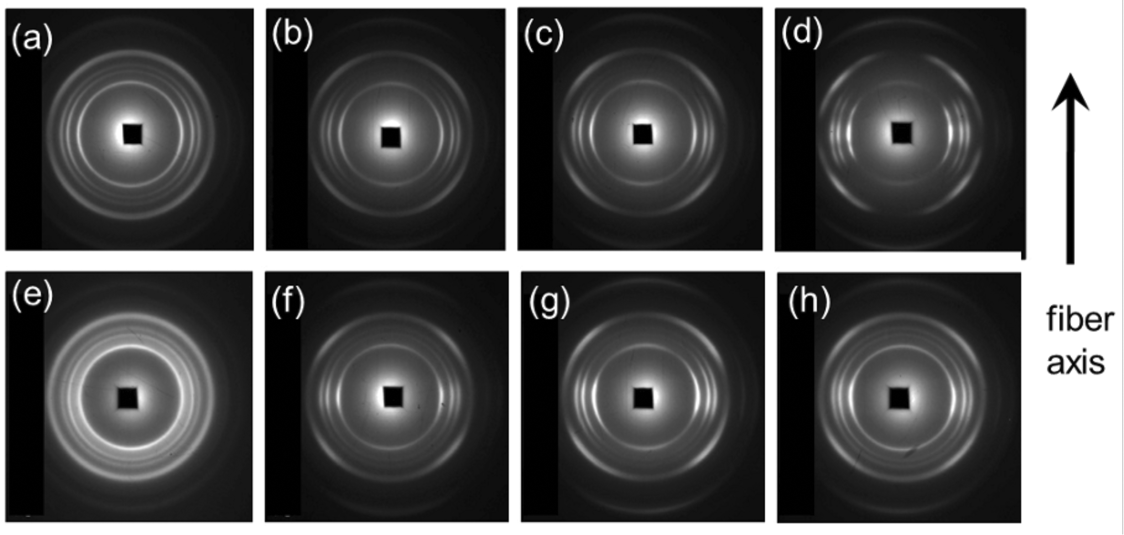
Two dimensional wide-angle X-ray scattering patterns of metallocene isotactic polypropylene (miPP) filaments (without photochromic pigments): (a) DR-2; (b) DR-2.5; (c); DR-3; (c) DR-3.5. miPP filaments with pigments (2.5%): (e) DR-1; (f) DR-2; (g) DR-2.5; (h) DR-3.
Figures 9 and 10 summarize the evaluation of structural parameters based on the results of SAXS/WAXS measurements. Overall, the behaviors as a function of the draw ratio were found to be almost the same for both cases with and without pigment particles. The long period increased because of the effect of elongation. The progress in the lamellar orientation and the crystallite orientation due to the drawing is also reasonable. Error bars (+/–0.1) are indicated for all the data points of the crystallite orientation factor to recognize their monotonically increasing tendency, regardless of the existence of the pigment particles, although all the data points themselves exhibit huge dispersion from the representative curve. In contrast, the behavior of the degree of crystallinity was complicated, showing a parabolic shape for the case of the filaments without pigments or almost constant with a slightly decreasing tendency in the higher draw ratio for the case of the filaments with pigments (see Figure 10). Such decreasing tendency of the degree of crystallinity with the draw ratio may suggest a decrease in the width of the crystalline lamellae (decrease in “w” in Figure 6). This means that the formation of the “Kebab” structure, which was overgrown on the surface of the “Shish,” was more suppressed when the draw ratio was higher. The diminishing of the “Kebab” contribution results in the improvement of the mechanical properties, such as increased tensile modulus and ultimate strength, so this scenario seems to be reasonable. Meanwhile, the degree of crystallinity in the lamellar stacks can be considered by evaluating the ratio of lc/D, which is plotted in Figure 10, showing a monotonical increasing behavior with the draw ratio, except for the initial filament. Thus, the increase in the crystallinity inside the “Kebab” structure (lamellar stack) is suggested as an effect of drawing the filaments, which is in good accord with common knowledge. It is to be noted here that the slightly higher value for the undrawn filament may be ascribed to the co-existing lamellar stacks (“Kebab” without “Shish”), and the value of lc/D is considered to decrease along with the transformation into the “Shish-Kebab” structure upon drawing (the decrease in its value from DR-1 to DR-2 in Figure 10). Note also that the evaluated values of the lamellar thickness were much thinner than those evaluated from the DSC Tm results through the Gibbs–Thomson equation, as plotted in Figures 3(e) and (f). This may be due to the uncertainty in the value of the surface free energy (φe) of the lamellar surface on which the polymer chain folds are located, because this value is very sensitive to the state of the polymer chain folds.
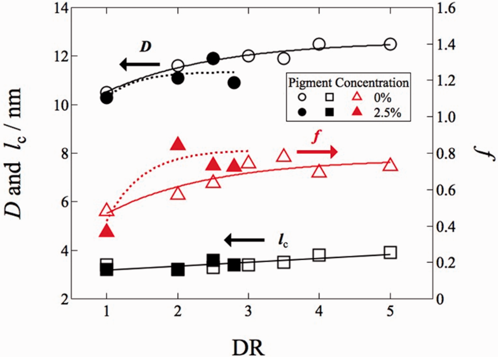
Long period (D), lamella thickness (lc) and lamellar orientation factor (f) versus drawing ratio of metallocene isotactic polypropylene filaments based on the one-dimensional correlation function method.
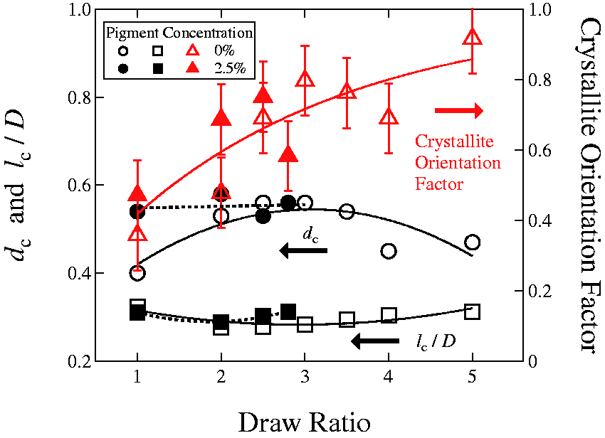
Degree of crystallinity (dc), lc/D and crystallite orientation factor versus drawing ratio of the ultraviolet sensible metallocene isotactic polypropylene filament.
Now, we discuss the features in the behaviors for the specimens with pigments, as compared with those for the specimens without pigments. The lamellar orientation and the crystallite orientation were more easily attained in the specimens with pigments. Furthermore, the degree of crystallinity is almost unchanged in the range of the draw ratio less than 3 because of its higher value as compared with those for the specimens without pigments. These features suggest that the drawing fibers worked more effectively for these specimens, although the size of the primary particle of pigment (∼2–5 μm) is much larger than the “Shish-Kebab” structures, as schematically illustrated in Figure 11, where some of the primary particles form a secondary particle by their aggregation. This situation looks similar to that of the case of the carbon black particles in an elastomeric polymer matrix, 41 although the size of the pigment particle is similar to the “agglomerate” in the carbon black case.Nevertheless, the structural similarity reminds us that such a filler effect may exist in the pigment inclusion case.
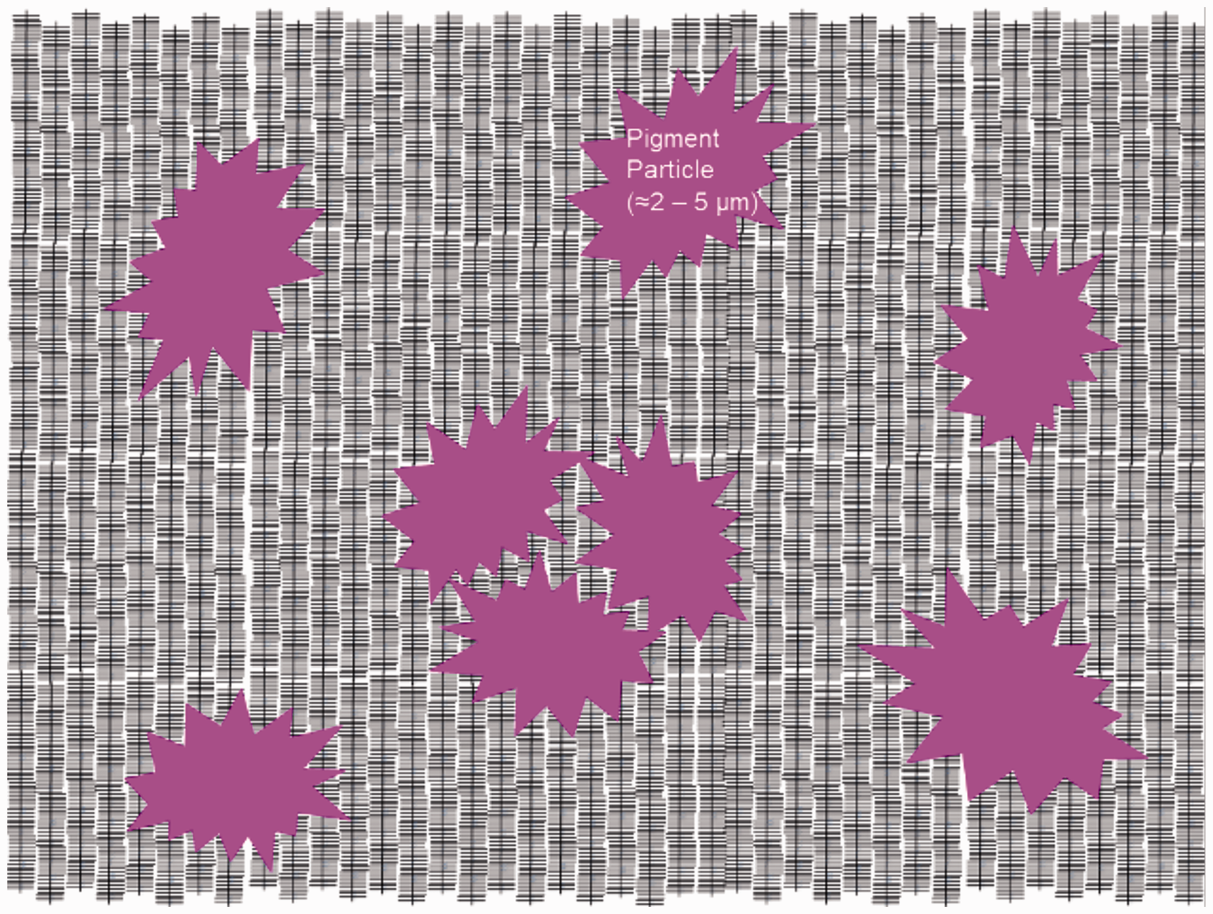
Schematic illustration of the structure in a filament with pigment particles.
So far, we have discussed the results of the miPP fibers with 2.5% addition of the pigment. We also have conducted SAXS/WAXS measurements on several undrawn filaments with different inclusion percentages of the pigment. The results are plotted in Figure 12. Note here that the error bars (+/–0.1) indicated for all of the data points of the crystallite orientation factor exhibit the same extent of the data dispersion as shown in Figure 10 (+/–0.1). Then, the monotonically decreasing tendency of the crystallite orientation factor (as shown with the red broken line) can be recognized within the error bars in Figure 12. Thus, all of the structural parameters decreased with increasing the pigment inclusion percentage. This is because the pigment particles or their network aggregation act as an impurity to degrade the crystallizability of miPP.
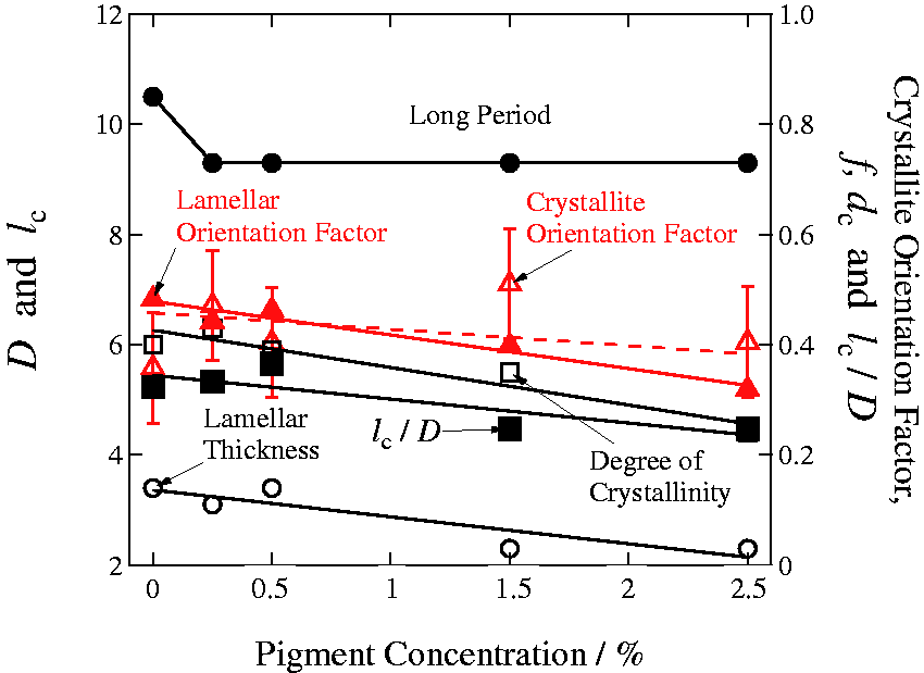
Interrelation of long period, lamellar thickness, lamellar orientation factor, degree of crystallinity and long period on the pigment concentration. (Color online only.)
Conclusions
miPP fibers were colored to varying depths of a magenta shade with the photochromic pigment used in this study. The optical color density of miPP colored by UV irradiation is directly proportional to the concentration of UV sensible pigment, and it is necessary to point out that the mass-colored fibers were darker than fibers coated by the screen-printing method in our previous studies. The increasing heat of fusion for the miPP filament is observed with the drawing ratio. The results confirm that lamellar thickening leads to an increase in the melting temperature, which is directly proportional to the drawing ratio of the miPP filament. The addition of UV sensible pigment causes a change in the higher-order structures. The DSC, as well as X-ray (1D correlation function method) technique, is one of the most frequently utilized techniques to estimate the thickness of the lamella and degree of crystallinity and other structural parameters for crystalline polymers. Analysis of X-ray data using the 1D correlation function method allows us to estimate the actual values of the lamellar thickness of the miPP and UV sensible miPP filaments. Overall, all the structural parameters were found to decrease with increasing the pigment inclusion percentage. This is because the pigment particles or their network aggregation act as an impurity to degrade the crystallizability of miPP.
Supplemental Material
sj-pdf-1-trj-10.1177_00405175211053394 - Supplemental material for Differential scanning calorimetry/small-angle X-ray scattering analysis of ultraviolet sensible polypropylene filaments
Supplemental material, sj-pdf-1-trj-10.1177_00405175211053394 for Differential scanning calorimetry/small-angle X-ray scattering analysis of ultraviolet sensible polypropylene filaments by Martina Vikova, Shinichi Sakurai, Aravin Prince Periyasamy, Hidekazu Yasunaga, Miroslava Pechočiaková and Anna Ujhelyiová in Textile Research Journal
Supplemental Material
sj-pdf-2-trj-10.1177_00405175211053394 - Supplemental material for Differential scanning calorimetry/small-angle X-ray scattering analysis of ultraviolet sensible polypropylene filaments
Supplemental material, sj-pdf-2-trj-10.1177_00405175211053394 for Differential scanning calorimetry/small-angle X-ray scattering analysis of ultraviolet sensible polypropylene filaments by Martina Vikova, Shinichi Sakurai, Aravin Prince Periyasamy, Hidekazu Yasunaga, Miroslava Pechočiaková and Anna Ujhelyiová in Textile Research Journal
Supplemental Material
sj-jpg-3-trj-10.1177_00405175211053394 - Supplemental material for Differential scanning calorimetry/small-angle X-ray scattering analysis of ultraviolet sensible polypropylene filaments
Supplemental material, sj-jpg-3-trj-10.1177_00405175211053394 for Differential scanning calorimetry/small-angle X-ray scattering analysis of ultraviolet sensible polypropylene filaments by Martina Vikova, Shinichi Sakurai, Aravin Prince Periyasamy, Hidekazu Yasunaga, Miroslava Pechočiaková and Anna Ujhelyiová in Textile Research Journal
Supplemental Material
sj-pdf-4-trj-10.1177_00405175211053394 - Supplemental material for Differential scanning calorimetry/small-angle X-ray scattering analysis of ultraviolet sensible polypropylene filaments
Supplemental material, sj-pdf-4-trj-10.1177_00405175211053394 for Differential scanning calorimetry/small-angle X-ray scattering analysis of ultraviolet sensible polypropylene filaments by Martina Vikova, Shinichi Sakurai, Aravin Prince Periyasamy, Hidekazu Yasunaga, Miroslava Pechočiaková and Anna Ujhelyiová in Textile Research Journal
Footnotes
Acknowledgements
The SAXS/WAXS experiments were performed at BL-6A in the Photon Factory, KEK, Japan (under Approval No. 2017G527). We thank Ing. Marcela Pechova and Ing. Tereza Pocova for their assistance during the microscopical measurements.
Declaration of conflicting interests
The author(s) have no conflicts of interest to declare.
Funding
The author(s) disclosed receipt of the following financial support for the research, authorship and/or publication of this article: This work was supported by the Ministry of Education, Youth and Sports of the Czech Republic and the European Union - European Structural and Investment Funds in the framework of the Operational Programme Research, Development and Education - project Hybrid Materials for Hierarchical Structures (HyHi, Reg. No. CZ.02.1.01/0.0/0.0/16_019/0000843).
Supplemental material
Supplemental material for this article is available online.
References
Supplementary Material
Please find the following supplemental material available below.
For Open Access articles published under a Creative Commons License, all supplemental material carries the same license as the article it is associated with.
For non-Open Access articles published, all supplemental material carries a non-exclusive license, and permission requests for re-use of supplemental material or any part of supplemental material shall be sent directly to the copyright owner as specified in the copyright notice associated with the article.