






Abstract
To enhance the coloration performance and light stability of anthraquinone natural dyes on cotton fabrics, cochineal was incorporated onto the surface of cationic cross-linked polymethyl methacrylate (PMA) colloidal microspheres via electrostatic adsorption. This process led to the formation of an external surface-absorbable dye structural element (cochineal/PMA microspheres), which serves as a functional colorant. In this study, cochineal composite microspheres with uniform particle size and clear morphology were prepared by mixing cochineal with polypropylene nanospheres, which were prepared via soap-free emulsion polymerization. Their characteristics were examined using thermogravimetric analysis, infrared spectroscopy, zeta-potential analysis, and particle size distribution analysis. In addition, their dyeing properties on cotton knitted fabrics were investigated. The results show that under certain laboratory conditions, the average microspheres size increased from 112 to 192 nm, the polydispersity index (PDI) changed from 0.028 to 0.052, and the zeta potential decreased from +52.8 to +29.3 mV. With the dye concentration of 2 mg/mL, a temperature of 50°C, a mixing rate 200 rpm, and 10 minutes of adsorption, cochineal was optimally absorbed by the microspheres. The adsorption kinetics and isotherm studies indicate that the adsorption of cochineal onto polypropylene nanospheres is mainly an electrostatic interaction process. With the increasing concentration of dye microspheres, the narrower bandgap within the fabric exhibits significantly enhanced light stability. The dry rubbing fastness of the cotton knitted fabrics dyed with cochineal composite microspheres ranged from 4 to 5, while the wet rubbing fastness, staining, and color fastness to soaping all ranged from 3 to 4.
Colored polymer microspheres have attracted significant academic interest due to their potential applications in ink-jet printing on textile substrates,1,2 biomarkers and detection, drug delivery systems, 3 clinical diagnosis, 4 and photon crystals. 5 These microspheres are valued for their large specific surface area, bright color, strong light fastness, and chemical stability. While colored polymer microspheres, which are frequently produced with synthetic dyes and exhibiting high reflectivity similar to photon crystal, are utilized in digital printing and retro-reflective fabrics, there has also been considerable research on microspheres made with natural dyes. 6 Liqin et al. 7 used an ink-jet printer to print a photonic crystal pattern on white fabric using colored microspheres, which were made by coating the only structural component of the microspheres, poly(styrene-butyl acrylate-methacrylate) microspheres, with black dispersion dye. Bright, brilliant structural colors may be seen in the produced pattern.
The majority of natural dyes exhibit poor light stability and dyeing performance as a result of their unique molecular structure. To enhance light fastness and color stability, various approaches have been explored, including mordant treatments, modifications of the dye’s molecular structure, or alterations to the fabric itself. For example, Vankar et al. 8 demonstrated that cotton and silk fabrics dyed with a mixture of protease and tannic acid exhibited superior dye uptake compared with traditional mordanting methods. Furthermore, Cristea and Vilarem 9 improved the light fastness of fabrics dyed with madder, mignonette, and woad by adding UV-absorbing agents and antioxidants to the dye solution, thereby preventing UV damage. These solutions, however, turn out to be ecologically unfriendly and costly to implement. A more recent study found that combining polymer microspheres with disperse dyes can produce a satisfactory dyeing effect. Yavuz et al. 10 dyed polyamide fabric with a mixture of poly(styrene-methyl methacrylate-acrylic acid) and dispersion red 343 to boost the saturation and brightness and improve the reflectivity of the coated cloth. Although much work has been put into this area, more extensive and in-depth research is still required.
Natural dyes, derived from animals, plants, or minerals, offer better environmental compatibility, faster degradation, and antiseptic properties, making them the main kind of colorants in ecological textiles.11,12 Cochineal, also known as carmine acid,13,14 is a natural dye extracted from female cochineal insects parasitizing cacti and is considered the safest of natural dyes and is thus widely used in textiles.15-17 However, cochineal is only suitable for dyeing wool and silk in strong acid and is not suitable for cellulose, limiting its application.
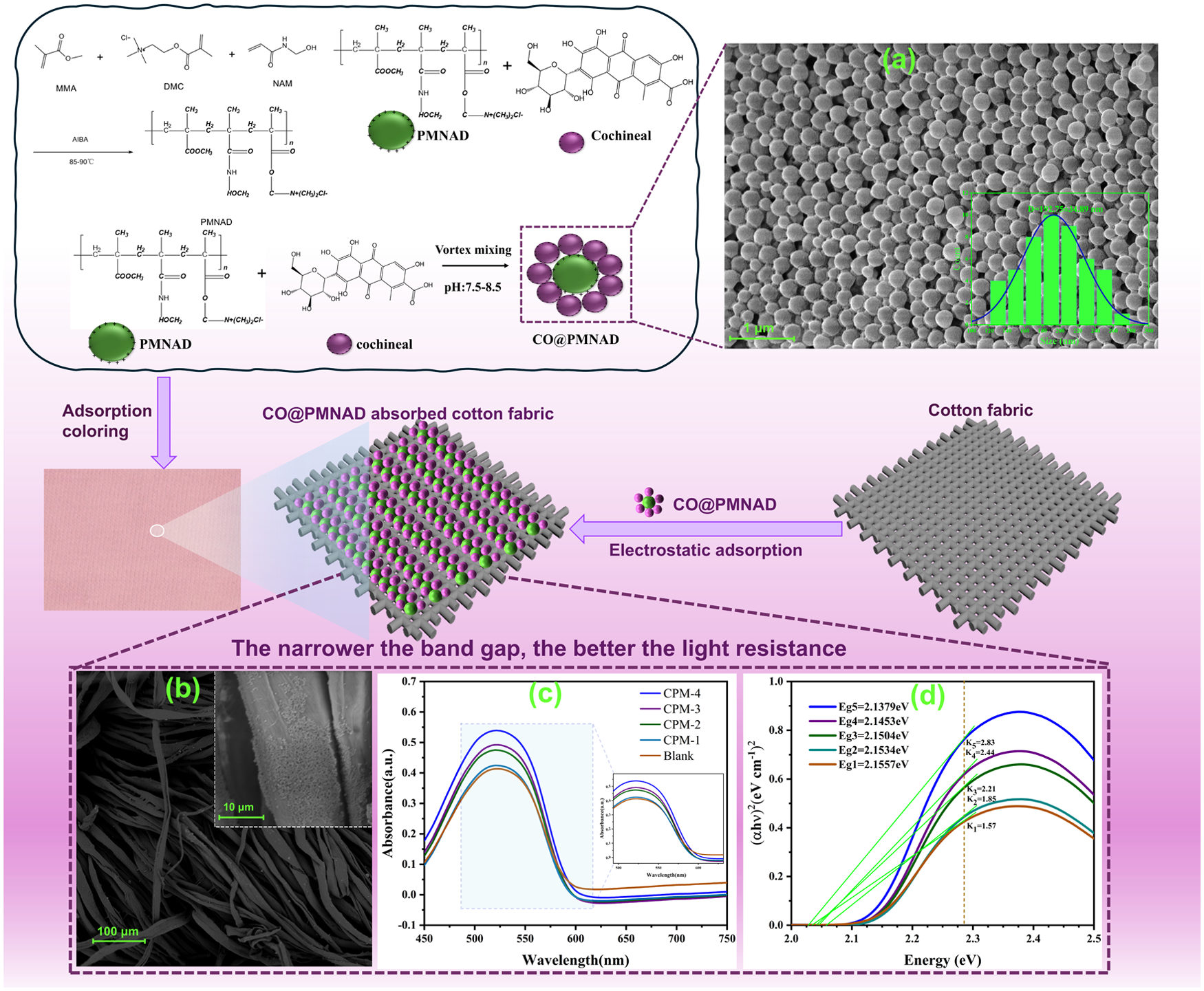
In this study, poly[methyl methacrylate-(methacryloxyethyl)trimethylammonium chloride-N-hydroxymethylacrylamide] (PMNAD) microspheres18,19 were synthesized via the soap-free emulsion method (Figure 1). These microspheres were subsequently loaded with cochineal through electrostatic adsorption to prepare cochineal composite microspheres (Figure 2), which were then used to dye cotton fabric.
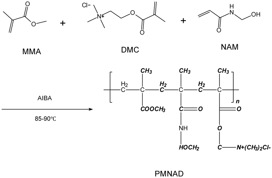
Microsphere synthesis reaction equation.
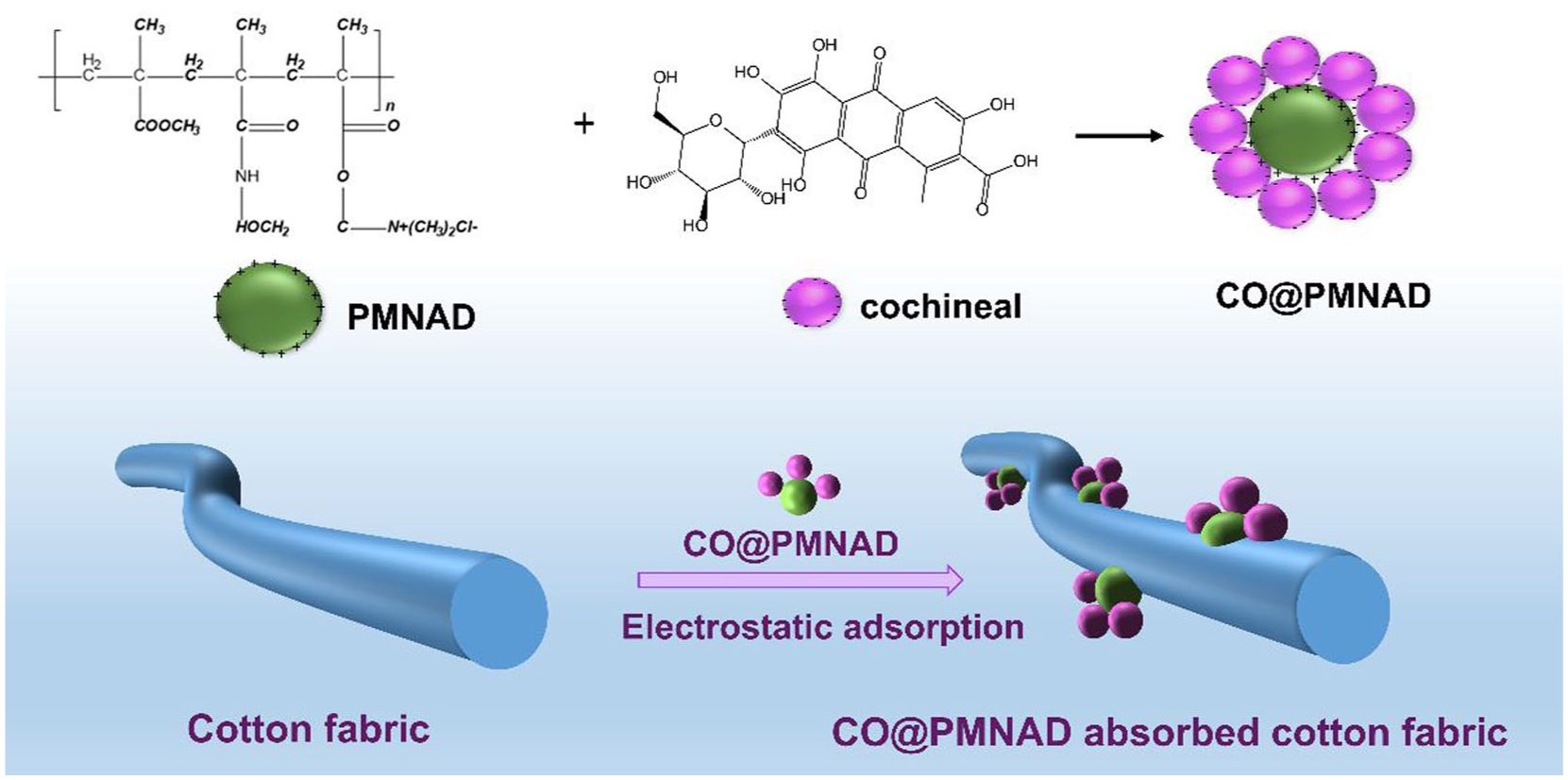
Microsphere adsorption process for cotton fabric.
The nanospheres obtained were characterized by scanning electron microscopy (SEM), Fourier-transform infrared (FTIR) spectroscopy, dynamic light scattering (DLS), and thermogravimetric (TG) analysis. Additional analysis, including ultraviolet–visible (UV-Vis) spectroscopy, diffuse reflectance spectroscopy (DRS), and CIE colorimetric characterization (CIE) colorimetry, was performed to evaluate the photostability and dyeing properties of the composite microspheres.
Experimental materials and methods
Materials
Pure cotton plain weave fabrics (8×18 tex yarn, 160 g/m2, white) were supplied by Lu Tai Textile Co., Ltd., (Zibo, China), cochinia red dye (Suzhou Baiyixin Biotechnology, China), methyl methacrylate (MMA), dimethylethyl trimethyl ammonium chloride (DMC), N-hydroxymethyl acrylamide (NAM), and azobis (isobutyronitrile) hydrochloride (AIBA) were all purchased from Aladdin Reagent Co., Ltd., Shanghai, China. Sodium dihydrogen phosphate, sodium carbonate, citric acid, and sodium sulfate were purchased from Shanghai Zhongbenfen Chemical Reagent Co., Ltd., China.
Fabric microsphere self-assembly process
Synthesis of microspheres
We added 65 g of deionized water and 12 g of purified MMA to a three-neck round-bottom flask equipped with a blender and a thermometer, then continuously stirred. DMC, NAM, and AIBA were put into the liquid separation funnel according to a total solid content of 10%, followed by the addition of an appropriate amount of deionized water to ensure complete mixing. After the dispersion liquid in the three-neck round-bottom flask was heated to 80–85°C for 30 min, the solution in the separating liquid hopper was slowly dropped into the three-neck round-bottom flask, with the dropping acceleration controlled. The reaction continued for 1 h, and the product was cooled down and filtered by a 0.45 μm hydrophilic microfiltration membrane to obtain a P(MMA–DMC–NAM) microsphere emulsion. Dialysis was performed with deionized water at 25°C for 72 h. P(MMA–DMC–NAM) microspheres were obtained by freeze-drying the postdialysis emulsion, abbreviated as PMNAD.
The adsorption process of PMNAD by cochineal
We weighed 10 g of PMNAD emulsion with a solid content of 10% into the conical flask and added 80 ml of distilled water was added. After 10 minutes, the pH of the system was adjusted to 7.5–8.5 using disodium hydrogen phosphate and citric acid, then 10 ml of dissolved cochineal red dye solution was added. After ultrasonic oscillation, the conical flask was placed in a constant temperature metal mixing instrument (HX-20T, Shanghai Analysis Instruments Co., Ltd., China) at 50°C for 10 min. The cochineal red@PMNAD microsphere emulsion was dialyzed with deionized water at 25°C for 72 h, then freeze-dried to obtain cochineal red@PMNAD microsphere powder (CO@PMNAD).
The standardized curve of cochineal red was measured at 440 nm through an UV-vis spectrophotometer (UV-1800PC, JINHUA Instruments Co., Ltd., China) weakly alkaline conditions. The adsorption performance of the PMNAD for cochineal was evaluated via a batch adsorption experiment that had the following phases: mixed solution, centrifugation, detection, and analysis. The mass of cochineal on the surface of PMNAD has been calculated using the standard curve. The dye fixation amount qe (mg/g) in nanospheres can be calculated using

where C0 (mg/L) and Ce (mg/L) are the initial and equilibrium concentration of cochineal, respectively, V (L) is the solution volume, m (g) is the mass of the PMNAD, and qe is the adsorption capacity of PMNAD for cochineal red dye. This value (C) is the measured result, distinct from the theoretical qe.
Coloration of cotton fabrics with microspheres
Solutions of various concentrations have been produced to investigate the coloring effectiveness of dye microspheres on the cotton fabric; specifics are given in Table 1. The preparation process involved adding 1 g of dye microspheres to 100 ml of deionized water. After bringing the pH down to between 8 and 9, the mixture was ultrasonically sonicated for 10 minutes to create the coloring dispersion. Notably, the dye concentration in the blank was identical to that in sample CPM-1.
Coloration formula for CO@PMNAD.

Cotton fabric (2 g) was treated with CO@PMNAD dispersion in a bath ratio of 1:50. The solution was subjected to ultrasonic dispersion for 10 minutes, followed by coloring in a constant-temperature oscillating dyeing machine at 30°C for 45 minutes. The fabric was then dried at 60°C and subjected to high-temperature heat setting at 120°C for 5 minutes. The dyed fabric was washed and soaped using a neutral soap agent (0.5 g/L) at 80°C for 10 minutes, then dyed at 50°C.
Measurements
CO@PMNAD microsphere characterization
The morphology of freeze-dried PMNAD and CO@PMNAD microspheres was observed by SEM (VEGA3, TESCAN Co., Ltd., China). A TG analyzer (NETZSCH•TG209F1, Germany) was employed to determine the thermal stability and composition of the microspheres and cochinense microspheres. The infrared spectra of PMNAD microspheres and CO@PMNAD microspheres were measured by a FTIR Spectrometer (Thermo Fisher Scientific Nicolet iS20, USA). A liquid nuclear magnetic resonance (NMR) spectrometer (Bruker 400 MHz, Germany) was used to measure the chemical structures of PMNAD and CO@PMNAD microspheres. The PMNAD microsphere emulsion and CO@PMNAD microsphere emulsion were diluted to 0.1% in deionized water. Particle size and zeta potential were determined by the Zetasizer nano ZS90 instrument (Malvern, UK).
Colorability and light ability of CO@PMNAD microspheres
The diffuse reflection of colored fabric was measured using air as a blank sample with a UV-Vis reflective spectrum (Electron Optics Corporation, Japan) that is outfitted with a lab sphere diffuse reflectance accessory. Based on the light reflectance of dyed fabrics, the light stability of dye microspheres is studied using the Tauc plot method, which can be described as follows:

where
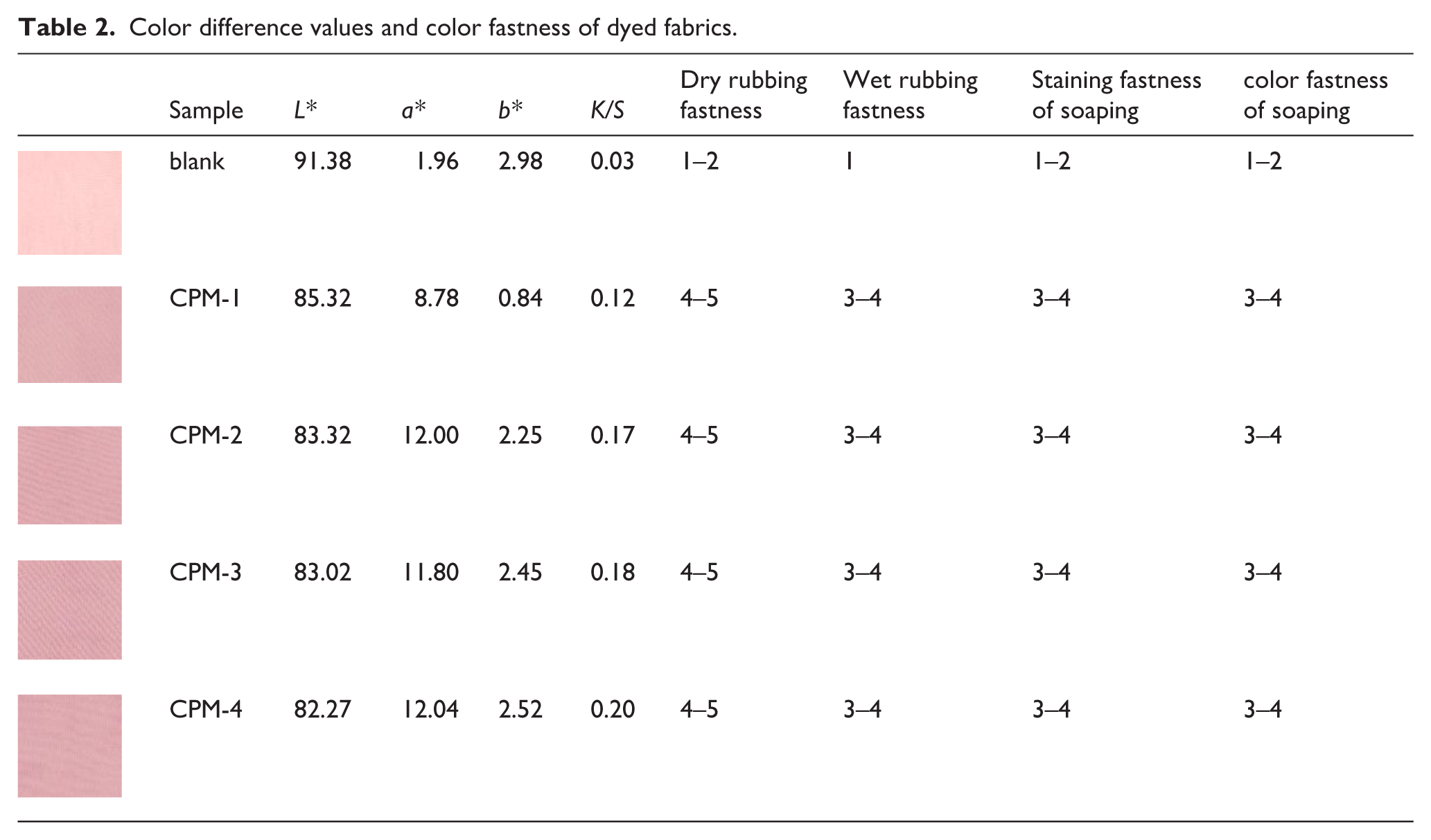
The K/S values of dyed fabrics (Table 2) were determined using an SF400 colorimeter (Datacolor, USA). The color fastness against rubbing and soaping of fabrics was assessed by ISO 105-X16-200, GB/T 21898-2023, GB/T 8424.1-2001, and ISO 105-C10-2006 under D65 illumination and 10 standard observers.
Color difference values and color fastness of dyed fabrics.
Results and analysis
Microspheres characterization
SEM
The SEM images of the freeze-dried PMNAD microspheres are shown in Figure 3.
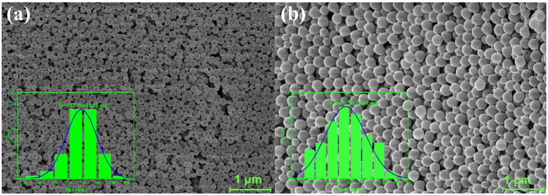
SEM images of PMNAD and CO@PMNAD microspheres.
Figure 3(a) illustrates that microspheres maintain a regular spherical shape without adsorption, with an average particle size of 112.36 nm. In contrast, the surface of composite copolymer microspheres (CO@PMNAD) after dye adsorption exhibits smoothness and a discernible core–shell structure, revealing a distinct contrast between a dense inner core and a dense outer layer. This difference in material density between the two regions characterizes the core–shell structure. Figure 3(b) illustrates the morphology of the composite copolymer microspheres after the adsorption of cochineal red dye. The dyed composite copolymer microspheres (CO@PMNAD) exhibit smooth surfaces, and the particle size maintains excellent homogeneity, with an average diameter of 192.75 nm. In comparison with the undyed microspheres, the mean particle size was augmented by 80 nm. Electrostatic interactions between the –OH groups of carmine red and the –NH4 groups in the nanosphere shell layer increase the distance between macromolecular chains, subsequently leading to an increase in particle size. 20
Comparison of particle size between colored (cochineal composite) and noncolored microspheres
Figure 4, which was obtained after the dispersions of colored and noncolored microspheres (both with 0.1% solid content) analyzed using a Zetasizer Nano ZS90, shows the particle size distribution of the microspheres.
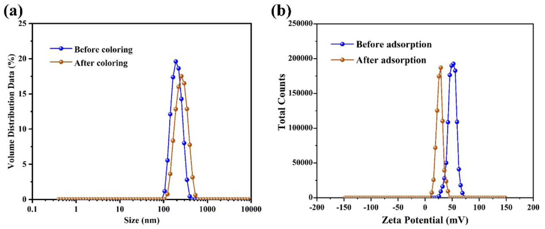
Distribution of (a) particle size and (b) zeta potential of microspheres before and after dye adsorption.
The particle sizes of non-colored PMNAD microspheres average 112.36 nm with a PDI of 0.028, while that of CO@PMNAD, with PDI being 0.052, increases by 80 nm to 192.75 nm, indicating that cochineal adsorbed on the surface of microspheres enlarges the particle size. While the potential of noncolored microspheres is measured at +52.8 mV, the potential of the colored microspheres drops to +29.3 mV, because cochineal’s electronegativity reduces the overall potential after it is absorbed by the microspheres.
TG analysis
As shown in Figure 5, cochineal dehydrates drastically after being heated to 100°C, then begins to decompose at 200°C or higher, and decomposes almost completely at 350°C. PMNAD microspheres decompose slightly between 200 and 350°C, and decompose drastically above 350°C, with none left at 430°C, because NAM and DMC, both acting as reactive emulsifiers, have participated in the polymerization to obtain a more stable soap-free emulsion with excellent heat resistance. The difference in thermal performance between CO@PMNAD (colored) and PMNAD (noncolored) microspheres is demonstrated by the steeper stage (200–250°C) of the CO@PMNAD curve due to thermal decomposition of cochineal covering the microspheres. As a result of electrostatic attraction between the microspheres and the dye, as well as the higher specific surface energy of the microspheres, the colored microspheres are more thermostable than cochineal.
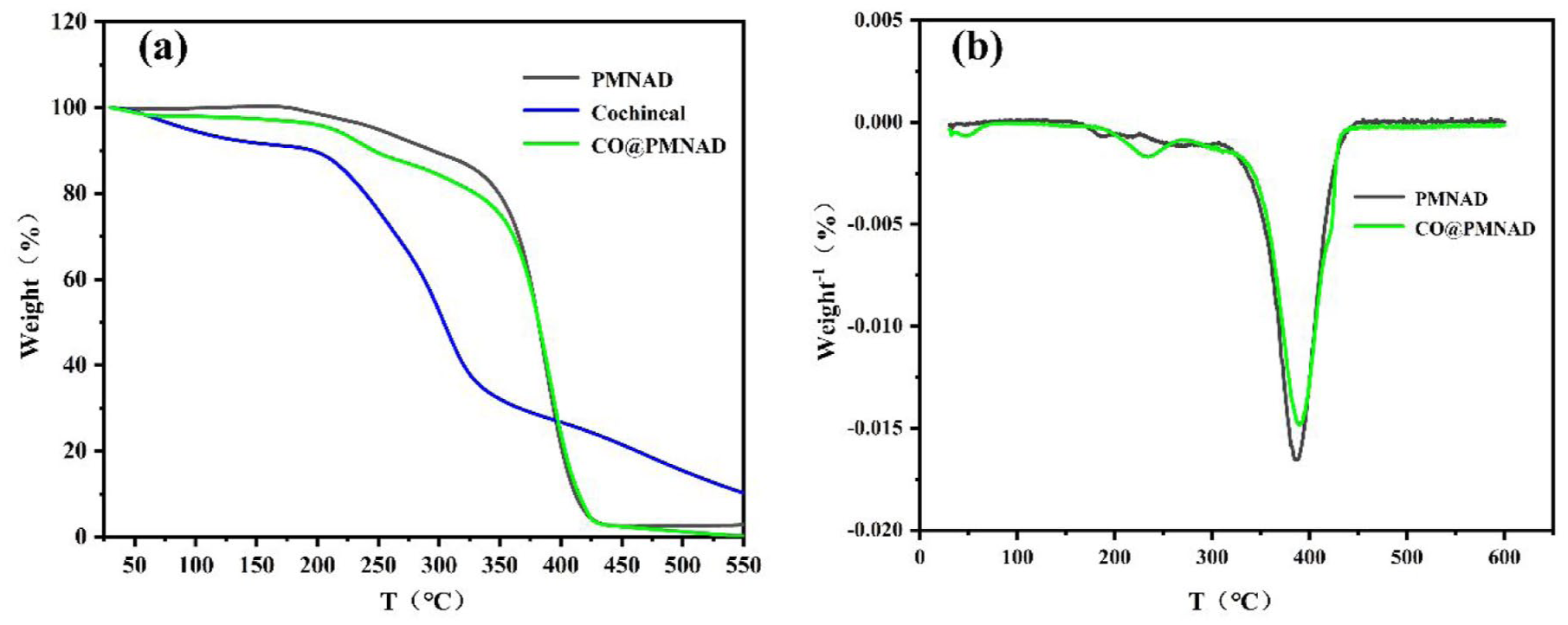
(a) TG and (b) DTG curve of cochineal red composite microspheres.
Infrared analysis
As shown in Figure 6(a), the peak of cochineal at 3421 cm−1 corresponds to the O–H stretching vibration. In PMNAD microspheres, the peak at 1449 cm−1 is assigned to the stretching vibration of the quaternary ammonium group of DMC, the peak at 1731 cm−1 to C=O stretching vibration of NAM, the peak at 1598 cm−1 to N–H stretching vibration of NAM (a cationic monomer), and the peak at 3445 cm−1 to O–H stretching vibration of NAM, indicating the success of the reaction to polymerize PMNAD microspheres. Figure 6(a) also shows that, compared with PMNAD, CO@PMNAD presents a much smaller peak at 1447 cm−1, because the stretching vibration of the quaternary ammonium group is covered up due to the adsorption between the microspheres and cochineal. The chemical structure of the synthesized PMNAD microspheres was further confirmed by ¹H NMR spectroscopy (Figure 6b). The spectrum exhibited a characteristic signal at δ 3.43 ppm for PMNAD, corresponding to the proton peak of the [–C(CH3)–] cationic group (e.g., quaternary ammonium salt –N+(CH3)3). In the CO@PMNAD spectrum, the sharp aromatic signals of free carmine (δ 6.4–7.1 ppm) exhibited significant broadening and weakening. This phenomenon characteristically reflects restricted molecular motion due to adsorption onto a larger solid surface. This directly demonstrates the successful immobilization of carmine dye onto the PMNAD microsphere surface, corroborating the electrostatic adsorption mechanism proposed based on FTIR and zeta potential analysis.
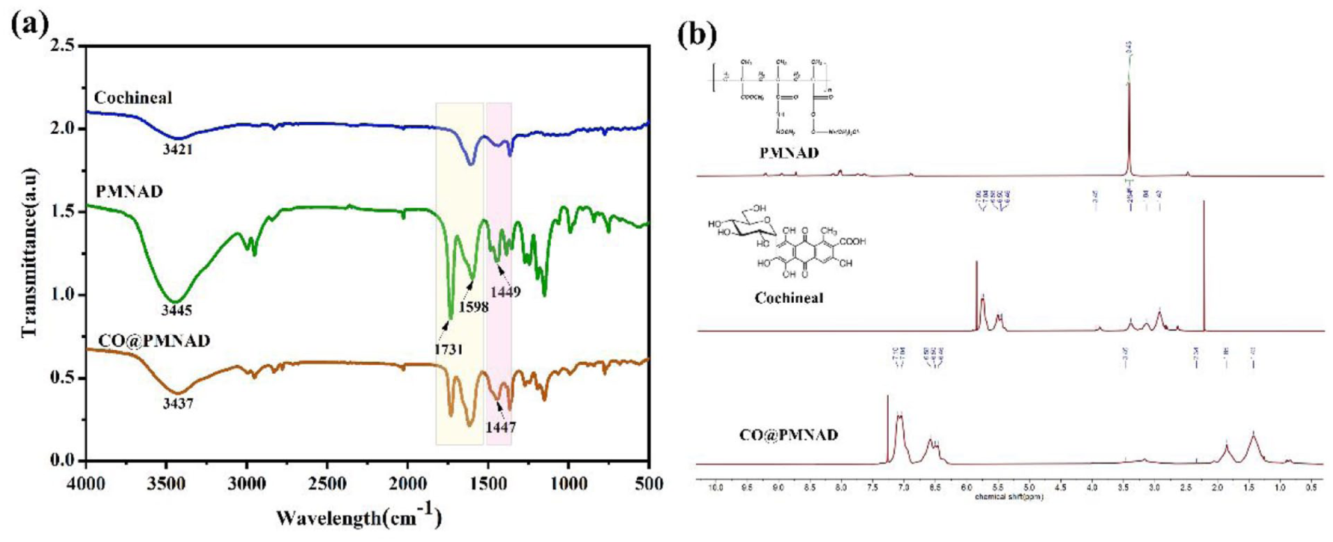
(a) Infrared spectrum and (b) NMR analysis of cochineal, PMNAD, and CO@PMNAD.
Adsorption of cochineal
Single factor for adsorption of cochineal
When dissolved in acid solution, carminic acid, the main element of cochineal, presents varied colors depending on different pH values, from yellow (pH 4.8), deep purple (pH 6.2),18,19 to red as a pH above 8 would ionize carboxyls and hydroxyl in the molecule, causing a redshift in its maximum absorption wavelength, and then to gradually deepened blues when the pH continues to rise and changes its hue. 21 Thus, the optimum pH is 7–8. The optimal experiment conditions for adsorption were acquired by varying factors such as the dye concentration, time, and temperature of the reaction, and mixing rate while keeping the solid content of microsphere emulsion, deionized water, and the pH of the working liquid unchanged.
Due to the cationic nature of DMC, the colloidal PMNAD microspheres are electropositive, absorbing carmine anions. As can be seen in Figure 7(a), the absorption between the microspheres and the dye rises sharply as the cochineal concentration goes up from 0 to 2000 mg/L. However, once the concentration reaches approximately 2000 mg/L, the absorption reaches a saturation point, with little further increase even when the concentration exceeds 2000 mg/L, and a decrease is observed beyond this level. Figure 7(b) shows that, at the concentration of 2000 mg/L, the absorption increases as the temperature goes up from 30 to 50°C, but decreases when the temperature is above 50°C, revealing that the optimal temperature is around 50°C. This is because higher temperature accelerates not only the diffusion of carmine anions to the microspheres’ surface, increasing the absorption, but also the desorption of the reactive dye from the surface. The mixing rate has little influence on absorption, which is shown in Figure 7(c), because the strong electrostatic attraction between the microsphere and the dye weakens the impact of the centrifugal force, which, at a high rate, would slow down the absorption. Therefore, given relevant factors, the best mixing rate is about 200 rpm. As can be in Figure 7(d), at the same concentration, temperature, and mixing rate and with the same volume of cochineal, the absorption increase significantly as reaction time is prolonged from 0 to 3 minutes, and reaches a plateau after 10 minutes of reaction, and shows little change from 10 to 60 minutes. The main reason is that at the initial stage, the strong affinity between the electropositive microspheres and the carmine anions makes the latter quickly occupy the former’s surface. Over time, more anions gradually reduce the electropositivity of the microspheres, slowing down the rising absorption. Once saturation is reached, further absorption ceases. Thus, we can conclude that the absorption progresses fast and ends in 10 minutes.
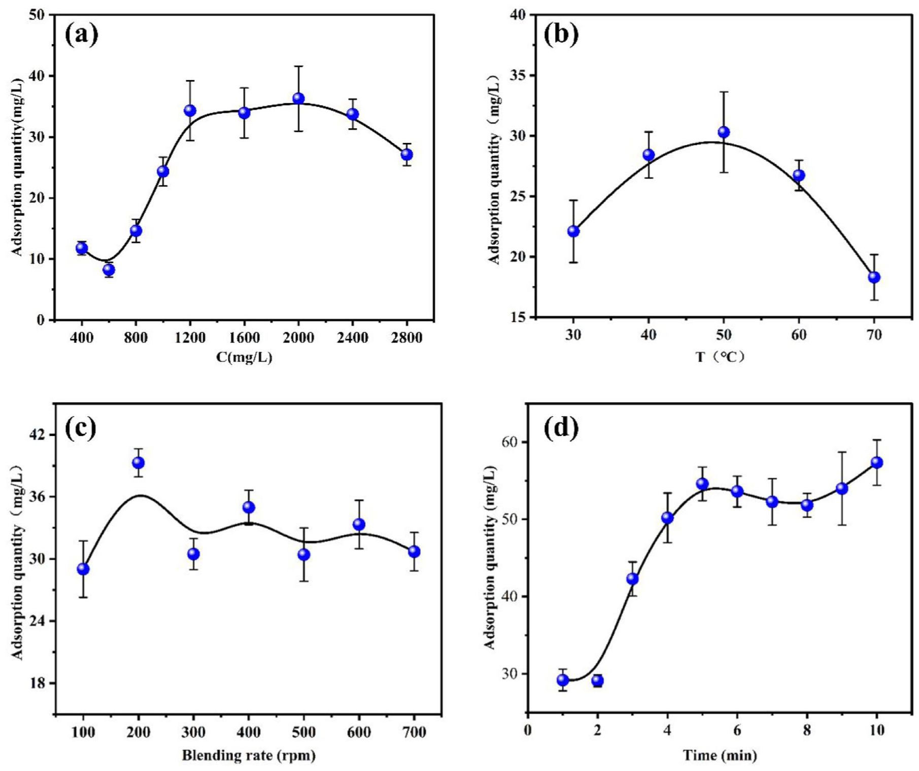
Factors that influences the absorption quantity: (a) dye concentrations, (b) reaction temperatures, (c) mixing rate, and (d) reaction time.
Adsorption isotherm
The adsorption behavior was examined utilizing the Langmuir and Freundlich isotherm models. The nonlinear representations of these isotherm models are articulated, respectively, in the following equations:


where: Ce (mg/L) and qe (mg/g) denote the equilibrium concentrations of the adsorbate in the aqueous and solid phases, respectively; qm represents the saturation (maximum) adsorption capacity (mg/g); KL is the Langmuir constant (L/mg), associated with the adsorption energy; and KF is the Freundlich equilibrium constant (mg(1-1/n) L1/n-1 G-1) pertains to adsorption capacity, where n is a constant representing the Freundlich isotherm.
The Langmuir and Freundlich isotherm models were employed to ascertain the chemisorption of the dye with the nanospheres. The loading of gardenia onto nanospheres at 323 K is illustrated in Figure 8. Table S1 in the supplemental material presents the constants of the two models utilized to simulate PMNAD adsorption. In terms of the R2 value of cochineal adsorption, the Langmuir isotherm model significantly outperforms the Freundlich model, suggesting that the equilibrium data align well with the Langmuir isotherm model. 22 This implies that the adsorption of cochineal on PMNAD nanospheres takes place as a monolayer of homogeneous adsorption. At 323 K, the Qmax achieved was 14.0153 mg/g, respectively, indicating that the uncolored particles remained in the upper solution.
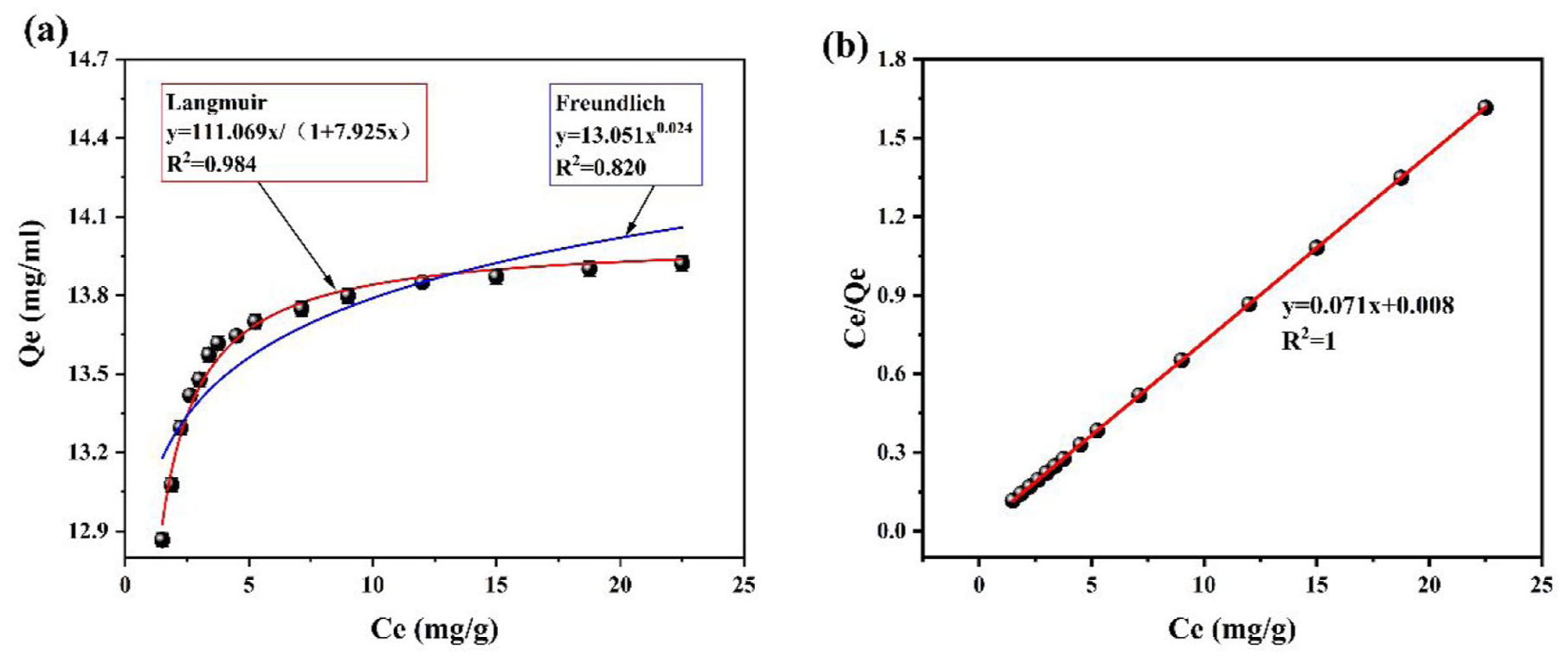
(a) The adsorption isotherms and fitting curve of the Langmuir isotherm model and (b) of cochineal onto PMNAD microspheres at 323 K (volume, 50 mL; m (PMNAD), 100 mg; C0 (cochineal), 0.4 mg/mL; pH 8–9).
Adsorption kinetics
The pseudo-first-order and pseudo-second-order models were utilized for analyzing the adsorption kinetics of cochineal on microspheres; the models used can be summarized as follows:


where k1 (min−1) and k2 (g/mg•min) correspond to the pseudo-first-order and pseudo-second-order adsorption rate variables, respectively, and qe and qt are the amounts of cochineal immobilized on the PMNAD microspheres (mg/g) at a state of equilibrium and point t (seconds).
Under weakly alkaline conditions, 10 mg of PMNAD was added to a 10 mL solution of cochineal aqueous extract with a concentration of 20 mg/L. Subsequently, the values of qt at various time points were measured, and the results are shown in Figure 9.
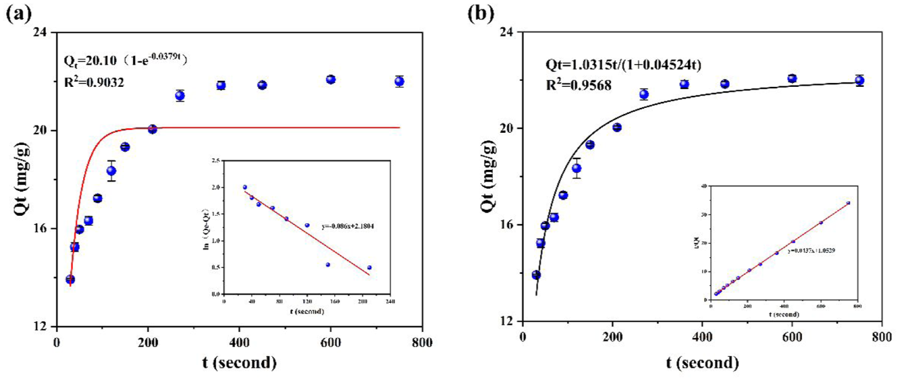
Fitting plots of the (a) pseudo-first-order and (b) pseudo-second-order model for cochineal onto PMNAD microspheres (volume, 50 ml/L; m (P(MA-DMC), 10 mg; C0 (cochineal), 20 mg/L).
The experimental data were fitted using (5) and (6), and the resulting fitted constants are summarized in Table S2 in the supplemental material. The R² value of the pseudo-second-order model (0.9551) is higher than that of the pseudo-first-order model (0.7786). This indicates that the adsorption of cochineal onto PMNAD follows a chemisorption process. Furthermore, it can be inferred that chemical adsorption occurs between the cations of the microspheres and the anions of the dye, which enhances dyeing fastness.
Dyeing performance of colored microspheres on cotton fabrics
Gap length, an important characteristic parameter of semiconductors, depends on the energy band structure of a semiconductor, that is, its crystal structure and atomic linkage.
Figure 10 shows that the bandgap of the cotton fabric added with colored microspheres reaches 2.15 eV. Thus, a conclusion can be drawn that there is an electronic transition in the combination between the colored microspheres and the fabric, and that their bonding is electrostatic. Figure 10(a) illustrates the UV diffuse reflectance spectra of materials subjected to various treatments. The blank sample devoid of nanoballs exhibits more reflectance than the samples containing nanoballs CPM-1–CPM-4. As the concentration of cochineal rises under the same conditions, the light reflectance of the fabric progressively diminishes. The nanoscale dimensions of CO@PMNAD and the surface roughness of the nanospheres post-dye-absorption result in reduced light reflection of the cotton fabric. Cotton fabrics exhibit lower reflectance after CO@PMNAD dyeing, essentially due to the photonic crystal structure acting as a “light trap.” By suppressing reflection in specific wavelength bands and significantly extending the light propagation path within the structure, it maximizes the light absorption efficiency of cochineal red pigment molecules, ultimately resulting in less reflected light energy. As illustrated in Figure 10(b), different concentrations of CO@PMNAD nanospheres can augment the photonic band gap to capture increased light intensity. When natural light meets the surface of CO@PMNAD, a tiny amount of the incident light of a specified wavelength is absorbed by cochineal red, while the other portion of the reflected light is prevented by the nanospheres. Since the CO@PMNAD nanospheres on the fiber surface have truly been enriched, the remaining reflected light may be reflected and blocked, thereby effectively boosting the stability of natural light reflection.
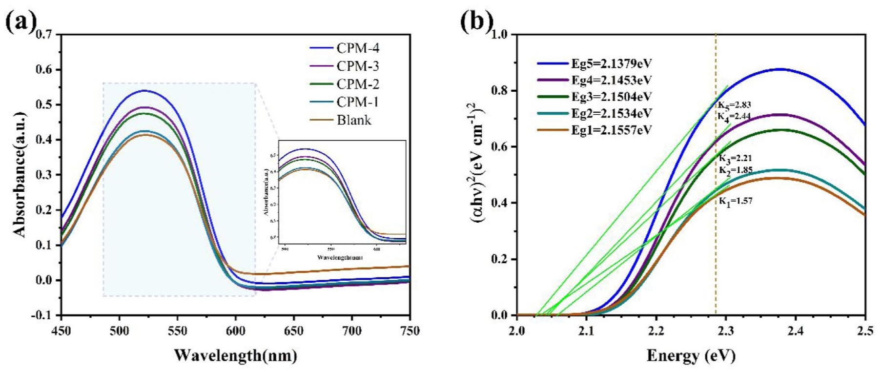
(a) ATR-UV analysis and (b) the UV-reflected photonic bandgap of the fabric at different concentrations.
Direct dyeing effect of the colored microspheres on cotton fabric
The cloth specimens that were washed, soaped, and dried after they had been directly dyed are shown in Figure 11.
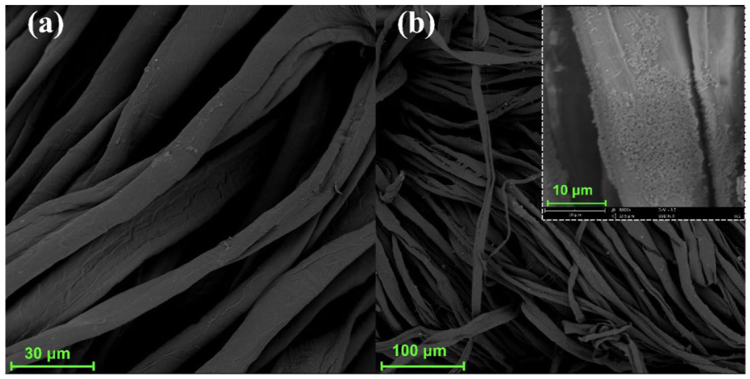
Fabric electron micrographs (a) before and (b) after dyeing.
Figure 11 demonstrates the direct dyeing effect of colored microspheres on cotton fabrics. As depicted in Figure 11(b), microspheres are clearly attached to the fiber surface, indicating successful coloring of the cotton fabric by the colored microspheres. Since PMNAD microspheres retain a strong positive charge after binding with cochineal red dye, they can interact with anionic cellulose groups on the fabric surface under alkaline conditions, thereby enhancing the dye uptake rate of cochineal red. Simultaneously, during the dyeing process, the dye microspheres assemble in layers on the fiber surface, leading to improved wash fastness and lightfastness of the dyed fabric to a certain extent.
The color strength was evaluated using the K/S value, which is derived from the Kubelka–Munk theory. This value represents the ratio of absorption (K) to scattering (S) of light by the dyed fabric and is a crucial parameter in the textile industry for predicting color strength and ensuring production consistency. Table 2 lists the dyeing properties of the colored microspheres. CO@PMNAD dispersion was employed to examine the dyeing characteristics of cotton fibers. The dyeing characteristics were evaluated against the sample using the color indices (L*, a*, b*, and K/S).
Table 2 indicates that the K/S value of the cochineal red blank sample is extremely low, indicating that the dye was barely absorbed. This occurs because the dye chromophore carries a negative charge, while cotton fibers also possess a negative charge due to the hydrolysis of their side-chain hydroxyl groups, making dye absorption difficult. However, from CPM-1 to CPM-4, the samples exhibit progressively lower L* values, higher a* values, and increasingly larger K/S values. This is because CO@PMNAD microspheres exhibit significantly higher K/S values due to strong electrostatic attraction with the fibers: postdyeing, the microspheres typically retain a positive charge, attracting the negatively charged fibers. The table also indicates that dry and wet rubbing fastness improves by 2 to 3 levels.
Conclusion
The PDI of the PMNAD microspheres, prepared via soap-free emulsion polymerization, is measured at 0.028, with the average size of the microspheres at 112.36 nm. After the coloring of cochineal, the PDI rises to 0.052, and the average size of microspheres grows to 192.75 nm. In addition, the zeta potential decreases from +52.8 to +29.3 mV. The microspheres are spherical with uniform particle size and good dispersity. Under optimal conditions, at a dye concentration of 2000 mg/L, a temperature of 50°C, a mixing rate of 200 rpm, and 10 minutes of absorption, cochineal is efficiently absorbed by the microspheres. The dry rubbing fastness of the cotton knitted fabrics dyed by cochineal composite microspheres ranges from 4 to 5, while the wet rubbing fastness, staining, and color fastness of soaping range from 3 to 4.
Supplemental Material
sj-docx-1-trj-10.1177_00405175251397606 – Supplemental material for Preparation of cochineal/polystyrene composite colloidal microspheres and their self-assembly and coloration properties on cotton fabrics
Supplemental material, sj-docx-1-trj-10.1177_00405175251397606 for Preparation of cochineal/polystyrene composite colloidal microspheres and their self-assembly and coloration properties on cotton fabrics by Nan He, Xiaoqiang Ma, Ting Pan, Yuexin Gao and Huiyu Jiang in Textile Research Journal
Footnotes
Declaration of conflicting interests
The authors declared no potential conflicts of interest with respect to the research, authorship, and/or publication of this article.
Funding
The authors disclosed receipt of the following financial support for the research, authorship, and/or publication of this article: This work was supported by Hubei Provincial Engineering Laboratory for Clean Production and High Value Utilization of Bio-Based Textile Materials, Wuhan Textile University (grant number SWJ202212), the Jianghan Plain Textile and Garment Industry Technology Research Institute (grant number JPTG06), and the Science and Technology Project of the Hubei Education Department (grant number 2021047).

Supplemental material
Supplemental material for this article is available online.
References
Supplementary Material
Please find the following supplemental material available below.
For Open Access articles published under a Creative Commons License, all supplemental material carries the same license as the article it is associated with.
For non-Open Access articles published, all supplemental material carries a non-exclusive license, and permission requests for re-use of supplemental material or any part of supplemental material shall be sent directly to the copyright owner as specified in the copyright notice associated with the article.
