
Abstract
Background
To research the effectiveness of hyaluronidase in evisceration surgery.
Methods
A total of 34 patients were studied as three groups: conventional surgery group (n = 13); isotonic fluid group (n = 8); and hyaluronidase group (n = 13). Hyaluronidase and isotonic fluid were introduced into the uveo-scleral space. Intraocular content was removed, if possible by a one-scoop method, with the aid of a spoon.
Results
Removing all the intraocular content as a one-scoop method was achieved in 10 of 13 (77%) in the hyaluronidase group and four of eight (50%) in the isotonic fluid group. The one-scoop method was not possible in any case without the introduction of fluid. The mean durations for the evisceration procedure were 32, 36.6 and 40.5 min in the three groups, respectively (P < 0.05).
Conclusions
Using hyaluronidase in evisceration surgery greatly facilitates and speeds up the removal of all the intraocular content in ophthalmic evisceration surgery.
Introduction
Evisceration and enucleation are radical ophthalmic interventions. Enucleation involves removal of the entire globe with cornea, sclera, and a small part of the optic nerve, whereas evisceration involves the removal of all the intraocular content while leaving the sclera and optic nerve in place.
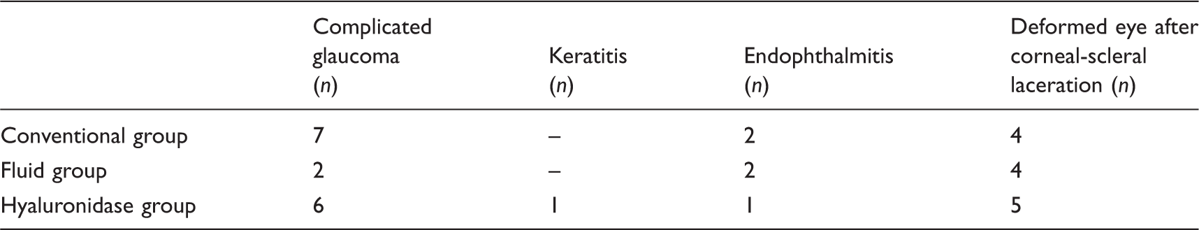
The indications for evisceration are penetrating trauma, complicated glaucoma, severe corneal disease, endophtalmitis and blind painful eye.1,2 The indications for enucleation most commonly were tumours such as uveal melanoma and retinoblastoma which are absolute contraindications to evisceration.
In this study we investigated the effect of hyaluronidase in facilitating evisceration surgery.
Hyaluronidase is a preparation of purified testicular protein enzyme with known effects on the connective tissue. Its action is to depolymerise and hydrolyse hyaluronic acid, a component of mucoprotein ground substance or tissue cement. Hyaluronidase appears to increase the permeability of the membranes and fibrous septa that compartmentalise the orbital contents.3,4 It augments the spread of local anaesthetic solutions in ocular anaesthesia and has a widespread acceptance in clinical practice.5–7 Its use is well documented in reduction of paraphimosis, reduction of prolapsed haemorrhoids or rectum, diffusion of extravasated chemotherapeutic medication, hypodermoclysis (subcutaneous fluid infusion – where intravenous access is impossible) and in beauty treatments such as in face lifts and skin tucks.4,8,9
The most troublesome and time-consuming part of evisceration surgery is the separation of all the intraocular content from the sclera. In this study we evaluated how hyaluronidase affects this part of the procedure.
Materials and Methods
We retrospectively evaluated 34 consecutive patients who underwent evisceration surgery in an ophthalmology department of a tertiary referral hospital between April 2012 and June 2014. All patients were operated under general anaesthesia by the same surgeon (AG). The patients were divided into three groups according to the agents applied introduced: none (conventional surgery) (n = 13); isotonic fluid (n = 8); and hyaluronidase (n = 13). Isotonic fluid was introduced in eight patients after the initial 13 conventional procedures, and subsequently hyaluronidase was used in the last 13 patients. Age, duration of surgery, duration of removal of intraocular content and surgical indications were recorded.
Conventional ophthalmic evisceration surgery begins with a 360° conjunctival peritomy. Tenon’s fascia is bluntly separated from the underlying sclera in all four quadrants. A full-thickness incision around the corneal limbus is made with a sharp scalpel blade and the entire corneal button removed. 8 Blunt dissection is made between uveal and scleral tissues.
Fluid or hyaluronidase (Hylase-Dessau 150 IE, Germany), diluted with 2 mL distilled water, when used, was introduced by a cannula at the four quadrants between the uveal tissue and sclera. After 3 min, the opposing edges of the anterior uvea were grasped with a forceps and the evisceration spoon positioned under the uveal tissue. An attempt was made to remove all the uveal tissue as a one-scoop method with the spoon (Figures 1 and 2). If this was not possible, the remainder of the uveal tissue was dissected away from the scleral wall back to the attachment around the optic nerve. Cotton-tipped applicators soaked in 95% alcohol were used for denaturation of any remaining pigmented uveal tissue. Subsequently, insertion of a silicon spherical implant into the scleral cavity, and the closing of sclera, Tenon’s fascia and conjunctiva were performed. A conformer was then placed on conjunctiva.
(a) anterior dissection of uvea and sclera, (b) giving hyaluronidase by a cannula into the uveal-scleral space, (c) grasping opposing edges of anterior uvea and positioning the evisceration spoon under the uveal tissue, (d) removing of all uveal tissue with one manoeuvre. (a–d) The inner parts of the sclera just after the removal of all uveal tissue without any intervention.

The time it took for removal of all the intraocular content was measured by a resident. The overall duration of the evisceration surgery starting from conjunctival peritomy to conjunctival closure was taken from the report of the anaesthetist.
Statistical analyses were done with independent samples t-test, Kruskal-Wallis, and Mann-Whitney U tests. Informed consents were obtained from all patients. The study adhered to the principles outlined in the Declaration of Helsinki.
Results
The mean ages of the patients were: 59 years (age range, 41–80 years) in the conventional surgery group; 53 years (age range, 28–75 years) in the isotonic fluid group; and 51 years (age range, 24–83 years) in the hyaluronidase group. The male to female ratio was 4:13, 5:8 and 6:13 in the three groups, respectively.
Evisceration surgery was performed in the right eye of six patients and in the left eye of seven patients in both the hyaluronidase and conventional surgery groups and in the right eye of five patients and in the left eye in three patients in the isotonic fluid group.
Indications for ophthalmic evisceration surgery.
Removal of intraocular content by a one-scoop method was achieved in 10 of 13 (77%) in the hyaluronidase group, four of eight (50%) in the isotonic fluid group, but in none of the cases in the conventional surgery group. The difference between the groups was statistically significant (P < 0.05).
Removal of intraocular content as a one-scoop method could not be performed in three eyes in the hyaluronidase group: one with complicated glaucoma which had undergone multiple cyclo-destructive procedures previously, and two deformed eyes due to corneal-scleral lacerations. Both the deformed eyes had had primary repair of the lacerations in our clinic. Likewise, failure in the isotonic fluid group was seen in three patients who had undergone surgery for deformed eyes due to corneal-scleral lacerations and one with complicated glaucoma.
In the conventional surgery group, uveal tissue was always removed with the spoon using multiple manoeuvres.
The total duration of the evisceration procedure was 32 min (range, 27–44 min), 36.6 min (range, 29–50 min) and 40.5 min (range, 33–50 min) in the hyaluronidase, isotonic fluid and conventional surgery groups, respectively. There was significant difference between the groups in terms of the duration of surgery (P < 0.05).
No complication such as orbital inflammation, allergic reactions, nor delayed healing were encountered in any patient.
Discussion
One of the hazards of ophthalmic evisceration is the risk of sympathetic ophthalmia. In our opinion, the most difficult part of the procedure is cleaning all the uveal tissue from the scleral shell. Improving total uveal tissue removal appears to reduce the contralateral eye to the exposure of antigens and so reduces the risk of sympathetic ophthalmia. 10
Introduction of fluid to enlarge the uveo-scleral space was considered to be a reasonable option to facilitate the separation of uveal tissue from the sclera. Ideally we sought to affect this in a single one-scoop procedure with an evisceration spoon. With isotonic fluid to improve the separation of uveal tissue from scleral, we had good results in four out of eight patients. But using hyaluronidase, which has long been used in cataract surgery to enhance the effect of local anaesthesia,11,12 we hypothesised that the separation of uveal from scleral tissue, the most time-consuming and troublesome part of the procedure, would be even further facilitated.
This was indeed achieved 10 of 13 (77%) patients. Where removal of uveal tissue by a one-scoop method was not achieved (three in the hyaluronidase group and four in the isotonic fluid group, the cause appeared to be multiple adhesions (synechiae) found as a result of complicated glaucoma, who had had multiple trans-conjunctival cyclo-destructive procedures previously. Furthermore, in deformed eyes after corneo-scleral repair, uveal inflammation and uveal tissue incarcerated in the sclera may further contribute to difficult separation of these tissues.
The difference between the hyaluronidase and isotonic fluid groups, in terms of ease of removal of uveal tissue, was statistically significant. Hence, it is proposed that the ease of extraction of uveal tissue in the hyaluronidase group was improved by the enzymatic action of hyaluronidase rather than due to its simple mechanical hydrodissective effect.
Furthermore, there was significantly shorter mean operating times (32 min) in the hyaluronidase group, compared with the others (36.6 min in the isotonic fluid group and 40.5 min in the conventional surgery groups).
Thus our small study suggests that, by facilitating the procedure, and shortening operative exposure, the risk of sympathetic ophthalmia may be significantly reduced, quite apart from the other benefits of reducing the surgical operating time.
Hyaluronidase for ophthalmic surgery has rare adverse reactions. It may lead to corneal toxicity and postoperative orbital inflammation.13,14 We did not see any enzyme-related complications in our few cases. A possible explanation may also be that we introduced hyaluronidase to the relatively avascular sclera rather than the periorbital area.
We cannot of course exclude the possibility that hyaluronidase may increase the diffusion of antigens and paradoxically increase the risk of sympathetic ophthalmia. However, a cleaner, neater and more accurately performed procedure usually predicts a better result than a messy dissection.
Conclusions
Using hyaluronidase in ophthalmic evisceration surgery greatly facilitates the removal of all intraocular content. It shortens operating time by 20%. It may have a significant effect in reducing the risk of sympathetic ophthalmia by improving total uveal removal, though a much larger trial would be needed to demonstrate this convincingly.
Footnotes
Declaration of conflicting interests
None declared.
Funding
This research received no specific grant from any funding agency in the public, commercial, or not-for-profit sectors.