
Abstract
Congenital syphilis occurs due to trans-placental transmission of Treponema pallidum or rarely, intrapartum contact with infectious lesions. Even though preventable, congenital syphilis occurs sporadically in India, owing to lack of antenatal screening as well as the lack of awareness among clinicians about the burden of syphilis in the community. Since a significant overlap of clinical manifestations exists with many systemic diseases, awareness among clinicians is crucial for an early diagnosis. Renomegaly, nephrotic syndrome and nephritis can all be the signs of renal involvement in congenital syphilis, which can provide clues of the diagnosis. Direct invasion by spirochetes, hypersensitivity reactions and immune complex deposition in glomeruli contribute to the pathogenesis. We report a case of congenital syphilis characterised by delayed diagnosis with renal as well as cutaneous manifestations from missed maternal syphilis during the antenatal period and owing to the lack of antenatal screening.
Case report
A two-month-old term male South Indian baby of a 32-year-old mother (gravida 5, para 4, spontaneous abortion 1, living children 3) and a 38-year-old father was born with a weight of 3 kg and developed papulovesicular and pustular lesions at the age of two months, all over the body sparing palms and soles (Figure 1). The lesions healed spontaneously and reappeared in new sites over several weeks. At the age of 3.5 months, the baby was hospitalised with gross haematuria, severe pallor and hepato-splenomegaly. Laboratory tests showed severe anaemia (Hb 60 g/l), leukocytosis (65 × 109/m3), low platelet levels (60 × 109/m3), reticulocyte count 1.4%, raised erythrocyte sedimentation at 40 mm/1st h and high C reactive protein (40 mg/l). The coagulation profile, liver and renal function tests were normal. Urinalysis showed the presence of albuminuria and microscopic haematuria. Bone marrow aspiration, performed since the peripheral smear showed a leucaemoid reaction and 14% blasts, was normal. The baby received a blood transfusion and was then referred to our clinic. Examination revealed bipedal oedema, pallor, skin lesions as described and firm hepato-splenomegaly. Blood pressure, growth and development were normal. No persistent nasal discharge was noted. Urine protein creatinine ratio (uPCR) was 3.9 mg/mg, serum albumin 31 g/l, cholesterol 2.42 g/l with normal complement 3 and 4 levels. Repeated peripheral blood smear showed normocytic normochromic anaemia, leucocytosis and mild thrombocytopenia with no blasts. An ultrasound examination confirmed splenomegaly and enlarged kidneys, the right kidney being 6.4 × 3.2 cm and the left 6.4 × 2.6 cm compared to the mean bipolar length at this age of 4.7 cm ± 0.7 SD. Renal echoes was elicited bilaterally. A kidney biopsy showed normal cellularity of vessels, tubules and interstitium with a negative immunofluorescence for IgA, IgG, C3 and IgM.
Vesiculobullous lesions with scars.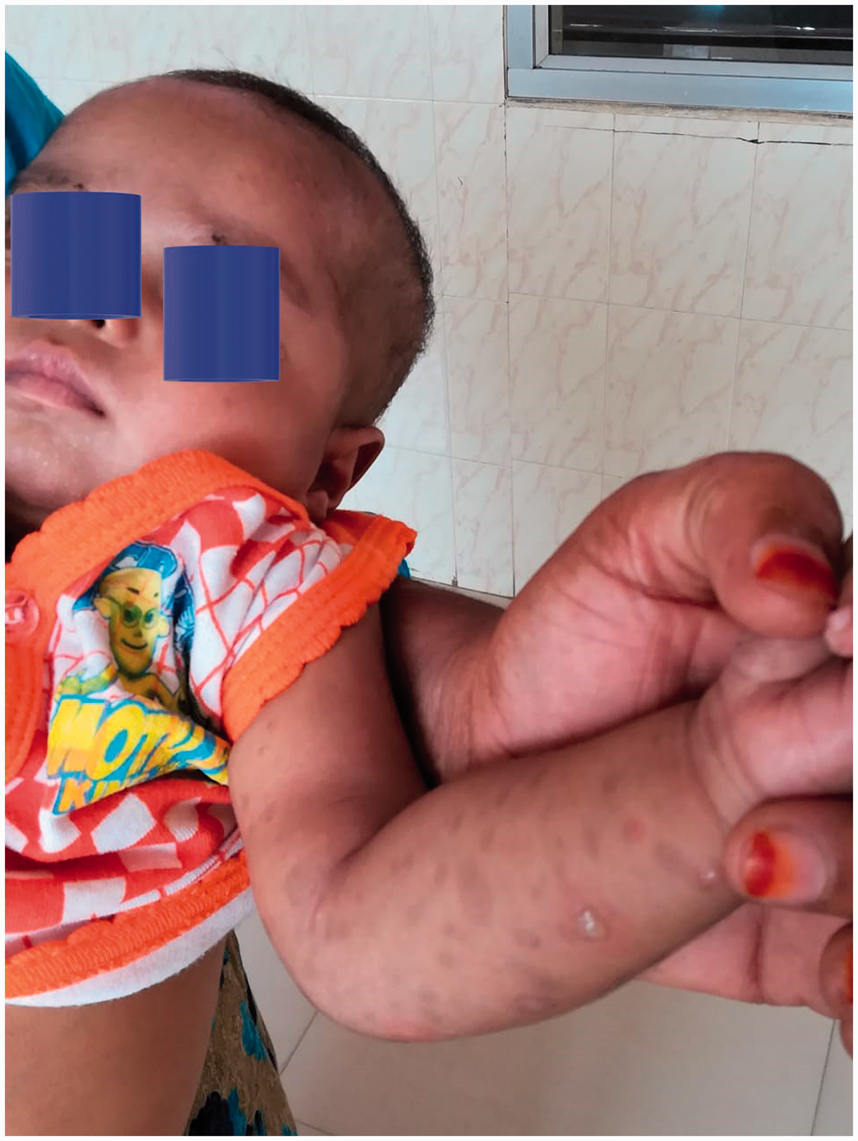
As there was nephritis with nephrotic range proteinuria, the mother was screened for chronic infections and was found to be venereal disease research laboratory test (VDRL) reactive, but HIV, hepatitis B and C seronegative. Antenatal screening with VDRL or rapid plasma reagin (RPR) had not been performed during the present or previous pregnancies. The mother’s VDRL screen was positive up to 1:30 and the treponema pallidum haemagglutination (TPHA) reactive up to 1/1280 dilutions; the baby’s being 1:128 and 1:250, respectively. The baby’s VDRL titre showed greater than four fold rise compared to the mother’s. A painless genital ulcer was detected on mother’s vagina which was apparently present during both pregnancies. Cerebrospinal fluid cytology and biochemistry were normal and non-reactive to VDRL. Radiography of the long bones showed a significant subperiosteal reaction (Figure 2). This had not been initially carried out, as congenital syphilis had not been considered, and moreover, no paradoxical crying by the baby had been noted.
X-ray right forearm–periostial reactions.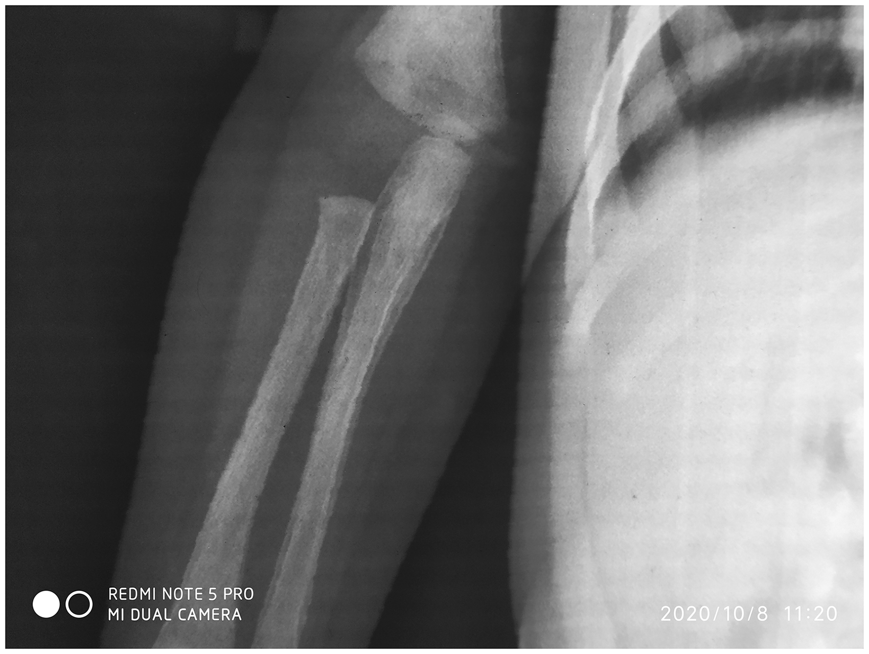
The baby was treated with crystalline penicillin 50,000 IU/kg qds for 14 days covering for neurosyphilis as well, because cerebrospinal fluid analysis cannot reliably exclude the central nervous system involvement in CS. Haematuria and proteinuria resolved in two weeks and skin lesions gradually also resolved. On follow-up after six weeks, his anaemia had corrected, hepatosplenomegaly was resolving and he was satisfactorily gaining weight. All other siblings screened negative for syphilis. The father was not screened, as he had travelled abroad, but counselling and follow-up has been ensured at the sexually transmitted disease (STD) clinic in our institution. The mother was treated with a 14 days course of ceftriaxone on an ambulatory basis which was the regimen opted by her instead of penicillin.
Discussion
Globally, the estimated prevalence of CS is 473 per 100,000 live births. 1 Based on the WHO tool, in 2015, seroprevalence of syphilis among pregnant mothers was 0.38% in India. 2 WHO introduced the elimination of mother-to-child transmission of HIV and syphilis initiative in 2014. 3 About 89.6% of pregnant women seek antenatal care at least once, 2 but in India during 2016–2018, the syphilis testing rate during the antenatal period was only 20% compared to 59% for HIV testing. 4 It is necessary to examine whether the decreasing seroprevalence of syphilis among adults engenders complacency among clinicians, resulting in lower rates of antenatal screening. Enhanced awareness, meticulous planning, implementation and monitoring of programmes at the national level are imperative. Lack of proper gynaecological examination resulted in missing the mother’s genital ulcers for years. Although the baby had several features of CS, the correct diagnosis was nonetheless delayed. The absence of a paradoxical cry and snuffles, as well as unusual distribution of his rash, also contributed to the delay. Mucocutaneous manifestations occur in about 70% of CS. 5 Although vesiculobullous or maculopapular lesions of palms and soles are classic, our case illustrates the variegated dermatological manifestations of syphilis. Periostitis of long bones, if identified earlier is, however, a classic sign. Nonetheless, renomegaly and nephritis with nephrotic range proteinuria associated with the obvious skin lesions were our clues. Our blood and urine investigations were consistent with infection-related glomerulonephritis rather than nephrotic syndrome, although there was increased uPCR and cholesterol level. Haematuria, oedema and normal serum albumin support the former. The kidney biopsy was, however, normal in our case, may be because it was performed during the second week of hospitalisation, by which time nephritis had virtually resolved.
Obviously, partner management is a crucial element of preventive care and one of the important tools for global CS elimination.
Our learning points were that (i) phenotypic features of CS vary greatly among patients; (ii) in the evaluation of multisystem involvement in young infants, intrauterine infections should be considered and (iii) CS should still be considered even in the context of low prevalence, although in Western nations, it is rising, particularly amongst the homosexually active population.
Footnotes
Acknowledgments
Authors acknowledge Dr.Ajithkumar VT, HOD Pediatrics, Dr.Rajesh TV, DR.Vinodkumar MS and Dr. Priya Velayudhan for their contributions in the diagnosis and management of this patient.
Consent
The mother has given consent for publishing the clinical photographs and X-rays of the baby.
Authors’ contribution
KC conceptualised the article and prepared the article. MGG critically analysed the data and article. MPS and SAPR were involved in the diagnosis and management.
Declaration of conflicting interests
The author(s) declared no potential conflicts of interest with respect to the research, authorship, and/or publication of this article.
Funding
The author(s) received no financial support for the research, authorship, and/or publication of this article.