
Abstract
Metanephric adenoma is a rare benign mass that may be radiologically indistinguishable from renal cell carcinoma. The clinical presentation of this entity is similar to a malignant renal mass. Only 200 cases have been reported worldwide till now. Furthermore, there are no strongly predictive clinical and radiological criteria. It, therefore, poses a diagnostic challenge. Since post-operative therapy may vary significantly if the lesion turns out to be malignant, it is crucial to distinguish it from other malignant renal masses.
Case report
A 65-year-old woman complained of seven days of left flank pain. The pain was subtle, dull, non-radiating and unaffected by anything. Fever, vomiting, haematuria and other urinary symptoms were absent. She had tachycardia and pallor on inspection. Physical examination showed left lumbar discomfort but no other abnormalities. The postero-superior left kidney had a 27 × 41 × 45 mm heterogeneously enhancing tumour with perinephric fat stranding and delayed renal excretion on CT scanning. It was unclear if this was a kidney abscess, an inflammatory lesion or a malignancy. An MRI scan indicated a 4 cm exophytic left renal tumour with intralesional and perinephric haemorrhage.
At operation, the whole kidney appeared oedematous and enlarged with dense adhesions around it with loss of peri-nephric planes. A left nephrectomy was carried out (Figure 1). Histopathological report suggested a metanephric adenoma (MA). Immunohistochemistry confirmed the diagnosis with positivity for WT1, AMACR, EMA, CK7 and negativity for CD10.
Discussion
The diagnosis of MA is most often made post-operatively. It is closely related to pure stromal lesions and metanephric adenofibromas. 1 It is usually a benign and well-differentiated tumour with female predominance and presenting usually in the fourth decade of life. 2 MA is asymptomatic in many patients but may present similarly to a malignant renal mass. Polycythaemia is seen in 10% and appears to be due to the production of erythropoietin and other cytokines by the lesion. 3
Radiographically these tumours may have peripheral or central calcifications in 20% and may be hypo-vascular on CT scanning and hyperechoic on ultrasonography. However, calcification is not diagnostic. 4
On histopathology, MA shows small cells with scanty cytoplasm, without mitosis, having small round acini and are phenotypically similar to nephroblastomas. 2 The Wilms tumour marker WT1 is frequently expressed. Α-Methylacyl-CoA racemase (AMACR) is poorly expressed in MA but well expressed in papillary renal cell carcinoma, whereas S-100 protein expression is the reverse (though weak in Wilms tumour). 5 These markers may improve the diagnostic yield of percutaneous biopsy and fine-needle aspiration, but the index of suspicion needs initially to be high for biopsy to be considered.
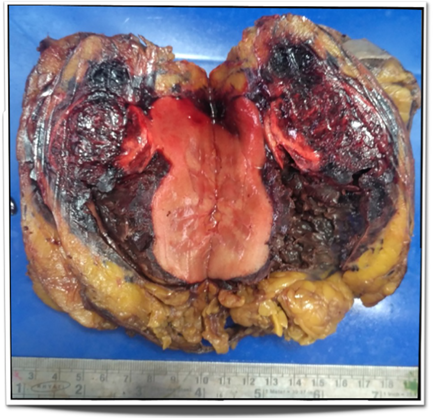
Cut open specimen of Lt kidney showing tumour 6.1×3.6×3.6 cm with extensive areas of haemorrhage and peritumoral extension.
Footnotes
Declaration of conflicting interests
The author(s) declared no potential conflicts of interest with respect to the research, authorship, and/or publication of this article.
Funding
The author(s) received no financial support for the research, authorship, and/or publication of this article.
Informed consent
Written informed consent for patient information and images to be published was provided by the patient.