






Abstract
This brief communication details the clinical, macroscopic, histological, and immunohistochemical features of a spontaneous case of uveodermatologic syndrome (UDS) in a laboratory beagle dog. A bilateral and symmetrical panuveitis, rich in macrophages and T lymphocytes, with distinctive extracellular clumped or phagocytised melanin pigment granules, and Dalen-Fuchs nodules was diagnosed following occurrence of ocular symptoms and subsequent blindness in a female peripubertal 9.5-month-old beagle dog. Although no macroscopic lesions were visible in the skin, microscopic examination revealed a histiocytic and lymphocytic lichenoid dermatitis with pigmentary incontinence. UDS has not been described as a background finding in laboratory beagle dogs before, although it is a well-known immune-mediated disease in certain canine breeds. Knowledge that UDS can occur in laboratory beagle dogs involved in preclinical studies, especially ocular studies, is essential for toxicologic pathologists.
Keywords
Beagle dogs used in preclinical toxicity studies are typically peripubertal to young adults (the length of canine preclinical studies does not generally exceed 39 weeks) and, therefore, less prone to develop immune-mediated disorders, which occur more frequently in older animals. The only idiopathic and potentially immune-mediated condition currently described as a background finding for laboratory beagle dogs in the International Harmonization of Nomenclature and Diagnostic Criteria (INHAND) guide 15 is canine juvenile polyarteritis (also known as Beagle Pain Syndrome).
Canine uveodermatologic syndrome (UDS) is an immune-mediated disease that targets tyrosinase or tyrosinase-related proteins within melanocytes,13,17 and that partially resembles human Vogt-Koyanagi-Harada syndrome (VKHS), both clinically and histologically. Besides the dog, this syndrome has not been reported to spontaneously occur in other laboratory animal species, although experimental animal models have been successfully developed in the rat, the Akita dog, and the monkey. 16
UDS causes a bilateral, severe, granulomatous/lymphohistiocytic panuveitis that leads to retinal detachment and blindness. 10 In the skin, a lichenoid or interface-like dermatitis occurs most often in the face or head with variable clinical manifestations such as leukoderma, leukotrichia, erosions/ulcers, alopecia, and crusts.10,14 In most cases ocular signs precede those in the skin, however less frequently, these can develop concurrently, or the dermatologic signs can appear first. In some canine breeds, including Akitas, Samoyeds and Siberian huskies, a predisposition has been reported; however, only for the American Akitas a higher risk to develop UDS has been connected with a specific dog leukocyte antigen haplotype: (DLA)-DQAI*00201.2,17 The mean age of onset of disease is 3.6 years and female to male ratio is 0.6 to 1.13,17
In this article, we describe the clinical, macroscopic, histological and immunohistochemical features of a spontaneous case of uveodermatologic syndrome in a peripubertal beagle dog; to the authors’ knowledge this is the first time that this syndrome is reported as a spontaneous finding in a laboratory beagle dog.
The affected dog was obtained from a USA-based commercial supplier and was part of a regulatory 39-week toxicity study, which was approved by the local ethical committee (AWERB), and conducted, in accordance with the UK Home Office “Guidance on the Operation of the Animals (Scientific Procedures) Act 1986,” at Labcorp, Harrogate, an AAALAC-accredited animal facility.
All dogs were subjected to a full clinical examination on arrival to the facility and ophthalmic examinations were conducted during the predose phase. At that time, no remarkable observations were recorded for any of the dogs on study.
The affected dog was a female allocated to the control group and was aged just over 6 months at the beginning of the study. The vehicle control article was NaCl 0.9%, administered at 10 ml/kg once daily by oral gavage. On day 90 of the dosing phase, bilaterally red and partially closed eyes were observed for this dog and following clinical examination by a veterinary surgeon, conjunctivitis was suspected and topical eye treatment was administered twice daily in the form of Isathal (Fusidic Acid) between days 90 and 92, and Maxitrol (Dexamethasone, Neomycin sulfate and Polymyxin B sulfate) and Viscotears (Carbomer (polyacrylic acid)) between days 92 and 96 of the dosing phase. However, despite treatment, no improvement was observed on the following days and on day 96, the animal appeared suddenly unable to see, was unable to detect movement and exhibited staring behavior, with dilatation of both pupils. Indirect ophthalmoscopy was attempted, however it was not possible to identify the fundus or vessels; therefore, due to the severity of the clinical observations, the dog was euthanized on day 96 of the dosing phase, aged 9.5 months and a full necropsy was performed post euthanasia.
Macroscopic findings noted during necropsy included stomach distention and colon mucosal surface striations for which no correlating findings were detected during microscopic evaluation. Additionally, mottled liver and dark cecum were observed, which microscopically correlated with venous congestion. The observations of bilateral red eyes and presence of a 10 x 5 mm red area in the right eye were also recorded, the former correlated with bilateral inflammation, however no microscopic correlating findings were noted for the latter. No macroscopic findings were observed in the skin at necropsy.
Hematology, coagulation profile, clinical chemistry and urinalysis results were examined for this dog during the predose phase, on day 90 of the dosing phase and on day 96, prior to necropsy. Hematology and urinalysis results did not show any abnormalities at any time points; however, a mild increase in urea (8.5 mmol/L; range 2.6-6) and creatinine (77 µmol/L; range 32-59) values was recorded on day 96 compared to results recorded on day 90 and to historical control data. Coagulation profile could not be assessed on day of necropsy due to the sample being clotted.
Eyes were fixed in Davidson’s fixative for 10 days before a full longitudinal section of the central region of the eye, perpendicular to the ciliary artery, was sampled for both eyes. All organs that form part of a standard tissue list for a regulatory toxicity study were fixed in 10% neutral-buffered formalin, then trimmed, routinely processed, and embedded in paraffin wax prior to sectioning (4-6 µm) and staining with hematoxylin and eosin (H&E) for routine histological examination.
Additional histochemical stains (Gram and PAS) were used to exclude presence of infectious agents in the eye and skin samples. Sections of the globe and skin were also subjected to immunohistochemistry (IHC) to examine expression of lymphocytic (CD3, CD20) and histiocytic (IBA-1) markers (Supplemental Table 1). Positive controls for formalin fixed and Davidson’s fixed tissues were run in parallel with each sample for each antibody. No vehicle control or test article-related histological findings were recorded for any dog on this study.
On H&E slide examination, a diffuse, bilateral, and symmetrical, moderate thickening of all components of the uveal tract by a granulomatous/lymphohistiocytic inflammatory infiltrate was noted (Figure 1A). The infiltrate was composed of numerous macrophages, often containing aggregates of cytoplasmic brown punctate granules consistent with melanin (melanophages), lymphocytes and fewer plasma cells, observed within the choroid, where it was effacing the retinal pigment epithelium (Figure 1B), and to a lesser extent in ciliary body and iris. The inflammation extended to the trabecular meshwork with obliteration of the filtration angle, to the limbus, and the sclera. Perivascular cuffing of scleral and episcleral blood vessels, and of the posterior ciliary arteries in the peripapillary region was evident (Figure 1B). A distinctive feature was the presence of granular melanin pigment dispersed within the uveal tract. Nodular aggregates of epithelioid macrophages engulfing melanin, and occasional lymphocytes were observed between the retinal pigment epithelium and Bruch’s membrane, consistent with Dalen-Fuchs nodules (Figure 1C). Similar nodules were also noted at the level of the iris.
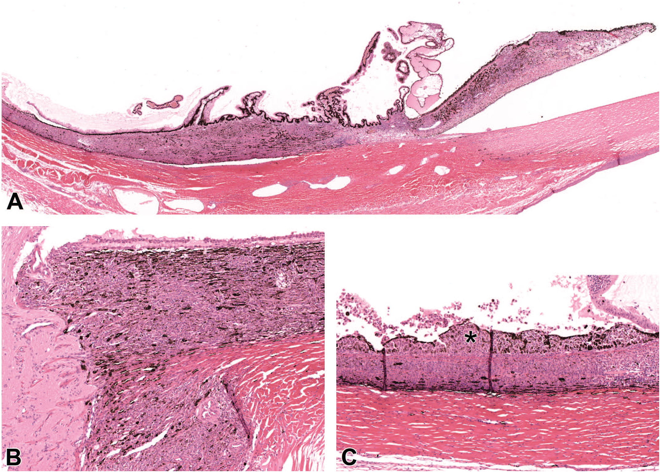
Uveodermatologic syndrome, beagle dog, eye (H&E). (A) The uveal tract is markedly and diffusely expanded by a moderate number of inflammatory cells (panuveitis). (B) Infiltration of the choroid by numerous lymphocytes and macrophages containing brown pigment (melanin), and inflammation of the posterior ciliary arteries (peripapillary). (C) Higher magnification of the choroid. Dalen-Fuchs nodules (nodular aggregates of macrophages, engulfing melanin, and lymphocytes) (asterisk) are located between the retinal pigment epithelium and Bruch’s membrane. Secondary detachment of the retina is present.
Moderate edema of the ciliary processes was noted in the pars plicata of the ciliary bodies.
Bilaterally, diffuse retinal detachment was observed (Supplemental Figure 1), with minimal hypertrophy of the retinal pigment epithelium exhibiting the typical tombstone-like appearance.
Bilaterally, within the vitreous, pale eosinophilic proteinaceous material admixed with few melanophages was observed, whilst within the anterior and posterior chamber no abnormalities were detected. The lens, cornea and optic nerve were unremarkable.
In the protocol-specified skin section, sampled from the ventral abdomen to include the teat, a moderate, locally extensive, dermatitis characterized by a lichenoid and perifollicular pattern was observed (Figure 2A). The inflammatory population consisted of numerous macrophages, fewer lymphocytes and occasional neutrophils and extended from the dermo-epidermal junction, with scarce intraepithelial aggregates, to the dermis and hair follicles. Melanophages engulfing melanin dispersed within the dermis (pigmentary incontinence) was a remarkable feature (Figure 2B). Mild focal epidermal hyperplasia and follicular hyperkeratosis were present.
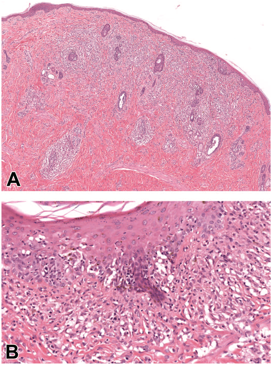
(A) Uveodermatologic syndrome, beagle dog, skin (H&E). The dermis is expanded by multifocal coalescing aggregates of macrophages and lymphocytes multifocally abutting the dermo-epidermal junction and infiltrating into the epidermis and the follicular epithelium. (B) Higher magnification of the skin. Macrophages, containing brown granular pigment (melanin), and lymphocytes infiltrating into the dermo-epidermal junction and forming aggregates in the basal and spinous layer of the epidermis. In the superficial dermis, some macrophages are engulfing melanin granules (pigmentary incontinence).
Similarly, at the anorectal mucocutaneous junction, a focal, superficial histiocytic-rich dermatitis with melanophages associated with pigmentary incontinence was observed.
Bilaterally, the mandibular lymph nodes were enlarged by mild lymphoid hyperplasia and increased histiocytes and melanophages within the paracortical area (reactive lymphadenopathy).
Gram and PAS stains on ocular and cutaneous sections did not highlight any bacteria or fungi.
IHC confirmed that the inflammatory cell population in both the eye and skin was mostly represented by T lymphocytes (CD3+) and macrophages (IBA+), with only few B lymphocytes (CD20+) (Figure 3A-C).
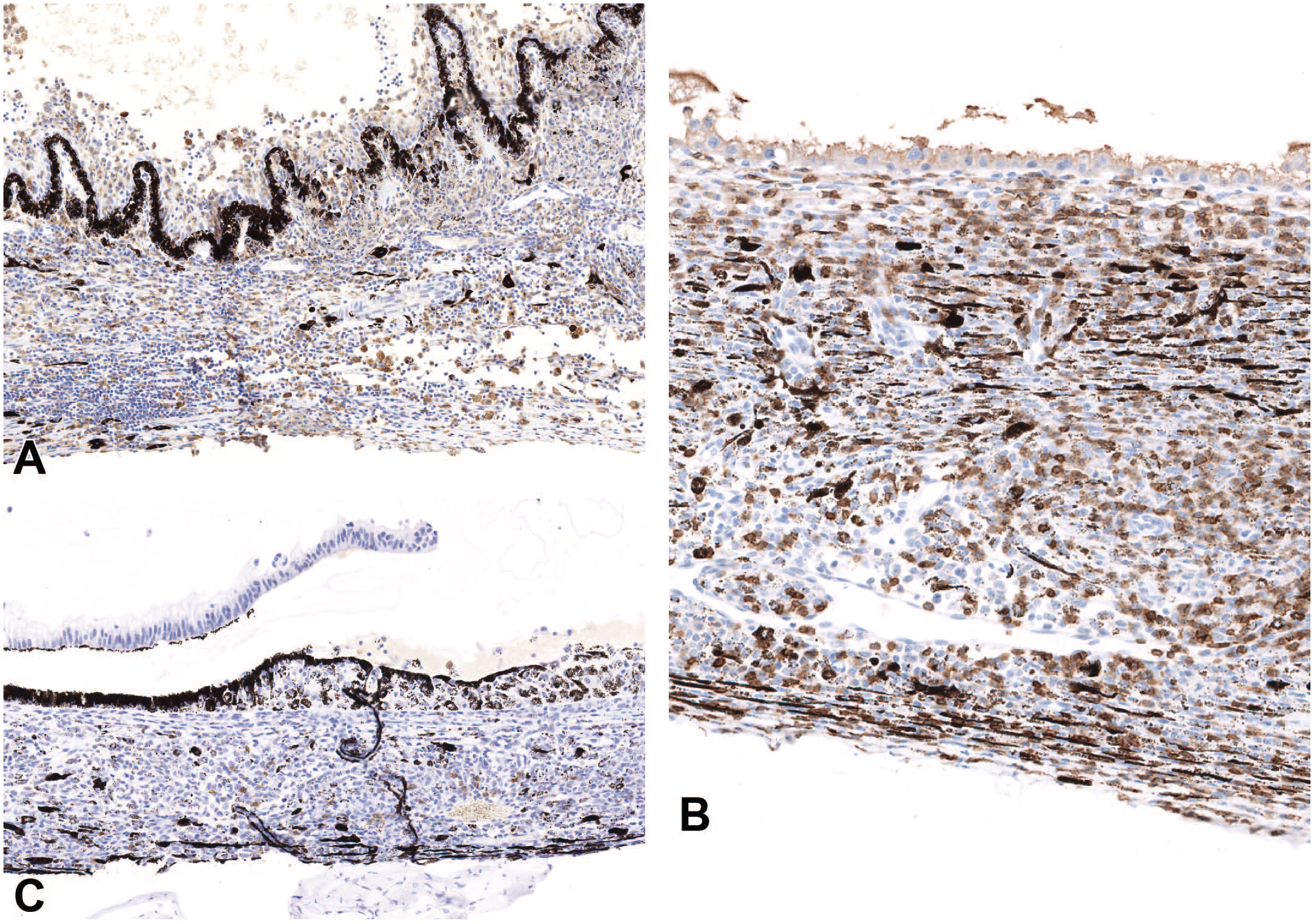
Uveodermatologic syndrome, beagle dog, eye (IHC). (A) IBA1 IHC. There are multiple aggregates of IBA1-positive phagocytic cells, showing brown cytoplasmic positivity, often containing black granules of melanin pigment at the level of the ciliary body. (B) CD3 IHC. The choroid is infiltrated by numerous CD3-positive lymphocytes showing brown cytoplasmic to membranous staining. Phagocytic cells containing black granules of melanin pigment are also present. (C) CD20. Few lymphocytes infiltrating the choroid showing CD20-positive brown membranous staining. Phagocytic cells containing black granules of melanin pigment are also present.
There are no specific tests for UDS, and the diagnosis is generally based on the combination of signalment, clinical observations and histopathological findings.5,12,13 In this case, the ocular clinical features and the microscopic morphologic presentation of the eye and skin lesions were highly indicative of UDS.
Asymmetric uveitis is histologically similar to UDS and represents its main differential diagnosis 17 (which is also true for human sympathetic ophthalmia and VKHS). 8 In this case, as there was no history or evidence of ocular trauma and the ocular symptoms developed bilaterally and concurrently, asymmetric uveitis was considered unlikely.
In UDS, the severity of inflammation varies within the uvea; however, the choroid always seems to be affected, 11 and in some previous reported cases all parts of the uvea were equally impacted.3,5 The other ocular anatomic sites are typically unaffected. 11 In this case the inflammation was most prominent in the choroid compared to the iris and ciliary body, with no significant abnormalities affecting other regions of the globe, apart from the retinal detachment which is a common secondary finding of UDS.
Other histological features associated with UDS include glaucoma, synechiae, retinal atrophy or degeneration,3,12,17 and in some cases the granulomatous inflammation had a neutrophilic component.3,6 These findings, considered secondary to the chronicity of the inflammation and/or opportunistic secondary infections, were absent in our report as the affected dog was euthanized within days of the onset of clinical manifestation.
The presence of aggregates of macrophages between the Bruch’s membrane and the retina pigment epithelium, consistent with Dalen-Fuchs nodules, have only been observed in the eyes of dogs with spontaneous or experimentally induced UDS,3,11 and they are a typical feature of sympathetic ophthalmia and VKHS in humans. 1 In the present case, similar nodules were also observed beneath the iris pigment epithelium.
Skin lesions at the anorectal junction and in the abdominal skin around the teat were only identified histologically and not macroscopically in this case. Mucocutaneous junctions are a common site of skin lesions in uveodermatologic syndrome7,17; however, the teat area has not been previously reported as an affected area. The lymphohistiocytic lichenoid dermatitis with macrophages containing melanin pigment described in this report is consistent with the typical UDS skin lesions.5,12
In UDS, T helper lymphocytes target and destroy melanocyte self-antigens present in the skin and the eye, 17 similar to human VKH syndrome. 8 In our case the majority of lymphocytes in the eye and skin were CD3 positive with only a small number of CD20-positive B lymphocytes. This was partially in contrast with findings from a paper describing immunohistochemical features of UDS in two Akita dogs, in which most lymphocytes observed in the eyes were of B lineage; 5 however, the same paper also reports that T lymphocytes were prevalent in the skin, similar to this case. 5
Other causes of panuveitis in the dog are fungal (histoplasmosis, blastomycosis, aspergillosis) or bacterial (leptospirosis) infections or are associated with neoplasia. 4 In this case, the absence of bacteria or fungi in H&E-stained slides was supported by negative Gram and PAS-stains in the ocular and cutaneous sections.
Regarding the clinical management of the case, at the onset of the ocular symptoms, topical antibiotics and corticosteroids were administered for a few days; however, the clinical picture worsened. This is not unexpected, as in UDS, long-term systemic treatment with immunosuppressive agents is required to achieve clinical remission of the disease, and topical treatment is not effective on its own. 13
The mild increase in urea and creatinine noted in the clinical chemistry was believed to be the result of dehydration, attributed to blindness and pain. The remainder of hematology and clinical chemistry showed no abnormalities, which was consistent with previous reports.6,7
A case of UDS in a pet beagle dog has been previously reported, 9 and since this disease has been regarded to be likely hereditary in other breeds where it frequently occurs, a possible genetic predisposition for beagle dogs is considered likely, and investigations into the genetic components of this disease should be carried out for affected individuals of this breed.
Bilateral, symmetrical, granulomatous/histiocytic panuveitis, which is the hallmark of UDS, 11 along with the presence of abundant melanin laden macrophages, dispersed extracellular melanin granules, presence of Dalen-Fuchs nodules and the relative absence of lesions in other parts of the eye, accompanied by lichenoid histiocytic-rich dermatitis, led to the diagnosis of UDS in the current report. In this dog, UDS consisted of a naïve, early presentation form of the disease, lacking some of the features associated with a later stage/chronicity of the disease or secondary infections (ie, glaucoma, synechiae, retinal atrophy or degeneration and more extensive skin lesions).
The presence of histological cutaneous lesions, undetected macroscopically, may suggest that they arise concurrently with the panuveitis, but it takes longer for the skin lesions to become clinically evident. Where canine uveodermatologic syndrome is suspected based on ocular signs or signalment, the absence of visible skin lesions should not prevent the veterinary clinician from obtaining biopsies of the skin and/or mucocutaneous junctions as these may be a critical aid in confirming the diagnosis.
In the context of toxicity studies, the pathologist should be aware that beagle dogs can develop UDS even at a young age (approximately 9.5 months in this case) and distinguish these lesions from treatment-related effects, particularly in ocular studies. UDS should be included among the immune mediated disorders that may affect the laboratory beagle dog.
Supplemental Material
sj-docx-1-tpx-10.1177_01926233251339116 – Supplemental material for Uveodermatologic Syndrome in a Laboratory Beagle Dog: Histological and Immunohistochemical Features
Supplemental material, sj-docx-1-tpx-10.1177_01926233251339116 for Uveodermatologic Syndrome in a Laboratory Beagle Dog: Histological and Immunohistochemical Features by Giulia Tosi, Elisavet Karamanavi, Zuhal Dincer and Michela Levi in Toxicologic Pathology
Supplemental Material
sj-tif-1-tpx-10.1177_01926233251339116 – Supplemental material for Uveodermatologic Syndrome in a Laboratory Beagle Dog: Histological and Immunohistochemical Features
Supplemental material, sj-tif-1-tpx-10.1177_01926233251339116 for Uveodermatologic Syndrome in a Laboratory Beagle Dog: Histological and Immunohistochemical Features by Giulia Tosi, Elisavet Karamanavi, Zuhal Dincer and Michela Levi in Toxicologic Pathology
Footnotes
Acknowledgements
The authors would like to acknowledge Andrew Daunt and James Leblanc for the IHC and histology technical help, and Chris Lawrie and Jon Carter at Harrogate for their support.
Author Contributions
G.T., E.K., and M.L. contributed to conception, design, analysis and interpretation of the data, G.T. and E.K. drafted the original manuscript, M.L and Z.D reviewed and edited the draft. M.L. supervised all parts of the project.
Declaration of Conflicting Interests
The author(s) declared no potential conflicts of interest with respect to the research, authorship, and/or publication of this article.
Funding
The author(s) received no financial support for the research, authorship, and/or publication of this article.

Supplemental Material
Supplemental material for this article is available online.
References
Supplementary Material
Please find the following supplemental material available below.
For Open Access articles published under a Creative Commons License, all supplemental material carries the same license as the article it is associated with.
For non-Open Access articles published, all supplemental material carries a non-exclusive license, and permission requests for re-use of supplemental material or any part of supplemental material shall be sent directly to the copyright owner as specified in the copyright notice associated with the article.
