
Abstract
The 21st ESTP’s (European Society of Toxicology Pathology) Annual Congress (2024) included a 3-hour scientific session on developmental neurotoxicity (DNT) as applied to chemical safety assessments. Key concepts of this session were to provide an introduction to public concerns around this endpoint, a status update on practical aspects of DNT studies, insights into the use of DNT studies within a regulatory context, as well as some pointers on how to evaluate specific parameters. Understanding the biological and technical variability in performing neuropathology examinations (such as morphometric evaluation) is critical during the course of DNT evaluation. Using thyroid hormone disruption as an example, challenges and pitfalls impacting data interpretation were discussed. Results from the European Centre for Ecotoxicology and Toxicology of Chemicals (ECETOC) Thyroid Task Force regarding thyroid hormone–related neurodevelopmental toxicity in humans and rodents were presented. Histopathological findings in the brain and potential changes in the cochlea of pups associated with thyroid hormone imbalance in dams during pregnancy were shown. A case presentation from an Extended One-Generation Reproductive Toxicity Study (EOGRTS) showing histopathological findings in the absence of changes with morphometric endpoints was included. In conclusion, all session participants underscored the need for integrated data evaluation for DNT risk assessment.
Keywords
Introduction
The scientific symposium session on developmental neurotoxicity (DNT), chaired by Drs Deepa Rao and Sibylle Gröters, was designed to provide an update on the basic concepts of the experimental approach, recent developments, and emerging topics in the area of chemical safety assessment. Following an introduction on historical perspectives and a fundamental overview on the best practice approaches for the conduct of DNT studies (particularly in the context of developmental neuropathology), a focus on critical challenges and pitfalls was included. A major scientific focus (driven by regulatory requirements and public concerns) during the entire symposium was on the testing and assessment of effects of “endocrine disruptors,” with specific reference to compounds that affect the morphology and function of the thyroid glands. Here, the question of specific, pathomorphological patterns in context of respective endpoints in the DNT rat model was addressed, and their relevance for humans was discussed. Juvenile animal studies and specific experimental designs for the assessment of neurodevelopmental toxicity of pediatric pharmaceuticals were not in the scope of this session.
Developmental Neurotoxicology: Introduction and Overview
Dr Frederic Schorsch began the session with a general overview on the prevalence of neurodevelopmental disorders (NDDs) that currently include intellectual disabilities, communication disorders, autism spectrum disorder (ASD), Attention Deficit Hyperactivity Disorders (ADHDs), specific learning disorders (SLDs), and movement disorders as possible causes for a decrease of intellectual capabilities in the current population.3,11 Although there is a genetic component with such associated NDDs, 17 our understanding of the root causes of NDDs remains incomplete as our understanding of the interactions between environmental/chemical exposures with our genes remains fraught with data-driven knowledge gaps. For example, the implementation of next-generation sequencing (NGS) technologies in the diagnostic flowchart of NDDs has dramatically increased the percentage of patients who receive a molecular diagnosis. However, surprisingly, none of the genes discovered so far seem to be responsible for more than only a small proportion of cases, thus leaving unexplained etiologies for majority of cases. 14 On the contrary, evidence from poisoning disasters (eg, polychlorinated biphenyls, mercury, or lead) has demonstrated that chemicals can indeed interfere with developmental processes in the human brain. Such concerns have driven and led to the development of DNT Test Guidelines (TGs) by multiple regulatory authorities worldwide over the last few decades.
In our current understanding, there is a significant scientific debate around the gestational consequences of reduced thyroid hormones induced by exogenous compounds. One working hypothesis is based on serious effects on neurodevelopment even with slight reductions in thyroid hormone occurring at critical time windows during brain development in children. To date, only a limited number of chemicals have been clearly categorized as developmental neurotoxicants, although it should be noted that only few have been fully characterized and investigated. While it could be interpreted that DNT due to chemical impacts remains a rare hazard, it should also be emphasized that DNT assessments are elaborated from complex study designs, where implementation and evaluation are often time-consuming and resource-intensive, thereby limiting the numbers of chemicals evaluated for meaningful evaluations. Given that the absence of evidence is not the evidence for absence, without data, untested chemicals cannot not be presumed to be safe for brain development.
The above introductory overview was then followed by historical context of the various existing DNT testing guidelines and the basic underlying premise by Dr Schorsch. The test methodologies recommended in the DNT TGs initiated by the US Environmental Protection Agency (EPA) 20 have been extensively reviewed and evaluated over the last 35 years. The first DNT guideline 6 (US EPA OPPTS 870.6300) has its roots in scientific publications from the early 1960s, eventually resulting in an extensive scientific database by the mid-1980s. The US EPA DNT guideline was published in 1998 and has since served as the prototype for the Organisation for Economic Co-operation and Development (OECD) TG 426. 28 In the mid-90s, the OECD initiated the development of the DNT TG, with the final version being adopted by the OECD Council in 2007. In 2011, the evaluation of DNT endpoints was included in cohort 2 of the OECD TG 443, 29 ie, the Extended One-Generation Reproductive Toxicity Study (EOGRTS, OECD TG 443). More recently, in 2020, after 20 years of collaborative work on a global scale, the European Food Safety Authority (EFSA) published a paper on the use of DNT In Vitro Testing Battery (DNT IVB) for the assessment of DNT with human and rodent cell-based assays. 23
In general, the main characteristic features of the DNT Testing Guidelines include exposure of dams and/or offspring to the test substance during gestation and lactation time periods, but there is some difference in the two main guidelines. Recommended exposure is from Gestation Day (GD) 6 to Lactation Day (LD) 21 in OECD TG 426. In contrast, OECD 443 usually involves continuous exposure from GD 0 to Postnatal Day (PND) 75 which means direct administration to offspring after weaning. Parameters evaluated in offspring include clinical signs, functional observations, motor activity, auditory startle habituation, learning and memory, and both qualitative and quantitative neuropathology. Both behavioral and neuropathological parameters are evaluated at early (during or immediately following lactation, PND 11 or 22) as well as late (around PND 60-70) timepoints. From a regulatory context, DNT testing is triggered for agrochemicals if other studies or the mode of action of the substance indicate potential concerns. Thus, recently, for biocides, a DNT study is mandatory under Registration, Evaluation, Authorization and Restriction of Chemicals (REACH), while OECD 443 is the primary experimental design to assess DNT. Although the focus of this session was on chemical safety assessment, it should be noted that for pharmaceuticals, the International Council for Harmonisation (ICH) S11 safety guideline for nonclinical testing of pediatric pharmaceuticals finalized in 2020 requires a similar assessment of the developing nervous system developing.15,30
Although the OECD 426 TG represents the best available science for assessing the potential for DNT in human health risk assessment, there are still significant challenges and limitations with experimental design strategies. In general, OECD 426 studies are elaborately complex, resource-intensive, use high numbers of animals, and encompass multiple endpoints at specific timepoints across time duration to include gestation, lactation, and post-natal periods. Moreover, based on experience with the conduct of few studies and the heterogeneity of experimental study designs, it is important to note that dependence on expert knowledge in developmental neurotoxicology and neuropathology is imperative for meaningful interpretation of study data generated from these complex studies. Finally, it was stated that even such a current battery of complex in vivo animal screening toxicology studies still may not predict all relevant human DNT outcomes due to the inherent complexity of the developing nervous system and its interactions with the environment.
Dr Schorsch concluded his talk with reference toward increasing reliance on alternative new approach methods (NAMs) and the use of DNT in vivo battery (IVB) as promising approaches with an increasingly important role in the future of DNT testing.4,16,23,33 Although NAMs cannot not be considered as a completely reliable surrogate or stand-alone approach to determine whether a chemical is a DNT risk in humans, strategic adoption of NAMs within various phases of chemical safety assessment holds significant promise to continue addressing the gaps to improve better predictive outcomes. In the context of NDDs, toxicologic neuropathologists will continue to contribute toward the development of improved strategies to detect neurotoxicity during neurodevelopment.
Developmental Neuropathology in Developmental Neurotoxicity Studies: The Standard Approach, Rationale, and Procedure Considerations
Following the general introductory framework on DNT evaluation, Dr Wolfgang Kaufmann focused on the neuropathology components of DNT studies for chemical safety assessments.
This second talk began with revealing the impact of two classical neurodevelopmental toxicants, namely ethanol and cannabiol on the developing brain. Dr Kaufmann proceeded to define developmental neuropathology to introduce common pathomorphological lesions of the central nervous system (CNS) and peripheral nervous system (PNS) due to any kind of impact (physical, chemical, drug, etc) during neurodevelopment to birth, and thereafter until adulthood. Such neurohistopathology evaluations are typically a mandatory endpoint for experimental rat toxicology studies employed to identify chemical and pharmaceutical compounds with any potential to cause DNT. The standard/conventional approach for developmental neuropathology is outlined by OECD and EPA guidelines6,28,29 and includes at least two timepoints of examination. As a rule, the first timepoint selected is PND 21/22 at weaning. At this time point, gross brain weights, brain length, and brain width measurements, as well as a qualitative analysis of major brain regions by light microscopy are performed. Dr Kaufmann recommends selecting the major brain regions as described by Garman and colleagues in their 2016 publication 12 as a guide in implementing a practical approach of using external landmarks for brain trimming along appropriate planes to achieve highly homologous brain sections. In addition, a quantitative analysis (“linear morphometrics”) of homologous brain sections is required. The underlying rationale is based on the knowledge that brain development impacted by compounds may not be associated with classical reactive, inflammatory responses (eg, glia cell reaction) as often seen in adult brains. From a neuropathology perspective, the histological landscape in developing brains is starkly different from mature brains. During prenatal phases of neurodevelopment, only a size reduction may be observed. Because such changes cannot reliably recognized in the developing brain by a qualitative (histological) analysis of brain tissue sections alone, a full neurohistopathology panel of slides is examined at the end of the study, at around PND 70 which is the second timepoint for evaluation in DNT studies. Neuropathology examinations encompass the CNS and PNS, including spinal cord, spinal ganglia, peripheral nerves, skeletal muscle, and eyes with optic nerves. Some laboratories also include the nasal cavity with the olfactory epithelium. Perfusion fixation at necropsy is the fixation method of choice and required for the PND 70 offspring. Even if immersion fixation is accepted for the PND 21/22 pups, perfusion fixation should be preferred whenever possible to reduce artifacts enabling optimum histopathology evaluation. The scope of examination for the PND 70 offspring is comparable with the scope for an adult neuropathology examination. However, a quantitative analysis of major brain regions as done for the early time point of PND 21/22 is required for this study type to recognize lasting effects in the brain development. For diagnostic purposes, the routine basic stain remains Hematoxylin & Eosin (H&E). Additional special stains may be applied, but it should be kept in mind that at the early time points, specific stains may not reflect familiar histological landscapes due to an immature neurodevelopment stage (eg,. the myelination process with the Klüver-Barrea myelin stain). Also, immunohistochemistry (eg glial fibrillary acidic protein [GFAP] for astrocytes) may fail at the early stages of neurodevelopment given that gliogenesis occurs later in the postnatal period in rats. 18 In essence, although the histological landscape of developing brains is relatively different than that of adult brains, there are still typical pathomorphological features that reflect clear evidence for an impact on neurodevelopment. For example, findings impacting cell proliferation and cell migration are readily detected by light microscopy by the trained pathologist. Common findings with neurodevelopmental toxicants include altered migration process, such as heterotopiae and/or ectopiae of neurons, reflecting neurons that settle at atypical locations. For example, a single high dose of methylazoxymethanol (MAM) at 30 mg/kg given intraperitoneally to dams at gestational day 15 induces such heterotopiae, in particular at the CA1 layer of the hippocampus in the offspring. 19 Such heterotopiae may be pathophysiologically responsible for the hyperexcitability as noted by testing the motor activity of treated offspring noted in experimental rat studies. In humans, major heterotopiae are considered inducers of epileptic seizures. 34 Other neurodevelopmental findings with MAM treatment include cerebral hypoplasia and associated “hydrocephalus ex vacuo” due to the normal skull growth.
The interpretation of developmental neuropathology data in experimental DNT studies is often a challenging process. Dr Kaufmann emphasized the value of applying a “weight-of-evidence” process considering integrated assessments of all available data for a given study (neurobehavioral, neuroclinical) including knowledge and published data of the given test substance. For reference, 12 include data constellations for a clear, ambiguous or no evidence for “developmental neurotoxicity” and a “weight-of-evidence” checklist may be used as a helpful tool for a sound interpretation.
Developmental Neuropathology in Developmental Neurotoxicity Studies: Brain Morphometrics—Technical Challenges, Pitfalls, and Data Interpretation Issues
The third talk for the DNT session was given by Dr Heike Marxfeld who focused on the quantitative analysis (“linear morphometrics”) of homologous brain sections as introduced earlier by Dr Kaufmann. Although morphometric measurements of histological sections of the brain are required by OECD guidelines28,29 for in vivo DNT studies, it is generally accepted that these measurements can often be difficult to interpret.
Dr Marxfeld’s talk provided a review of the workflow involved to obtain morphometric measurements while highlighting preliminary data obtained from ongoing evaluations to critically assess associated potential biases.
To address the bias between two- and three-dimensional measurements, preliminary data calculating the correlation between length and width measurements (two-dimensional value) vs the absolute brain weight (brain volume as a three-dimensional value) were shown. The underlying rationale was to evaluate the relevance of two-dimensional measurements as an indicator of the size for complex three-dimensional structures, such as for the hippocampus. Preliminary data showed that the correlation was not high. So far, the concordance of two-dimensional measurements of brain regions to the three-dimensional measurements with a technique like magnetic resonance imaging (MRI) is, to the knowledge of the authors, not well established.
Because the technical aspects of slide preparation can be influenced by multiple factors, a comparison on the width of the perfusion-fixed brain and the width on the slide was examined. Preliminary data showed variations up to 12%. In addition, the temporal influence of varying times on the water bath was assessed in serial brain sections. When brain sections were taken off the water bath following 1, 3, 5, and 10 seconds, it was noted that sections expanded primarily within the first 3 seconds, thereby demonstrating that care should be taken to remove sections as soon as possible.
To estimate interindividual variability, measurements taken individually by three pathologists were compared, and the percent deviation of the minimum and maximum value was calculated. Preliminary data indicate a 2% to 10% interindividual variability.
Finally, the coefficient of variation (standard deviation/mean*100) was calculated when measurements were performed separately for each hemi-brain relative to the mean of bilateral measurements. This gave inconclusive results.
Thus, because consistent measurements in histopathology are technically challenging and the criteria for measured parameters are generally impacted by multiple variables, such measurements should ideally be investigated and documented by each laboratory to ensure reproducible results within acceptable limits. Dr Marxfeld stated that ongoing work is slated to assess whether other neuroanatomic sites for microscopic measurements may yield more reliable/reproducible results and whether relatively larger data sets can play a role.
The human relevance of neurotoxicity studies was discussed by Dr Marxfeld with thyroid hormone disruption as an example where the differences between findings in animals and humans were shown. In the context of thyroid hormone disruption, the differences and similarities of the pathomorphological finding of heterotopias in the brain were discussed, comparing the classical test substance propylthiouracil (PTU) with a proprietary BASF compound.
In overview, the factors impacting the interpretation of morphometric data from DNT studies are manifold, challenging meaningful interpretation. Specific factors include statistical significance that is not dose-related, effects that are limited to a single sex, findings that are limited unilaterally in microscopic measurements, or, in the case of macroscopic measurements, in the absence of changes in brain weights.
In addition, it was highlighted that the relevance can also be challenging to assess if no other endpoints in the entire study show differences from the control. In general, morphometric measurements are often interpreted without any knowledge about potentially influencing factors on the methodology and its sensitivity. As with all neurotoxicity evaluations, integrated evaluations of clinical, behavioral, and pathology data remain essential for data interpretation.
A comprehensive evaluation to understand technical variations and uncertainties is currently under investigation and expected to be complied for publication.
Special Focus: Chemicals Inducing Thyroid Function Changes and Underlying Concerns to Cause Human Relevant Neurodevelopmental Toxicity
The fourth topic talk was given by Dr Stephanie Melching-Kollmuss who provided an overview about the activities and achievements of the European Centre for Ecotoxicology and Toxicology of Chemicals (ECETOC) (a non-profit industry-sponsored scientific organization) Special T4 Task Force (https://www.ecetoc.org/task-force/special-t4-task-force/). The ECETOC Special T4 Task Force was formed in 2018 to investigate the relevance of thyroid hormone–related neurodevelopmental toxicity in humans and rodents with the ultimate goal to develop a testing and assessment scheme.
The foundation of the ECETOC Special T4 Task Force was motivated by perceived uncertainties and limited guidance regarding endocrine disruption assessments of compounds showing thyroid toxicity in animal (mainly rat) studies. Specifically, substances inducing histopathological changes in the thyroid and/or thyroid hormone changes in rodents are under the focus of the European Regulatory Authorities (EC 605/2018 for plant protection product 8 and EC 2100/2017 for biocide regulation 9 ). As defined in the criteria, a plant protection active ingredient or a biocide is to be considered an endocrine disruptor if it causes an adverse effect, if it shows endocrine activity, and if there is evidence for a plausible link between the adverse effect and the endocrine activity. Although further guidance has been provided in the European Chemicals Agency/European Food Safety Authority (ECHA/EFSA) Guidance Document, 2018, 7 actual guidance for testing and assessment of compounds targeting the thyroid gland is limited.
However, the issue at hand is that none of the existing guidance documents [ECHA/EFSA Guidance Document 7 (2018) and (ECHA, 2024, Version 5.0)] contain specific or comprehensive guidance on how to assess neurodevelopmental toxicity in rodents secondary to thyroid function impairments, based on either quantitative aspects or functional changes in humans. Nevertheless, EFSA has been conducting endocrine disruption assessments on pesticide active ingredients since 2019, and an overview of finalized assessments is periodically updated in the following table available at overview-endocrine-disrupting-assessment-pesticide-active-substances.xlsx. Moreover, starting in 2025, ECHA Committee for Risk Assessment will include endocrine disruption in their opinion building related to the risks of substances to human health and the environment (Committee for Risk Assessment—ECHA).
In order to overcome the scientific and regulatory uncertainties and to provide more guidance regarding testing and assessment of thyroid toxicants, the guiding questions of the ECETOC Thyroid Task Force were:
How is maternal thyroid hormone imbalance related to neurodevelopmental outcomes?
What are the most sensitive parameters for human/rat neurodevelopment?
Are there thresholds?
The overall goal of the Thyroid Special T4 Task Force was to develop a testing and assessment scheme. Thyroid hormone changes with subsequent thyroid histopathological changes in rodent could be caused by a wide variety of Molecular Initiating Events (MIEs) and a number of respective adverse outcome pathways (AOPs) as are included in AOPwiki (www.aopwiki.org), some of which are even endorsed by OECD.
The MIE can be either a direct effect on thyroid hormone synthesis such as thyroid peroxidase (TPO) or sodium-iodide-symporter (NIS) inhibition, or an indirect liver-mediated effect via liver nuclear receptor activation, which can lead to the key event (KE) phase 2 liver-enzyme induction and increased thyroid hormone glucuronidation and excretion. The interaction with serum binding proteins likewise can lead to increased thyroid hormone excretion. For other possible MIEs, readers are referred to a comprehensive overview by Noyes and colleagues published in 2019. 27
One of the outcomes resulting from the ECETOC Thyroid Special T4 Task Force evaluations is based on human epidemiological data that confirm the association between altered maternal serum-free T4 and/or Thyroid-Stimulating Hormone (TSH) and increased risk for child neurodevelopment. However, there is a broad variety of neurodevelopmental parameters investigated (histopathological and behavioral) in human studies. Most importantly, it was noted that a sensitive parameter has not been identified. 31 Comparatively, when evaluating rodent generation studies (treatment during gestation/lactation, investigating thyroid function, and brain-related parameters), one of the main outcomes was that thyroid hormone data generated from offspring are clearly more decisive in predicting neurodevelopmental toxicity, as that is normally not investigated. While in rodents total thyroid hormone is measured, maternal thyroid hormone data in humans are not, thus posing challenges in the correlation between humans and rodents. 22
The brain-related parameters investigated in rats included functional changes in late-stage KEs (eg, alterations in electrophysiology or auditory signaling), neurobehavioral effects on the organism level (motor activity, acoustic startle response, learning, and memory), structural changes in brain (eg, periventricular heterotopia, decreased volume or thickness of specific brain layers, altered glial cell labeling), and changes in the expression of brain genes and brain-related proteins. While some of the correlations between offspring thyroid hormone values and the observation of brain-related changes were good (eg, motor activity, learning and behavior), some were not good (eg, heterotopia). A phenomenological threshold of approximately 50% to 60% between the determination of offspring thyroid hormone decrement and the increased risk to develop at least one brain-related parameter in offspring was shown, 22 which suggests that a significant decrease of thyroid hormones is needed to induce neurodevelopmental effects in rats.
As mentioned earlier, the ultimate goal of the scientific project undertaken by the ECETOC Special T4 Task Force—together with toxicologists working together under the umbrella of the industry association Crop Life Europe (CLE), who joined the Task Force later—was to provide more guidance in how to assess thyroid-acting compounds. The principles for the CLE-ECETOC Thyroid-NDT-TAS (Thyroid Function-Neurodevelopmental Toxicity-Testing and Assessment Scheme) are to evaluate the available data set (be it large, as for the pesticide active ingredients, or smaller—for evidence of thyroid function changes [Tier 0]). 24 Follow-up testing/assessment can either be approached by conclusively demonstrating non-human relevance of the MIE/ KE having led to thyroid hormone changes in rats. This path (called Tier 1) is mainly foreseen for substances with an indirect liver-enzyme induction-mediated mode of action, but also for substances interfering with serum binding proteins. 24 In an alternative Tier 2 approach, additional testing is proposed to follow up with the generation of more decisive in vivo parameters, like, eg, the investigation of thyroid function in offspring (if the method is available: possibly also thyroid hormone in brain) and/or neurodevelopmental toxicity testing (in EOGRTS or DNT studies).
Following the Thyroid-NDT-TAS, a decision on the endocrine disruption potential for the thyroid modality of compounds can be made 24 ; however, also a differentiation between European Directive Human Health (ED HH) Cat 1 and ED HH Cat 2 is possible, when the weight of the evidence of the several decisive parameters for endocrine adversity, endocrine activity, and non-human relevance is made. The evaluation of a number of thyroid case studies using the proposed testing and assessment scheme and the development of a set of criteria to differentiate between Cat 1 and Cat 2 has been published 25 .
Dr Melching-Kollmuss concluded that in the future, assays and evaluation schemes to investigate species differences (preferably early in an AOP, at the MIE or early KE level) need to be developed and agreed among the scientific and regulatory community. The most developed assay type is a comparative liver-enzyme induction study 2 (published by Baze and colleagues in 2024), and work is ongoing to publish reference compound and historical control data. Also, the development of kinetic models to estimate rat vs human thyroid hormone concentrations at different timepoints and in different compartments is ongoing, taking into account species differences, like, eg, the presence of different serum binding proteins, having largely different binding properties and differences in thyroid hormone half-lives.1,10
Thyroid Hormone Imbalance in Pregnant Rats and its Impact on Neurodevelopment in the Pups: A Hunt for a Reliable Histopathological Marker
The final topic talk was given by Dr Babunilayam Gangadharan who presented potential reliable histopathological endpoints in rodent studies to relate maternal hypothyroidism and neurodevelopment in pups.
For background, evidence for neurodevelopmental effects in children of pregnant mothers with thyroid hormone disruption is well known. 5 Since 2018, market authorization in Europe for a pesticide or a biocide requires hazard assessment of endocrine-disrupting properties including the thyroid modality. 7 Specifically, substances inducing histopathological changes in the thyroid and/or thyroid hormone effects in rodent studies need to be further investigated to evaluate potential endocrine disruption, including possible neurodevelopmental toxicity in the pups.
More recently, Thyroid Task Force of the ECETOC and CLE has proposed a tiered testing and assessment scheme to identify thyroid hormone disruptors and neurodevelopmental effects in the progeny. 24 Histopathological assessment of the brain to identify a reliable histopathological endpoint to assess the neurodevelopmental effect is an important aspect in this testing scheme. Periventricular heterotopia in the corpus callosum (Figure 1) and persistence of external germ cell layer in the cerebellar cortex are being proposed as potential histopathological endpoints for some time.13,26 In addition, histopathological correlates in the cochlea associated with hearing impairments observed in the rat pups derived from hypothyroid dams are another potential reliable histopathological endpoint.
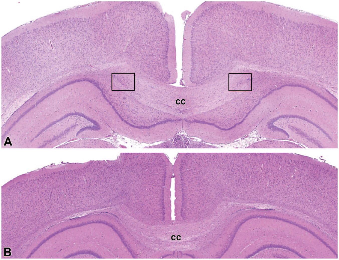
The above two images of the brain depict bilateral periventricular heterotopia (boxed areas depict the location of ectopic neurons within the corpus callosum [cc]) in a rat pup of a dam treated with PTU (Figure 1A). Figure 1B is provided as a comparable location from a control rat. Additional images and details are included in the publication by Gangadharan and Schorsch in this issue.
Dr Gangadharan concluded that identifying reliable histopathological endpoints is crucial for assessing the neurodevelopmental risks associated with thyroid hormone imbalance during pregnancy. Dr Gangadharan’s presentation included three histopathological endpoints in rodent studies to relate maternal hypothyroidism and neurodevelopment in pups, namely, periventricular heterotopia (corpus callosum), persistence of external germ cell layer (cerebellum), and potential changes in the cochlea (ongoing evaluation). For a more detailed review, readers are referred to the separate manuscript by Gangadharan and Schorsch is this symposium issue.
An Extended One-Generation Reproductive Toxicity Study Case Presentation of a Test Substance Affecting Function and Morphology of the Thyroid Glands
Finally, a case study narrative showing pathomorphological findings from an EOGRTS that was presented by Dr Kathrin Becker is included here. Dr Becker began her presentation with an overview of the study design and pathology data from an EOGRTS performed according to OECD guideline 443. The undisclosed (proprietary) test substance was administered via the drinking water to male and female Crl:Wl(Han) rats. There was no unscheduled or early mortality, and all animals were terminally sacrificed as scheduled. A whole organ spectrum was investigated by light microscopy, including the DNT cohorts of offspring animals. The DNT cohorts at both timepoints included perfusion fixation using Karnovsky’s fixative for optimal histopathology evaluation.
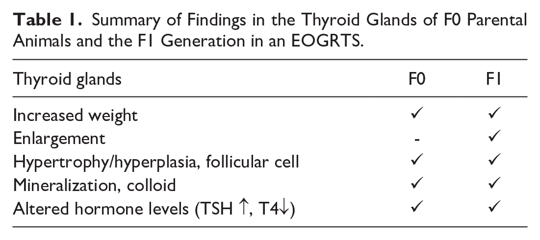
Compared with the control group, body weight data showed a decrease in the F0 parental males of the high-dose group as well as in both sexes of the F1 generation (comprising cohorts A and B) of the high-dose group. In the F0 parental animals, the weights of the thyroid glands were increased in both sexes of the high-dose group as well as in females of the mid-dose group. Similarly, the mid- and high-dose groups of the F1 generation showed a weight increase of the thyroid glands in both sexes, correlating with visible enlargement of the thyroid glands in a subset of animals of the high-dose group. Histologically, diffuse hypertrophy/hyperplasia of follicular cells was present in the thyroid glands. In the F0 generation, both sexes were affected in the low-, mid-, and high-dose groups, showing a dose-dependent increase in severity. In the high-dose group, 25/25 male and 21/25 female animals were affected, with the hypertrophy/hyperplasia graded mainly moderate to massive (grades 3-5) in male animals and slight to marked (grades 2-4) in female animals. In the F1 generation, the mid- and high-dose groups showed hypertrophy/hyperplasia of follicular cells in both sexes, with a dose-dependent increase in severity grade. An additional finding was small foci of mineralization within the colloid of affected thyroid glands, which also showed a dose-dependent increase in both F0 parental animals and the F1 generation.
Clinical pathology data revealed changes in thyroid hormone levels (decreased T4, increased TSH) in both sexes of the mid- and high-dose groups of both generations. The findings in the thyroid glands were considered a direct effect of the test substance. While histological treatment-related findings were limited to the thyroid glands in F0 parental animals, the hippocampus was an additional target organ in the F1 generation.
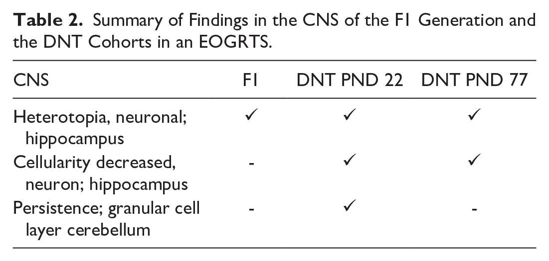
In the hippocampus, heterotopic neurons were present in 7/20 male and 4/20 animals of the high-dose group of the F1 generation. Hippocampal pyramidal neurons were scattered around their anatomically correct location in the stratum pyramidale, primarily affecting the CA2 and CA3 region, and to a lesser extent, the CA1 region. Hippocampal changes were also present and were more distinctly visible in the DNT cohorts for weanlings and adults of the F1 generation sacrificed on PND 22 and 77, respectively. In both sexes of the high-dose groups of PND 77 animals, hippocampal pyramidal neurons were scattered between the oriens layer (“striatum oriens”) and their anatomically correct place. In addition, the width of the layer of correctly placed neurons was also reduced in thickness.
Such findings were also present in both sexes of the high-dose group of PND 22 animals. In addition, persistence of external granular cell layer of the cerebellum was noted in this cohort. Furthermore, the myelinization of the mammillothalamic tract appeared slightly reduced in mid- and high-dose male animals. It is interesting to note that morphometric analysis did not reveal any treatment-related findings in both DNT cohorts. A tabulated summary is shown in Tables 1 and 2.
Summary of Findings in the Thyroid Glands of F0 Parental Animals and the F1 Generation in an EOGRTS.
Summary of Findings in the CNS of the F1 Generation and the DNT Cohorts in an EOGRTS.
In conclusion, Dr Becker considered the findings in the CNS to be a consequence of the proliferative changes of the thyroid glands. Similar findings were described in publications noting observations after thyroid hormone disturbance.21,32 It is hypothesized that the altered hormone levels in F0 parental animals and/or a direct effect of the test substance on the thyroid glands of the F1 generation animals could be underlying mechanisms for the CNS findings, potentially leading to a disturbance in neuronal migration.
The enhanced visibility of CNS findings in the DNT cohorts underlines the importance of these cohorts. The detailed and standardized processing of the CNS facilitates the detection and classification of pathomorphological findings for the pathologist.
Symposium Session Platform Discussion
In summary, this 3-hour symposium session highlighted several important topics that the practicing toxicologic pathologist may encounter during the design, data analysis, and interpretation of DNT studies. Topics included background and historical perspectives, basic experimental approaches, challenges with specific endpoints such as morphometrics, and reflections on the current scientific debates impacting experimental considerations and regulatory requirements in designing DNT studies. The objective of the session was not to focus on thyroid hormone disruption but rather to highlight the various challenges associated with the conduct of DNT safety assessments using thyroid hormone disruption as an example. The objective was to provide the session participants some overarching perspectives on the complexities and challenges involved with evaluating and interpreting data from DNT studies. In conclusion, considering the multiple variables impacting DNT, all session participants were in consensus that integration of knowledge was not only important to drive effective experimental study design implementation, but that integration of resulting data from all endpoints is also paramount to arrive at meaningful data interpretation.
Footnotes
Acknowledgements
The authors gratefully acknowledge Dr Peter Maslej for his diligent review of abstracts during preparation for Session V on DNT of the ESTP component of the CEP Congress 2024.
Authors’ Note
This document has been reviewed in accordance with policies of the authors’ respective institutions of employment. The opinions expressed in this paper solely represent those of the authors and should not be construed as official views or policies of the authors’ institution.
Author Contributions
All authors contributed toward the writing of this manuscript.
Declaration of Conflicting Interests
The author(s) declared no potential conflicts of interest with respect to the research, authorship, and/or publication of this article.
Funding
The author(s) received no financial support for the research, authorship, and/or publication of this article.