






Abstract
Timely tissue fixation is critical to prevent autolysis, which confounds histopathologic assessment. This study systematically mapped autolysis progression in ex vivo rat tissues under room temperature and refrigerated (4°C) storage conditions to establish an evidence-based, tissue-specific timeline for practical guidance. Thirty-six Sprague-Dawley rats were euthanized; major organs were rapidly collected and maintained in sterile saline at either room temperature or 4°C for intervals ranging from 0.5 to 16 hours before fixation in 10% neutral buffered formalin and H&E evaluation. The data provide a clear timeframe of autolytic change in rat tissues. At room temperature, most tissues exhibited minimal autolysis within the first 2 hours, and refrigeration at 4°C extended the time to autolysis. Immunohistochemistry for the endothelial marker CD31 suggested that CD31 antigenicity remained detectable in most tissues maintained at room temperature out to 16 hours with the exception of intestinal tissues demonstrating severe autolysis, indicating that advanced autolysis can compromise immunohistochemical interpretation. In conclusion, this work provides guidance for handling tissues that require temporary preservation in saline. The documented tissue-specific timelines serve as a valuable reference for pathologists.
Introduction
Autolysis—an enzyme mediated self-digestion process initiated immediately after animal death—is a critical consideration in histological evaluations,11,13 and rapid and adequate fixation after sampling is essential. However, transient ex vivo tissue preservation between tissue harvesting and formalin fixation is unavoidable in laboratory practice of toxicologic pathology studies if additional procedures are required. For instance, obtaining organ weights and collecting additional tissue samples for bio- and drug distribution studies can lead to delayed fixation.
The extent of autolysis is influenced by multiple factors, including tissue type, preservation temperature, and duration of ex vivo storage.3,10 Previous studies have primarily focused on postmortem autolysis;2,4,6,10 however, systematic data on time-dependent autolysis in ex vivo tissues that are directly relevant to preclinical histopathologic assessment are sparse. Specifically, the optimal time window for ex vivo tissue maintenance in saline before fixation and differences in autolytic susceptibility among major organs commonly evaluated in drug safety studies (eg, heart, liver, kidney, lung, intestine, and brain) have not been fully evaluated.
Inconsistent tissue handling protocols (eg, delayed fixation) may introduce variability in histopathologic features, potentially leading to misinterpretation of drug toxicity or experimental outcomes. This study aims to characterize the time-dependent histological changes in 9 key rat tissues (heart, liver, kidney, lung, spleen, pancreas, small intestine [jejunum], large intestine [colon], and brain) maintained ex vivo in saline at room temperature or at 4°C. The tissues were evaluated microscopically to compare the autolytic susceptibility among different types of tissue, elucidate the potential value of refrigeration, and propose evidence-based recommendations to optimize tissue handling and fixation protocols for nonclinical samples temporarily held in saline before histopathologic evaluation.
Materials and Methods
Experimental Animals
Thirty-six healthy Sprague-Dawley rats (male, 8-10 weeks old, body weight 195-220 g) were purchased from Zhejiang Vital River Laboratory Animal Technology Co., Ltd. (License No. SYXK(SU) 2023-0053). Animals were housed in a specific pathogen-free (SPF) environment with controlled temperature (20°C-26°C), humidity (40%-70%), and a 12h light/dark cycle, room ventilation of 10 to 15 air changes per hour, and free access to standard chow (The maintenance compound feed for SPF rats sterilized by cobalt-60 irradiation was provided by Beijing Keao Xieli Feed Co., Ltd., 30 g/animal/day, cafeteria feeding) and water (reverse osmosis). All experimental procedures were approved by the Institutional Animal Care and Use Committee (IACUC) of the authors’ institution (Approval No. IACUC-2025-r-265) and conducted in compliance with the Guide for the Care and Use of Laboratory Animals. All animals were euthanized by exsanguination under deep anesthesia with 40 mg/kg ketamine in combination with 5 mg/kg xylazine via intraperitoneal injection as per standard operating procedures.
Experimental Design
Within 15 minutes posteuthanasia, technicians carefully harvested the tissues and placed them in sterile 0.9% NaCl solution. Tissues were kept at room temperature (20°C-26°C) or in the refrigerator at 4°C for 0.5, 1, 2, 4, 8, or 16 hours. Harvested tissues included heart, liver, kidney, lung, spleen, pancreas, small intestine (jejunum), large intestine (colon), and brain.
Histopathologic Evaluation and Scoring
At the end of each timepoint, tissues were placed in 10% neutral buffered formalin (pH 7.4) at room temperature for 48 hours. All tissues were trimmed, processed, embedded in paraffin, sectioned at 3 μm, and stained with hematoxylin and eosin. All slides from a given organ were stained in a single batch to minimize inter-batch staining variation.
Microscopically, the degree of autolysis was evaluated using a semi-quantitative 5-point scale (commonly used in toxicologic pathology): 1 (minimal), 2 (mild), 3 (moderate), 4 (marked), and 5 (severe). 7 Scoring was performed independently by 2 board-certified veterinary pathologists who were blinded to the preservation time and temperature. Using a multiheaded microscope, the pathologists discussed and reconciled any scoring differences, arriving at a final agreed-upon score for each specimen.
The total score per organ per animal was used for descriptive presentation. Given the exploratory nature of this study and the sample size, scores are presented descriptively to illustrate trends rather than for definitive statistical comparison between organs. Autolysis was assessed based on tissue architecture integrity and cellular morphology, with the following diagnostic criteria:
Intracellular vacuolation.
Enlarged intercellular space.
Epithelium separation from the basement membrane.
Pyknosis, karyorrhexis and karyolysis.
Cytoplasmic lysis.
Cellular edema/swelling.
Loss of differential staining.
Altered architecture of tissue unrelated to a pathological process.
Complete cytoplasmic dissolution, absent or unidentifiable nuclei.
Immunohistochemistry
The IHC analysis was performed on the tissue samples maintained in room temperature. Tissue sections (3 µm) from formalin-fixed, paraffin-embedded (FFPE) samples were used for IHC analysis. Staining was performed using an automated immunostainer (CELNOVTE, CNT320 Full Automatic IHC Stainer) according to the manufacturer’s protocol to ensure consistency. Briefly, the protocol consisted of the following steps: Slides were baked, deparaffinized in xylene, and rehydrated through a graded series of ethanol to distilled water, heat-induced epitope retrieval (HIER) was performed using a Tris-EDTA target retrieval solution in a water bath at 95°C to 99°C for 20 minutes. Slides were treated with endogenous enzyme block (3% hydrogen peroxide) for 10 minutes to quench peroxidase activity, then the slides were incubated with the respective primary antibodies (CD31, ab281583, rabbit, Abcam, 1 : 1000 dilution) for 30 minutes at room temperature. The bound primary antibody was detected using the MicroStacker Plus Polymer Detection Kit (CELNOVTE) for 30 minutes, resulting in a brown precipitate at the antigen site. The negative control was prepared by using specimens that had not been incubated with the anti-CD31 antibody, and positive controls were prepared from specimens with confirmed CD31 expression, including freshly harvested lung tissue that was promptly fixed.
Image Acquisition
Photomicrographs were taken using an Olympus BX43 microscope and an Olympus DP74 camera using Olympus CellSens software.
Statistical Analysis
In light of the descriptive and exploratory nature of this study, coupled with the limited number of biological replicates (n=3 per group), the histopathology scores are reported as descriptive statistics, with mean scores serving as the primary measure of central tendency.
Result
Histomorphology Evaluation
The onset and characteristics of autolytic changes varied across tissues and organs (heart, liver, spleen, lung, kidney, small intestine [jejunum], large intestine [colon], pancreas, brain). As shown in the Figure 1, representative photomicrographs decipt the H&E-stained sections of each organ maintained in saline over time at room temperature (RT) or 4°C. The mean histology scores of each organ across the different groups are presented in Table 1. The following are the descriptive findings for each organ.

Representative photomicrographs of the H&E-stained sections of the heart, liver, kidney, small intestine, large intestine, and pancreas maintained in saline over time at room temperature (RT) or 4°C. Hash mark: enlarged intercellular space; asterisks: cytoplasmic lysis and/or pyknosis; arrows: intestinal villus epithelial sloughing, loss of tissue architecture and failure to take up stain; triangle: intracellular vacuolation in pancreas acinar cell. All images were captured using a 20×/0.50 objective.
Mean histopathology scores of different organs in each group (n = 3 per group).
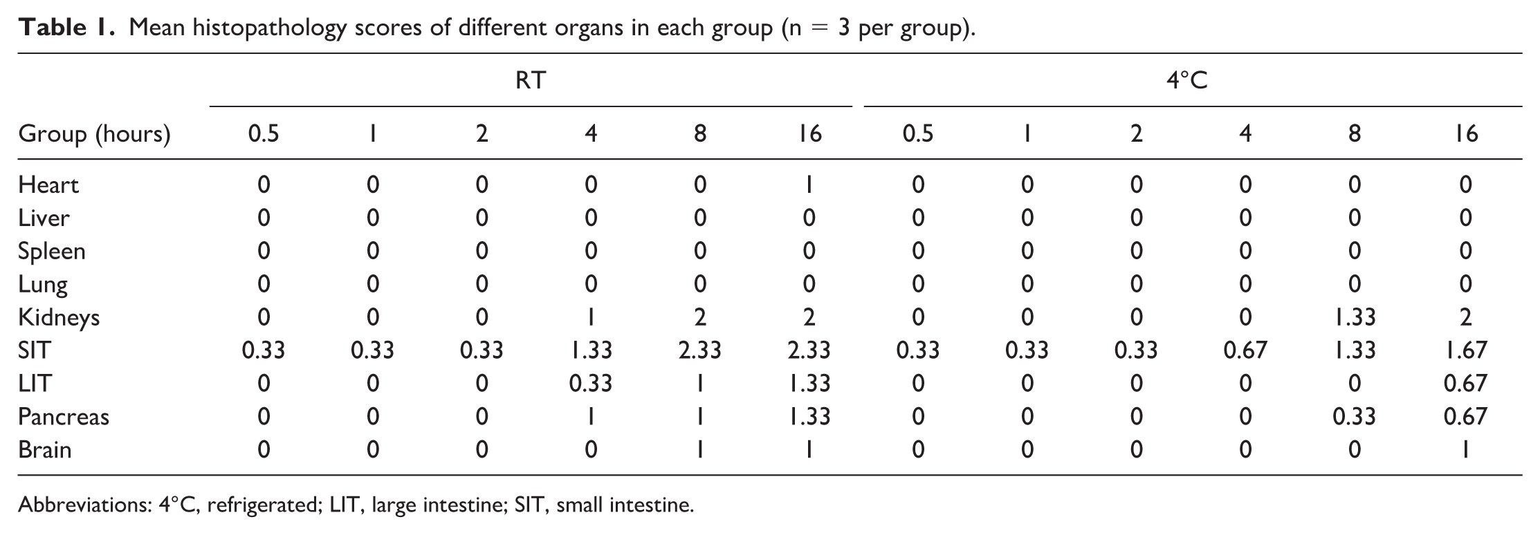
Abbreviations: 4°C, refrigerated; LIT, large intestine; SIT, small intestine.
Small Intestine (Jejunum)
At room temperature, minimal autolysis was observed at the tips of intestinal villi as early as 0.5 h, characterized by loss of differential coloration and epithelial detachment from the basement membrane. However, autolysis remained confined to the villus tips without significant progression until 2 hours. From 4 to 16 hours, autolytic changes gradually extended along the mid-villus to the intestinal crypt region. At 4°C, no autolysis was observed within 4 hours. From 8 to 16 hours, overall autolysis scores trended lower than those at room temperature, indicating that refrigeration will slow the progression of autolysis microscopically.
Large Intestine (Colon)
Samples maintained in saline at room temperature, minimal autolytic changes were evident in the superficial mucosa at 4 hours, progressing in a time-dependent manner down the crypt, with autolysis at the crypt base by 16 hours. This progression was slowed at 4°C, where initial superficial mucosal autolysis was not apparent until the 16-hour timepoint.
Pancreas
At room temperature, extensive microvesicular vacuolation of pancreatic acinar cells was evident as early as 4 hours. While cellular architecture and staining remained intact until 16 hours, expansion of intercellular spaces was observed at this time point. In contrast, at 4°C, no significant acinar vacuolation occurred within 16 hours, although intercellular space widening was noted by the end of this period.
Kidney
Autolytic changes were evident at 4 hours at room temperature, characterized by swelling, pyknosis, and cytoplasmic lysis of distal tubular epithelium. These changes were noted at 8 hours at 4°C. Later timepoints were associated with more widespread cell swelling, and cytoplasmic lysis became more widespread, eventually affecting all tubules. The mean autolysis scores at 8 and 16 hours were similar between the room temperature and refrigerated groups, suggesting that the benefit of refrigeration in mitigating renal autolysis after 4 hours may be limited.
Heart
Myocardial autolysis at room temperature was characterized by obviously widened intercellular spaces at 16 hours, while cardiomyocyte architecture and tinctorial properties remained largely intact. No such changes were apparent at 4°C.
Brain
Vacuolation within the cerebral cortex was first noted after 8 hours in saline at room temperature. Findings were characterized by the appearance of numerous vacuoles in nerve fibers and neurons extending from the superficial to deeper layers of the cerebral cortex, accompanied by loss of differential staining. This change was more evident 16 hours at room temperature, but was not observed until 16 hours under refrigerated conditions.
Liver, Spleen, and Lung
The parenchymal tissues of these organs remained free of autolytic changes and retained intact morphology throughout the 16-hour experimental period.
Immunohistochemistry
CD31 immunohistochemistry was performed to assess vascular marker integrity. In most organs examined (eg, heart and liver), staining intensity appeared largely comparable across the different time points (Figure 2), suggesting relative detectability of this antigen after delayed fixation in 10% neutral buffered formalin (NBF). However, a notable exception was observed in the small intestine, where areas of advanced autolysis, particularly at the villus tips, corresponded to markedly attenuated or absent CD31 immunoreactivity (Figure 2). These areas of diminished immunoreactivity corresponded precisely to regions exhibiting advanced autolytic changes, particularly at the tips of the intestinal villi. In the intestine, nonspecific staining was noted in goblet cells and enterocytes, and that advanced autolysis (particularly in small intestinal villi) can complicate CD31 interpretation.
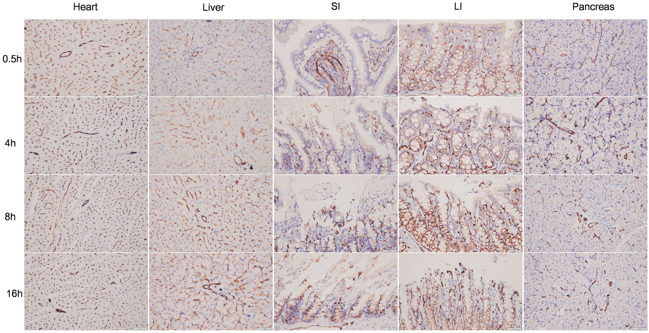
Immunohistochemistry staining for CD31 was performed on the heart, liver, small intestine, large intestine, and pancreas maintained at room temperature (RT) over time. All images were captured using a 40×/0.75 objective.
This finding suggested that while CD31 may be robust in well-preserved tissues, severe autolytic damage can lead to loss of antigen detection. Therefore, interpreting IHC results in tissues prone to autolysis requires careful correlation with the degree of morphological preservation.
Discussion
It is crucial to note that the following observations and guidance are derived specifically from ex vivo tissue maintenance in saline before routine fixation in formalin. Even at the latest time point (16 hours) at room temperature storage, autolysis did not reach moderate-to-severe levels in most tissues, which may not necessarily reflect autolytic change when tissue collection is delayed in found dead or euthanized animals.
The most prominent autolytic changes were observed in the small intestine, kidney, and pancreas, with late-onset changes in the large intestine, brain, and heart. The small intestine had the most rapid onset of autolysis, consistent with previous postmortem autolysis studies,6,10 and refrigeration did not dramatically alter progression. Autolysis at the tips of jejunal villi was present within 0.5 hours postdissection, characterized by cell disintegration and loss of differential staining. Autolytic change remained confined to the villus tips without significant expansion for 2 hours, with a clear boundary between autolyzed and nonautolyzed part. After 4 hours in saline, autolysis gradually progressed along the villus into the crypt, with partial or complete mucosal disintegration by 16 hours. Superficial mucosal autolysis in the large intestine was not observed until 8 hours under room temperature. Notably, the penetration rate of formalin into tissues at room temperature is approximately 2.4-5 mm/24 hours.1,12 Because intestinal villi are the last structures to be fixed during formalin immersion, their tips are particularly susceptible to autolysis. This delayed fixation likely explains the observed pattern. The contribution of resident putrefactive bacteria may also be a facilitating factor. 5 To avoid early autolysis in the small intestine, intraluminal perfusion of formalin immediately after intestinal dissection or incising the intestine before formalin fixation is recommended.
Cytoplasmic vacuolation of pancreatic acinar cells is considered either a spontaneous animal change 9 or an early autolytic change. 8 In this study, this change was first noted in pancreatic tissues stored at room temperature for 4 hours, with acinar cell structure and staining remaining normal. This finding persisted in all subsequent time points, confirming that such vacuolation represents an early autolytic change. In pancreatic tissues stored at 4°C, no notable vacuolation was observed within 16 hours, with only scattered individual cells showing this feature. Transmission electron microscopy studies 14 have indicated that early pancreatic autolysis is characterized by the accumulation of amorphous material within the endoplasmic reticulum, resulting in its dilation. During postmortem autolysis in animals, focal complete loss of pancreatic tissue structure (with a clear boundary from surrounding tissues) is often observed, which is attributed to the action of proteases in pancreatic tissue, particularly around pancreatic ducts.
Autolytic changes in the kidney were first observed after 4 hours at RT and 8 hours at 4°C saline, primarily affecting the distal convoluted tubules. The affected epithelial cells exhibited pale, flocculent cytoplasm and condensed chromatin. By 8 hours, diffuse swelling of renal tubular epithelial cells was noted, accompanied by pale staining and numerous fine intracytoplasmic vacuoles. Ultrastructural studies of renal changes by Yukari Tomita et al 14 showed that distal tubular epithelial cells exhibited mitochondrial swelling, chromatin margination, pale cytoplasm, and peripheral chromatin condensation 5 hours postmortem. Mild mitochondrial swelling and irregularly arranged microvilli were observed within 3 hours postmortem. These ultrastructural changes are consistent with the findings observed in our H&E-stained sections.
Microscopic evidence of autolysis was limited to expansion of the intercellular space and was noted only at 16 hours at room temperature, with myocardial cell structure and staining remaining unaltered. Notably, the cerebral cortex exhibited vacuolation after 8 hours at room temperature and 16 hours at 4°C saline. This finding is commonly observed in the brains from animals found dead in nonclinical studies.
Our results suggest that refrigeration of tissues in saline effectively delays the onset and progression of autolysis across most tissues, providing a crucial practical strategy for maintaining ex vivo samples temporarily in saline. However, the observation of significant changes in the kidney and intestine after 8 hours at 4°C serves as a critical caveat: for these sensitive organs, refrigeration alone will not preserve morphological integrity. This differential susceptibility may further implicate factors beyond the high intrinsic metabolic and enzymatic activity in these organs, such as the activity of commensal bacteria in the intestine. This underscores the necessity of prioritizing the fixation of these tissues even under cooled conditions and defines a more realistic, tissue-specific effective window for cold storage.
This study has several limitations. The primary constraint is the small sample size (n=3 per time point), which limits the statistical power and generalizability of inferential analyses. Therefore, our findings should be interpreted as providing descriptive trends and practical guidance rather than definitive quantitative thresholds. Secondly, the assessment relied solely on H&E morphology. Incorporating ancillary techniques such as molecular integrity assays in future studies could provide a more comprehensive understanding of early autolytic changes and validate the morphological scoring system. Despite these limitations, this systematic mapping of time- and temperature-dependent autolytic change across 9 key rat organs provides a valuable reference for toxicologic pathology practice and we hope will serve as a useful reference to distinguish early autolysis from drug-induced lesions.
Conclusion
To minimize confounding autolytic change in tissues temporarily maintained in saline before fixation, it is recommended that tissue fixation be initiated ideally within 2 hours after collection if maintained at room temperature. For longer delays, refrigeration (4°C) is strongly advised to decelerate the autolytic process, particularly for highly susceptible tissues like the intestine and pancreas.
Footnotes
Author Contributions
Methodology, Writing – original draft, Project administration (ZQ); Data curation (ZQ, JM); Software (ZQ, JM, XZ); Visualization (ZQ, JM, LW, XZ, XC, ZY, HK); Validation (ZQ, JM); Supervision (ZQ, LW, YZ); Formal analysis, Investigation (JM, XZ, XC, ZY, HK); Resources, Funding acquisition (LW, YZ); Conceptualization, Writing – review & editing (YZ).
Declaration of Conflicting Interests
The authors declared no potential conflicts of interest with respect to the research, authorship, and/or publication of this article.
Funding
The authors disclosed receipt of the following financial support for the research, authorship, and/or publication of this article: All work was supported by InnoStar Bio-tech Nantong Co., Ltd (Independent research project, Grant number: T25ZZYF02).
