





Abstract
Objective. To identify the presence of B lymphocytes in the rhinoscleroma granulomas as a possible precursor of plasma cells, whose presence has always been described but whose role is still unclear.
Study Design. Case series with chart review.
Setting. Sapienza University of Rome.
Methods. The study was carried out on 6 patients (3 women, 3 men). The following parameters were examined for each patient: clinical manifestations, number of leukocytes, lymphocytes and lymphocyte subsets (CD3+, CD4+, CD8+, and CD19+) in blood samples, time from the onset of symptoms, biopsies, and expression of CD3, CD4, CD8, and CD20 antigens in tissue samples with immunohistochemical techniques.
Results. In this study, the values of CD3+, CD4+, and CD8+ T lymphocyte subsets in venous peripheral blood are in line with previously reported data, whereas CD19+ lymphocyte cells tended to show an ambiguous behavioral pattern. In tissue samples, approximately one-third of the T lymphocyte population showed a CD3+/CD8+ immunophenotype (cytotoxic/suppressor), and two-thirds of the T lymphocytes expressed a CD3+/CD4+ immunophenotype (helper/inducer). The authors also identified an unexpected large amount of CD20+ non-plasmacellular B cells in addition to the plasma cells usually detected in rhinoscleroma biopsies.
Conclusions. This study demonstrates the presence of B lymphocytes in rhinoscleroma tissue. It is presumable that the mature B cells activated by antigenic stimulation of Klebsiella rhinoscleromatis are the precursors of the plasma cells typically found in the granuloma of rhinoscleroma.
Rhinoscleroma (RS) is a rare chronic granulomatous disease of the upper respiratory tract that in some cases may extend to the tracheobronchial tract. It is believed that RS is caused by Klebsiella rhinoscleromatis, an optional aerobic intracellular bacillus that in the host may produce a Th1-type chronic inflammatory reaction mediated by CD4+ T lymphocytes. 1-3 In the early stages, the histologic aspect of RS lesions is characterized by an aspecific submucosal inflammatory process with the presence of perivascular T lymphocytes, plasma cells, leukocytes, neutrophils, and a small amount of eosinophils. Subsequently, a granulomatous inflammatory reaction develops and Mikulicz cells appear; they have a histiocytic nature, as confirmed by the presence of CD68 antigen and α1-antitrypsin. They probably represent macrophages or regenerated plasma cells. An abnormal distribution of T suppressor/cytotoxic lymphocytes (CD3/CD8+) and helper/inducer lymphocytes (CD3/CD4+) is also present. They are distributed uniformly and in contact with the undifferentiated macrophages (histiocytes), lacking a CD8+ cell coating. For the features mentioned above, RS is characterized by an abnormal granuloma architecture. In fact, within a well-structured granuloma, T helper/inducer lymphocytes (CD4+) are in contact with epithelioid cells and T suppressor/cytotoxic lymphocytes (CD8+) at the periphery to form a mantle that circumscribes the reaction. 4-6
These features suggest an abnormal cell-mediated immune reaction, whereas the antibody-mediated immune reaction is considered normal. 7 In fact, plasma cells are the most represented cellular population in affected tissue. To the best of our knowledge, in the English literature, no studies have adequately explained the origin and the role of plasma cells and their precursors, the B lymphocytes. 8 The purpose of this study is to investigate the presence of mature B lymphocytes in venous peripheral blood and tissue specimens of patients affected by rhinoscleroma to hypothesize the origin of plasma cells commonly present in RS granulomas.
Materials and Methods
Patients
This research has been approved by the institutional review board of our university department. The study was carried out on 6 patients (3 women, 3 men) with active RS who were treated at the “G. Ferreri” ENT Department from January 1993 to August 2009. At the time of the first examination, a diagnosis of RS was suspected, and no patients received steroid or antibiotic treatment before confirming diagnosis. For each patient, the following parameters were collected: clinical manifestations and time (in months) from the onset of symptoms and histologic confirmation of the disease. Blood samples and biopsies were taken during the same admission, with an interval of a maximum of 3 days between the 2 procedures ( Figure 1 ).
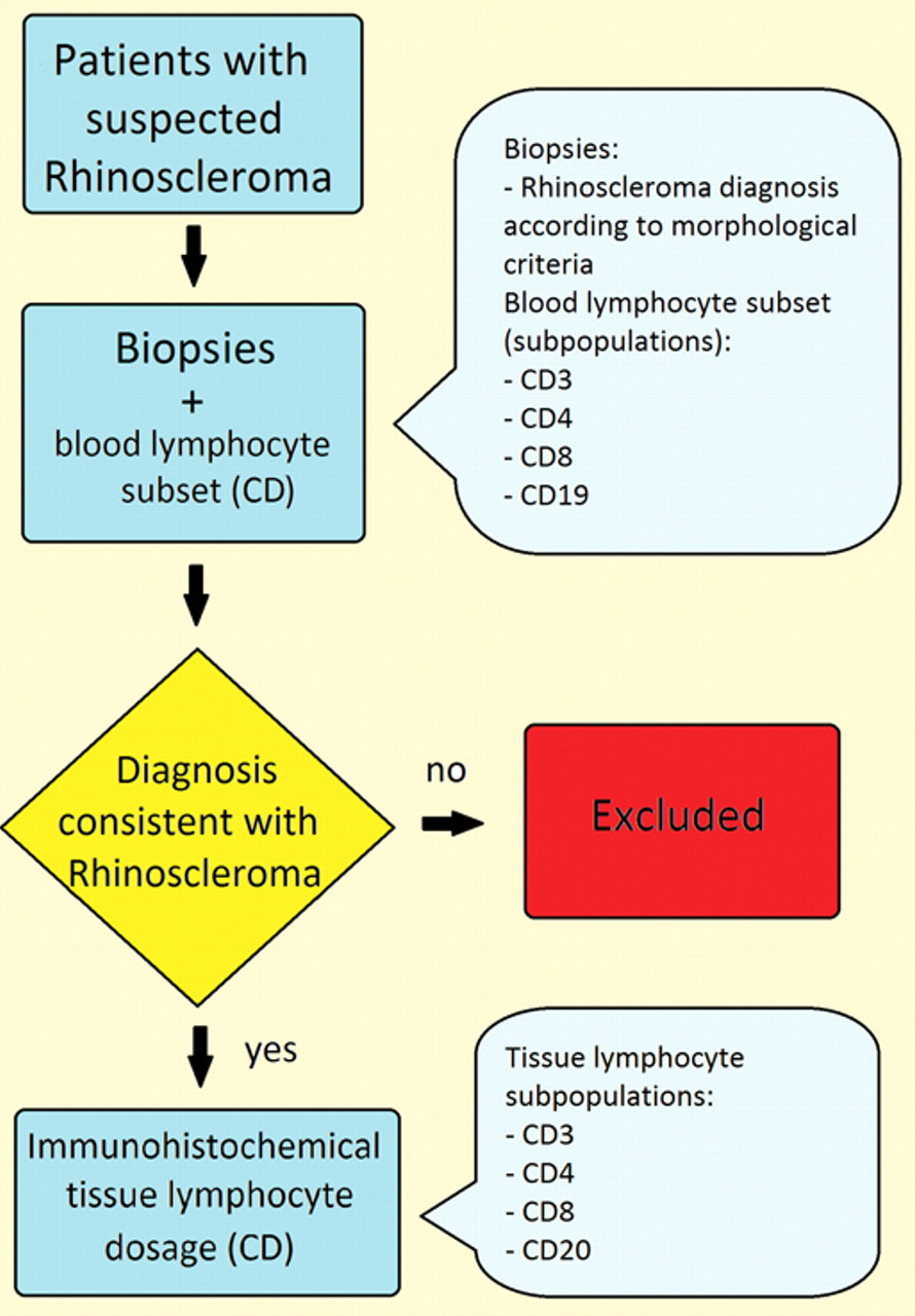
Study design: all patients underwent nasal biopsies. Each patient underwent blood sampling for the determination of lymphocyte subpopulations and biopsies for histological diagnosis according to morphological criteria. Patients with clinical and diagnostic evidence of rhinoscleroma were included in the study. Subsequently, immunohistochemical analysis was carried out on tissue samples. CD, cluster of differentiation.
Blood Samples
In peripheral blood, the following lymphocyte subsets were tested for all patients: CD3+, CD4+, CD8+, and CD19+ ( Table 1 ). CD3, CD4, and CD8 antigens were studied to identify T lymphocytes: CD3/CD4 positivity identifies helper/inducer T lymphocytes; CD3/CD8 positivity identifies cytotoxic/ suppressor T lymphocytes. CD19 antigen was used to identify B lymphocytes. These parameters were investigated after histologic diagnosis and before medical treatment. To determine the cluster of differentiation (CD), fluorescent monoclonal antibodies (Coulter Corp, Miami, Florida) were used. Mononuclear cells were separated according to Boyum’s method: 10 mL of heparinized venous blood was diluted at a 1:1 ratio in phosphate-buffered saline (PBS) and centrifuged on Lymphoprep (Nycomed Pharma AS, Oslo, Norway) at 1600 rpm for 15 minutes. The mononuclear cells were collected, rinsed twice with PBS, and suspended in PBS at 105 cells/mL. Then 400 mL was incubated with 10 mL of anti-CD4, anti-CD8, and anti-CD19 (B lymphocytes) monoclonal antibodies for 60 minutes at 48°C; subsequently, they were washed and suspended again in PBS and analyzed with an EPICS XL-MCL flow cytometer (Coulter Corp). Results are expressed as the percentage of positive cells.
Lymphocyte Subset Description

Biopsies
The morphologic study was carried out on paraffin sections and stained with hematoxylin and eosin. Using immunohistochemical procedures, we studied the expression of CD3, CD4, CD8, and CD20 antigens on paraffin tissue sections ( Table 1 ). CD3, CD4, and CD8 antigens were studied to identify T lymphocytes. CD20 was used to identify B lymphocytes. The sections were 3 to 4 µm thick, and monoclonal antibodies were used with the following dilutions:
CD3 (Novocastra) 1:100
CD4 (Novocastra) 1:100
CD8 (Kako) 1:25
CD20 (Novocastra) 1:100
Paraffin was removed from the sections and rehydrated with xylol, ethanol, and distilled water. Endogenous peroxidase activity was blocked with hydrogen peroxidase (3%) in methanol. The samples were incubated with the Streptavidin-Biotin-Peroxidase Complex (Dako LSAB-Kit; Dako, Carpinteria, California). Reactions were visualized with the use of diaminobenzidine (DAB).
Results
Patients
The time from the onset of symptoms and diagnosis was 21, 20, 25, 30, 10, and 18 months for patients 1, 2, 3, 4, 5, and 6, respectively (mean, 20.6 months).
In the 6 patients (3 women, 3 men), the disease was located in the nose and presented crusty rhinitis with chronic nasal bleeding. No patients had clinically appreciable locoregional adenopathies.
Blood Samples
In all patients, we noted normal amounts of leukocytes and lymphocytes ( Table 1 ). In CD4+ cells, we detected a normal absolute value (mean, 574 cells/lL) and a reduced percentage value (mean, 25.4%). In CD8+ cells, we noted an increase in the absolute (mean, 967 cells/lL) and percentage values (mean, 39.3%); therefore, there was an evident inversion of the CD4+/CD8+ ratio (0.72). The CD19+ cells tended to show normal values. In our series, in which the numerical value of lymphocytes was normal, a reduction of the CD3+ cell percentage value (mean, 53.1%), but not of the absolute value (mean, 1276 cells/lL), also emerged ( Table 2 ).
Patient Lymphocyte Subsets in Blood Samples

Lower-than-normal values are italicized. Higher-than-normal values are bolded.
Biopsies
A diagnosis of RS was confirmed for all patients included in this study. The reactive lymphoid and plasmacellular infiltrate, which was chronic, was mainly present at the mucosal and submucosal levels with nonextensive epitheliotropism. It was frequently perivascular and diffuse and sometimes had a follicular pattern. In at least 2 cases, the reactive lymphoid infiltrate showed focal reacutization with a modest neutrophilic, granulocytic component. Strong neoangiogenesis was also present.
Immunohistochemical studies revealed a heterogeneity of the lymphocytic population with a prevalence of CD3+ cells ( Figure 2a ) over CD20+ cells ( Figure 2b ). Approximately one-third of the T lymphocyte population showed a CD3+/CD8+ immunophenotype (cytotoxic/suppressor), and two-thirds of the T lymphocytes expressed a CD3+/CD4+ immunophenotype (helper/inducer) ( Figure 2c ).
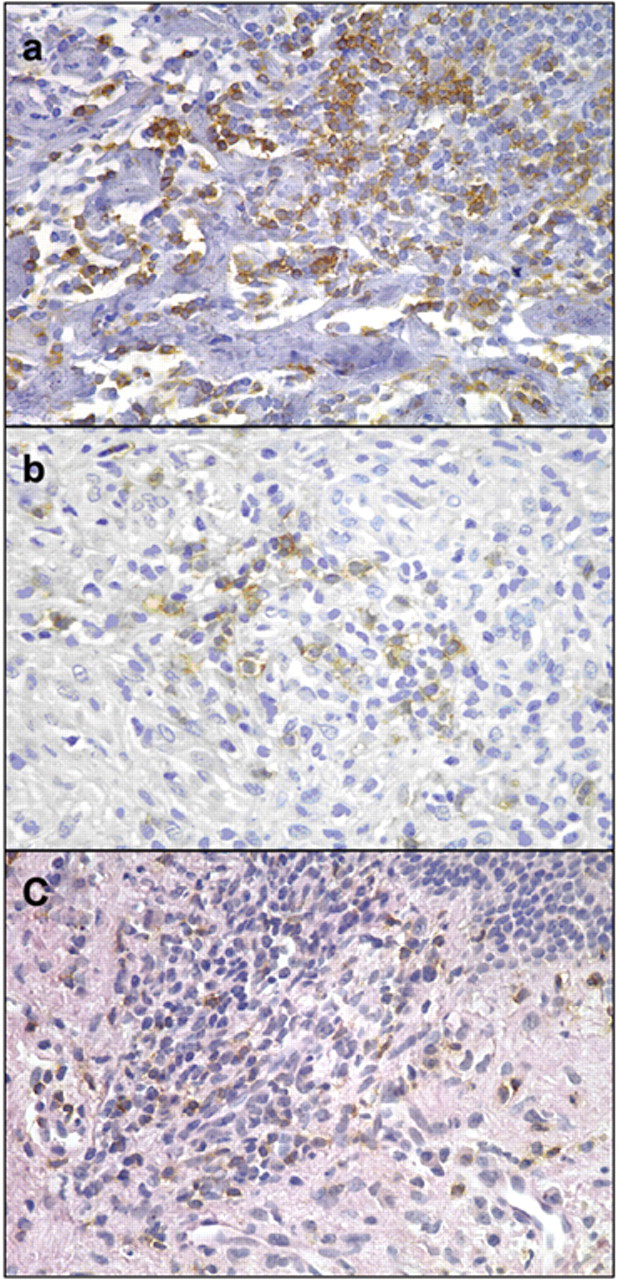
Lymphocyte cluster differentiation on tissue samples: (a) immunohistochemical staining for CD3+, 40×: diffuse presence of CD3+ T lymphocytes; (b) immunohistochemical staining for CD20+, 40×: discrete presence of CD20+ B lymphocytes; (c) immunohistochemical staining for CD8+, 40×: inflammatory infiltration with follicular architectural arrangements and scarce distribution of CD8+ T lymphocytes.
Discussion
Our series consists of only 6 patients, and thus the results obtained have only an indicative value. This reflects the epidemiological data that show RS as being a rare disease in Western Europe. For the same reason, it is impossible to achieve any statistical significance even when comparing the values obtained with a healthy sample of patients.
In our case study, no changes in the absolute count of leukocytes and lymphocytes were observed in venous peripheral blood. In all patients, we noted a relative reduction of CD4+ cells and an absolute increase of the CD8+ cells. A consequence of the increase of the CD8+ cells is the inversion of the CD4+/CD8+ ratio. A reduction of the percentage value (mean, 53.1%) but not of the absolute value (mean, 1276 cells/lL) of the CD3+ cells also emerged. These findings are similar to those reported by other authors. 4,6,9
Immunohistochemical analysis on tissue samples revealed that CD3+ lymphocytes were predominant; two-thirds of the T lymphocytes expressed a CD3+/CD4+ immunophenotype, and one-third was characterized by a CD3+/CD8+ immunophenotype. This result correlates with the preeminent role played by CD4+ lymphocytes in the formation of granulomas. However, it is not surprising to find that previous studies report findings that are in contrast with our experience; some authors report a predominance of CD3+/CD8+ lymphocytes over CD3+/CD4+ ones 10 or an equal distribution of CD3+/CD4+ and CD3+/CD8+ lymphocytes. 6 In fact, granuloma formation is a dynamic process involving the recruitment and modulation of many effectors. The numeric discrepancy of the cellular subsets may have occurred because the biopsies were taken during different phases of granuloma formation.
In venous peripheral blood, we have identified B lymphocytes through the CD19 surface antigen. It is expressed in all stages of B cell differentiation with the exception of pro-B cells. The CD19 antigen is also expressed by plasma cells, but in this stage of differentiation, its expression is downregulated. 11,12 Because pre-B cells and immature B cells are present only in bone marrow ( Figure 3 ) and plasma cells are not commonly present in venous peripheral blood, CD19+ cells indicate the presence of mature B lymphocytes in venous peripheral blood.
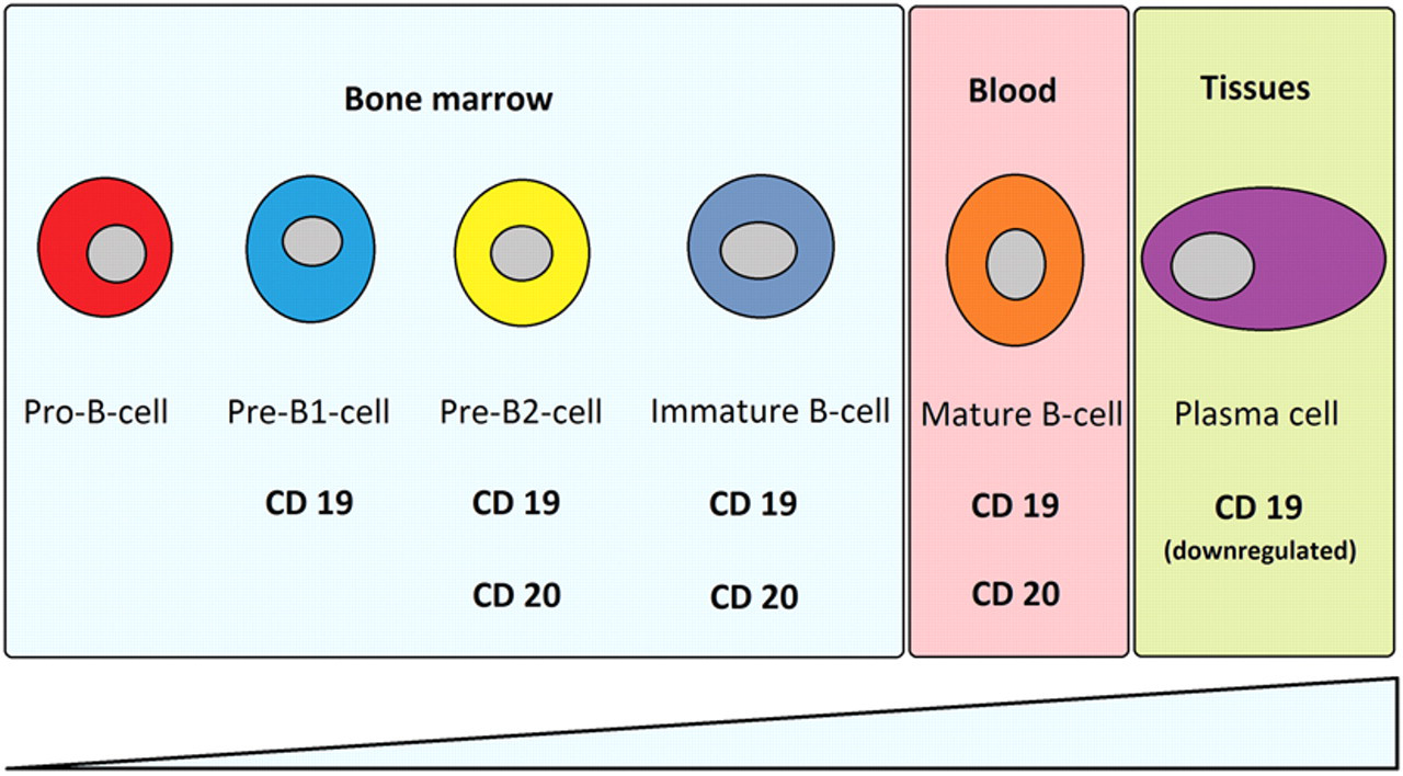
Expression of CD19 and CD20 according to maturation steps. CD19 is expressed on pre-B1, pre-B2, immature, and mature B cells. Its expression in plasma cells is downregulated. CD20 is expressed on pre-B2, immature, and mature B cells. Its expression in plasma cells is abolished.
In tissue samples we identified, by immunohistochemical analysis, the CD20+ B lymphocytes. CD20 antigen is coexpressed with CD19 in the same stages of B cell differentiation with the exception of plasma cells, in which CD19 was downregulated and the expression of CD20 was abolished. The data show that the CD20+ population is quantitatively significant in granulomatous inflammatory infiltrates. 13-15 Because pre-B cells and immature B cells are present only in bone marrow ( Figure 3 ) and CD20 is not expressed by plasma cells, these data indicate the presence of mature B lymphocytes in tissue samples. Therefore, plasma cells are not the only B-line cells of rhinoscleromatous tissue, as known until now. It is presumable that the mature B cells activated by antigenic stimulation of K rhinoscleromatis are the precursors of plasma cells typically found in RS granulomas. It may also be argued that B lymphocytes do not derive from blood vessels or lymph nodes because there was not a significant B lymphocyte increase in peripheral blood or clinically appreciable regional adeno-pathies in any of the patients. However, many studies performed on RS have shown a marked B lymphocyte antibody-mediated response against the M antigen of the mucous envelope, K antigen of the capsula vera, and O somatic antigen of the S phase of K rhinoscleromatis. 7-10
In support of a high B cell activity in RS, there is the presence of Russell bodies, which are atresic corpuscles derived from plasma cells detectable by optical microscopy in the majority of cases. They are not pathognomonic of RS but are also present in other morbid processes (cancers, granulation tissue of gastric ulcers, lymph nodes of laboratory animals treated with substances capable of determining plasma cell system hyperplasia, aspecific chronic inflammatory processes, chronic viral diseases), all characterized by a high plasma cell turnover. 1,8,10 Furthermore, in Mikulicz cells, cytoplasmic vacuoles containing bacilli covered with a coat of electron-dense mucopolysaccharides called “A granules” are demonstrable by electron microscopy. According to Toppozada et al, 16 these granules are antibodies against K rhinoscleromatis. Our findings, together with the evidence described above, suggest the strong activation of the B cell–mediated immune system, most likely oriented inefficaciously against an intracellular pathogen. Furthermore, because we enrolled patients without a clinically evident nodal reaction, as is usual in RS, and there was no significant expansion of CD19+ B lymphocytes in the peripheral blood, it is presumable that NALT (nose-associated lymphoid tissue) B lymphocyte activity is predominant compared to the systemic humoral response. In fact, antibodies against K and O K rhinoscleromatis antigens are detected only in the phases of reactivation of the disease.
Conclusions
Our study demonstrates the presence of mature B lymphocytes in RS tissue by CD20 positivity. We hypothesize that these cells belong to the NALT. These CD20+ B lymphocytes are presumably the precursors of the numerous plasma cells always described in RS.
Proof of the presence of B cells reinforces the view that in tissues affected by RS, 2 components of the immune system are involved: a qualitatively ineffective Th1 granulomatous reaction and a humoral response given by B lymphocytes. The latter turns out to be equally ineffective because K rhinoscleromatis is an intracellular pathogen. We believe that a consequence of this process is the chronicization of the disease. However, our study includes a small series of patients, and further investigations comparing blood and tissue samples of patients with RS versus normal patients are required.
Author Contributions
Disclosures
Footnotes
Acknowledgements
We are grateful to Maria Grazia Saladino for her valuable contribution in editing the manuscript.
Sponsorships or competing interests that may be relevant to content are disclosed at the end of this article.
This article was presented at the 2010 AAO-HNSF Annual Meeting & OTO EXPO; September 26-29, 2010; Boston, Massachusetts.