
Abstract
Objective
The purpose of this study is to determine if crawling wave elastography, a novel sonoelastography technique, can be used to provide quantitative measurements of thyroid tissue shear velocity (a measure of tissue stiffness) and distinguish between benign and malignant thyroid nodules.
Study Design
Diagnostic test assessment.
Setting
Academic university.
Subjects and Methods
Fresh thyroid specimens (n = 20) with 44 regions of interest were imaged ex vivo with crawling wave sonoelastography over a 9-month period in 2010 at a single institution. Using the sonoelastography technique, shear velocity estimations and contrast-to-noise ratios were calculated. The higher the shear velocity (SV) and contrast-to-noise ratio (CNR), the greater the tissue stiffness. Histological diagnosis was correlated with shear velocity and contrast-to-noise ratio values.
Results
Both the shear velocity and contrast-to-noise values of papillary thyroid carcinoma (n = 10, CNR = 5.29, SV = 2.45 m/s) were significantly higher than benign nodules (n = 22, CNR = −0.41, SV = 1.90 m/s). There is a maximum sensitivity and specificity of 100% and 90.9%, respectively, for differentiating papillary thyroid carcinoma from benign nodules using contrast-to-noise ratio values. There is a maximum sensitivity and specificity of 83.3% and 72.7%, respectively, for differentiating papillary thyroid carcinoma from benign nodules using shear velocity values. Insufficient samples were obtained for comparison with other histological types.
Conclusion
Crawling wave sonoelastography can provide quantitative estimations of shear velocity, thereby depicting the elastic properties of thyroid nodules. The shear velocity and contrast-to-noise ratio can differentiate between benign thyroid nodules and papillary thyroid carcinoma with high specificity and sensitivity.
Keywords
As more thyroid nodules are detected with the increasing use of thyroid ultrasound, there is greater need to improve the diagnostic specificity and sensitivity for malignancy through noninvasive techniques. With the use of ultrasound, it is estimated that 60% of women aged 75 years will have thyroid nodules. In addition, up to 35% of thyroid glands on autopsy contain papillary thyroid carcinoma. 1 Currently, features such as microcalcifications, increased Doppler flow, hypoechogenicity, and irregular borders lack sufficient sensitivity and specificity to differentiate thyroid carcinoma. 2 The American Thyroid Association (ATA) has recommended biopsy for most nodules 1 cm or greater or any nodule 5 to 9 mm with suspicious sonographic features in a high-risk patient. This, combined with the recommendation to biopsy up to 4 nodules per patient, highlights the need for noninvasive techniques to increase the pretest probability in detecting malignancy in fine-needle aspiration (FNA) biopsy. 3
Sonoelastography is a general term for the use of ultrasound in imaging tissue elastic parameters. Various sonoelastography methods have been explored, including strain variation, velocity, compression, and vibration amplitude. 4 Early vibrational elastography was pioneered by Lerner et al. 5 Since then, elastography has grown significantly as a field and is being used in liver imaging and to differentiate breast and prostate cancer.6,7
More recently, attention has been drawn to the utility of elastography in thyroid nodules. Multiple studies have demonstrated its effectiveness in differentiating benign and malignant nodules in vivo.8-18 However, the majority are qualitative elastographic images that depend on maintaining a highly standardized technique and a subjective grading system.19,20
Crawling wave (CrW) elastography is a method of shear wave sonoelastography that provides quantitative estimations of tissue elasticity properties. In particular, it provides estimations of tissue shear velocity (m/s), which is proportional to tissue stiffness. The higher the shear velocity, the greater the stiffness of the tissue. The reliability and accuracy of this technique already have been demonstrated in other tissues.7,21-23
The purpose of this study was to demonstrate feasibility and utility of using a novel technique, CrW sonoelastography, in thyroid tissue to differentiate between benign and malignant nodules and to compare the quantitative measurements with histopathology. We hypothesized that CrW sonoelastography can produce reliable quantitative results in ex vivo thyroid tissue and differentiate with adequate sensitivity and specificity between benign and malignant nodules.
Materials and Methods
The study protocol was approved by the Institutional Review Board of the University of Rochester, and signed consent was obtained for all tissue specimens. A priori power analysis determined that a sample size of 25 to 35 nodules was needed to obtain 80% power to detect a difference between the 2 groups. Twenty unselected adult patients (3 men, 17 women; mean age 52.9) undergoing total or hemithyroidectomy at the University of Rochester from January to December 2010 were recruited for study. Children, revision surgery, recurrent disease, or prior radiation patients were excluded. Fresh, unfixed whole and hemithyroid specimens were suspended in an agar gel phantom (
Figure 1
). In each case, regions of interest (ROIs) for scanning were defined as a nodule with surrounding homogeneous thyroid tissue on B-mode ultrasound. Normal tissue scans were also performed in lobes without nodules and with homogeneous-appearing ultrasound images. Two B&K Minishakers with piston vibration exciters (Model 2706; Brüel & Kjaer, Naerum, Denmark) were applied as the vibration sources. A dual-channel signal generator (Model AFG320; Tektronix, Beaverton, Oregon) produced 2 monochrome low-frequency signals that were slightly offset (typically less than 0.5 Hz). These signals were passed through a 2-channel amplifier before being input to the minishaker vibration devices. A LOGIQ 9 scanner (General Electric Medical Systems, Milwaukee, Wisconsin) modified for sonoelastography was used with a M12L linear array probe (5-13 MHz bandwidth) for real-time visualization of the propagating crawling waves (ie, shear wave interference pattern motion). Two-dimensional B-mode and sonoelastographic video images were recorded over a range of shear wave frequencies from 80 to 280 Hz. The corresponding 2-dimensional (2D) imaging plane was then marked with a needle for later direct histopathologic correlation. Imaging data were then analyzed with Matlab 7.6 (Mathworks, Inc, Natick, Massachusetts) creating 2D shear velocity maps. Shear velocity (SV) means and contrast-to-noise ratios (CNRs) were determined for scanned nodules. CNR, defined as (|SV(nodule) − SV(background)|)/σ(background), is commonly used in image analysis. Histologic slides corresponding to the 2D plane of scanning were created and photographed. Histologic findings of the needle-localized imaging planes were correlated with SV and CNR values (
Figures 2
and
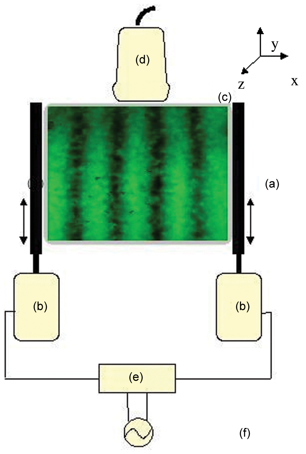
Experimental setup. (a) Vibrator extensions, (b) piston vibration exciters, (c) gel phantom with suspended specimen, (d) ultrasound transducer, (e) amplifier, and (f) function generator.

B-mode ultrasound imaging plane of suspended thyroid specimen with nodule (arrow) and histologic slide corresponding to 2-dimensional imaging plane and nodule.
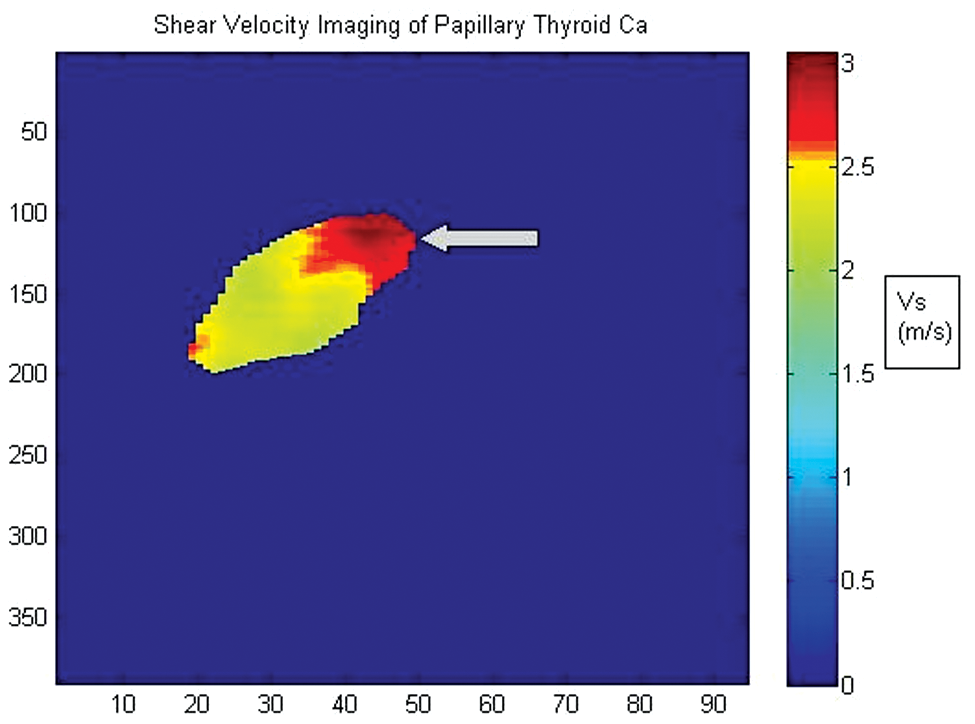
Two-dimensional quantitative shear velocity map (m/s) corresponding to ultrasound and histologic planes of a nodule (arrow).
Results
A total of 20 patients (3 men, 17 women; mean age 52.9 ± 14 years) were included in the analysis. Forty-four tissue regions were scanned (12 malignant, 4 adenoma, 22 benign nodules, and 6 areas of normal thyroid). The average nodule size was 2.26 ± 1.12 cm. There were a total of 5 nodules with reported calcifications. Four of 5 nodules with calcifications were malignant. No cystic nodules were included. Doppler characteristic data were incomplete and not included. Preoperative FNA was performed on 19 nodules (43%). Eight of the 12 (67%) malignant nodules were correctly identified by FNA. The remaining 4 were incidental papillary thyroid carcinoma (PTC) or follicular carcinoma. Two of 5 FNAs with findings of follicular neoplasm were malignant. Of the 2 follicular neoplasms on FNA, only 1 was follicular carcinoma. The other was determined to be a PTC on final pathology. The calculated shear velocities and CNR for all histologic types are in Table 1 . Outliers identified were the 1 medullary carcinoma and 1 benign nodule.
Quantitative Measurements of Shear Velocity and CNR
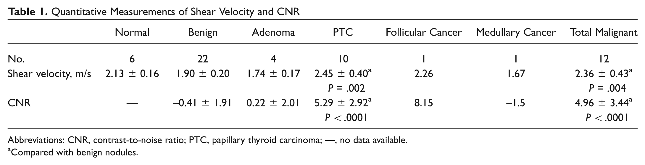
Abbreviations: CNR, contrast-to-noise ratio; PTC, papillary thyroid carcinoma; —, no data available.
Compared with benign nodules.
Normal Thyroid
Normal tissue regions were scanned for 6 patients (5 women, 1 man) with a mean age of 50.8 years. The mean shear velocity was 2.13 ± .16 m/s for the scanned regions. No patients had a diagnosis of Hashimoto disease. None had undergone ablation or thyroid hormone replacement.
Benign Nodules
Twenty-two benign colloid nodules were scanned from 12 patients (all women) with a mean age of 55.6 years. The mean shear velocity was 1.90 ± 0.20 m/s, with a mean CNR of −0.41 ± 1.91.
Adenomas
Four adenomas were scanned from 4 patients (1 man, 3 women) with a mean age of 50 years. The histologic diagnoses were 2 follicular adenomas, 1 Hurthle cell adenoma, and 1 hyalinizing trabecular adenoma. The mean shear velocity was 1.74 ± 0.17 m/s, with a mean CNR of 0.22 ± 2.01.
Malignant Nodules
Twelve malignant nodules were scanned from 11 patients (2 men, 9 women) with a mean age of 51.8 years. The histologic diagnoses were PTC, follicular carcinoma, and medullary carcinoma. The papillary thyroid carcinomas included classical, sclerosing, and follicular variants. No cystic variant of papillary carcinoma was identified. The mean shear velocity for all malignant nodules was 2.36 ± 0.43 m/s, with a mean CNR of 4.96 ± 3.44. The mean shear velocity for PTC alone was 2.45 ± 0.40 m/s, with a mean CNR for PTC of 5.29 ± 2.92.
Comparison
Comparison was between benign colloid nodules and malignancy. Adenomas were excluded from the analysis due to the insufficient numbers. Analysis demonstrated a significant difference for both the mean shear velocities and CNR between benign and malignant thyroid nodules (P = .004 and P < .00012, respectively). When considering differentiating between benign nodules and PTC, the difference was even more significant for both shear velocity and CNR (P = .002 and P < .0001, respectively) ( Figures 4 and 5 ). The box plots in Figure 4 show a small overlap that is predominately from a small handful of outlier measurements. Although we think that the variability within the box plots represents true variations in the shear velocities, it is possible that these differences may have arisen from variations in the measurements or other factors. It is known that certain conditions, such as Hashimoto thyroiditis, do increase the shear velocity of thyroid tissue, which could affect measurements obtained in that gland. However, there were not a significant number of patients with Hashimoto thyroiditis in this sample. It is also possible that a greater difference between the box plots might have emerged if all nodules scanned came from independent tissue samples.

Quantitative shear velocity estimations of benign versus papillary thyroid carcinoma. P = .0002.
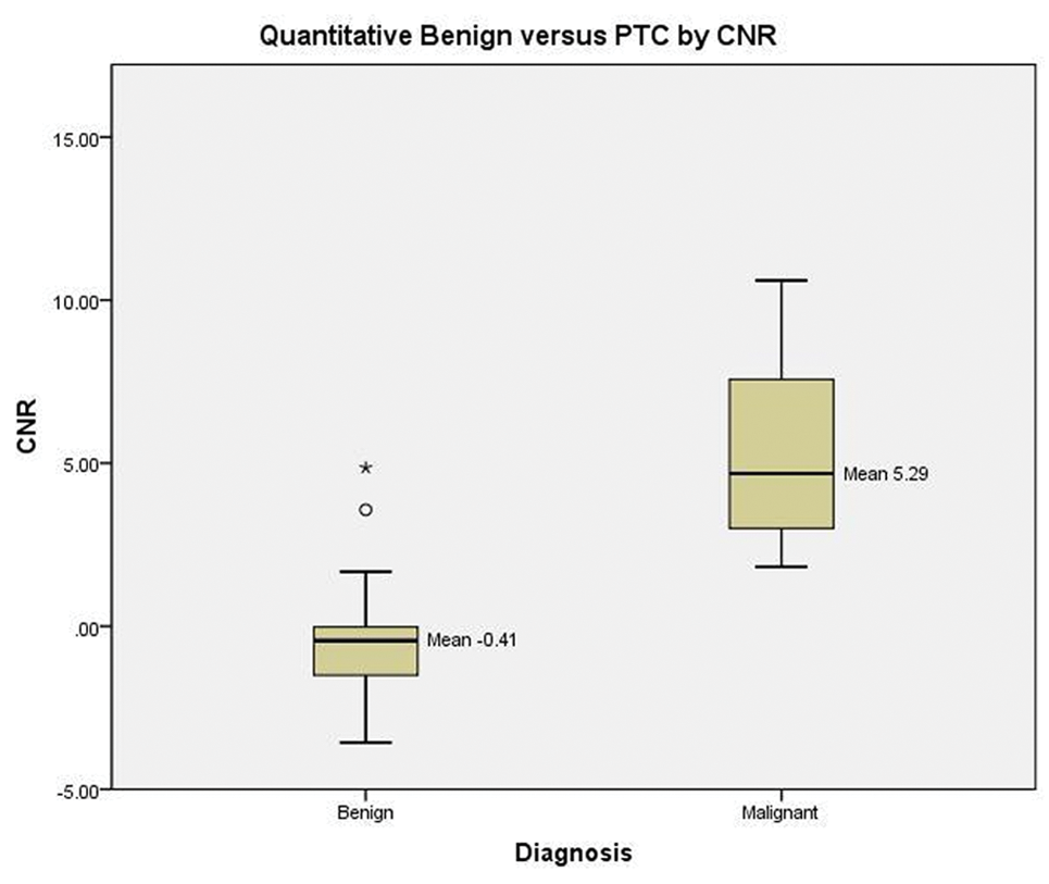
Quantitative contrast-to-noise ratios (CNRs) of benign versus papillary thyroid carcinoma. P < .0001. The circle represents a moderate outlier, and the asterisk represents an extreme outlier.
The ROC curves for both shear velocities and CNR were created. The area under the curve (AUC) for detecting benign and malignant nodules using a mean shear velocity cutoff of 2.02 m/s was 0.82 with a sensitivity and specificity of 83.3% and 72.7%. The AUC for detecting benign and PTC nodules using a mean shear velocity cutoff of 2.02 m/s was 0.88 with a sensitivity and specificity of 90.0% and 72.7%. The AUC for detecting benign vs malignant nodules using CNR was 0.90 with a maximum sensitivity and specificity of 91.7% and 90.9%, respectively, at a CNR of 1.75. The AUC for detecting benign vs PTC nodules was 0.96 with a maximum sensitivity and specificity of 100% and 90.9%, respectively, at a CNR of 1.75.
Discussion
Thyroid elastography is amassing supportive data for its utility in determining benign vs malignant nodules.8-18 However, there remains variability in the qualitative assessments that are obtained. Specifically, the interobserver agreement between these measurements has been called into question. 24 This highlights the importance of providing quantitative measurements of the elastic properties of thyroid nodules.
The current feasibility study demonstrates that crawling wave sonoelastography can provide quantitative measurements of tissue shear velocity. For example, the quantitative shear velocity measurements obtained of normal thyroid tissue (2.13 m/s) in this study correlated well with previously published mechanical testing and elastography measurements (2.07 m/s).16,25,26
This preliminary ex vivo testing allowed for a more standardized imaging protocol as well as definitive confirmation of the pathology that correlates with the scanned image. This protocol has been demonstrated to be reliable and reproducible in prostate specimens and prostate cancer. 7 Extrapolations of these results to future in vivo results may not be simple but are not insurmountable. In patients, the effects of overlying tissue, blood flow in the vasculature, and body temperature may alter the shear wave speed somewhat. Shear wave sources also need to be integrated into the handheld ultrasound transducer for patient studies. Nonetheless, early elastographic results in the thyroid seem to indicate that the contrast between malignant and benign lesions is present in vivo.8,9 In addition, this preliminary ex vivo study allows for direct tissue comparison that is lacking in previous in vivo studies.
The 1 medullary carcinoma demonstrated a much lower shear velocity and CNR than expected. It is unclear if this was due to error in measurements or is consistent with medullary carcinoma true shear velocity. Without additional specimens, this finding was unable to be confirmed. The 1 follicular carcinoma demonstrated a SV of 2.26 m/s. This might hint at a difference in calculated shear velocities of follicular carcinoma vs follicular adenoma. Unfortunately, during our study period, there were limited samples of adenomas to include in analysis. Histologically, there often is very little difference in the appearance of follicular neoplasms, so it seems unlikely that shear velocity imaging would detect differences between follicular neoplasms unless their histologic characteristics are very de-differentiated or invasive. Obviously, much greater numbers will be needed to determine if this difference exists. At this time, no definitive statement can be made regarding the ability to distinguish adenomas from carcinomas using elastography.
In addition, 1 benign nodule demonstrated a much higher shear velocity and CNR than other nodules scanned. This represented a specimen with a whole-tissue pathologic report of a diagnosis of papillary thyroid carcinoma in the gland, but the specific 2D imaging plane slide did not contain the malignancy. Therefore, it was included in the benign group but may represent a true malignancy. Although the study provided sufficient power to demonstrate differences based on a priori power analysis, additional samples would provide useful information with regard to specific histologic subtypes of benign and malignant diseases. Of important note is that no cases of a cystic variant of papillary thyroid carcinoma were identified in the study. With all techniques of elastography, the cystic variant would not demonstrate a shear velocity or stiffness that is suggestive of malignancy.
There was a significant difference between the shear velocities and CNR for benign and malignant nodules. This difference was even more significant when comparing benign lesions and those of papillary thyroid carcinoma alone. The utility of these findings are highlighted with the ROC curves for CNR demonstrating a high specificity and sensitivity of 91.7% and 90.9%, respectively. Current ultrasound features suggestive of malignancy, such as microcalcifications, irregular boarders, increased Doppler flow, hypoechogenicity, and absence of halo, are insufficient in predicting malignancy. The sensitivities and specificities of these features have been demonstrated to range from 42.3% to 77.2% and 61.2% to 91.2%, respectively. 2 Crawling wave sonoelastography could augment these ultrasound features to better detect malignancy without biopsy.
A question that remains is the range of nodule size in the effectiveness of all elastography techniques. In our study, the mean nodule size was 2.26 cm. It is known that SV estimations are affected by boundary conditions. Along boundaries of different tissue types, there is distortion and interference of the waveforms, as well as a greater likelihood of variability of readings. Therefore, the smaller the nodule, the greater the impact of boundary conditions. Crawling wave sonoelastography tends to becomes less reliable for ROI less than 0.5 cm. Also, in very large, cystic nodules, the waveforms become attenuated, and the reliability of readings also declines. Variations of these limitations are expected in all forms of elastography.
This study was unique in that it used surgical histopathologic diagnosis of the needle-localized 2D imaging plane instead of FNA diagnosis alone. This allowed for direct comparison of 2D structure with quantitative shear velocity images. Studies with FNA diagnosis alone are subject to biopsy sampling error and carry the possibility of missing occult thyroid malignancy. This study demonstrated the feasibility of the quantitative technique and provides encouraging data for in vivo applications. Variations of this technique are currently being applied in acoustic radiation force imaging (ARFI)25,26 and in clinical devices by SuperSonic Imagine (Aix-en-Provence, France). This study provided the initial step for proof of concept and application for further prospective in vivo studies that would include a clinically applicable device for scanning thyroid nodules prior to FNA biopsy. Until in vivo testing occurs, it is unclear how much of an interference there will be with trachea, carotid pulsations, and surrounding lymphadenopathy. Shear wave sources will also need to be integrated into handheld probes or be applied through external vibration sources. Despite these future obstacles, the current ex vivo study of shear wave estimation provides a basic model for all forms of shear wave imaging, including ARFI, for which a clinical device currently exists. Currently, all forms of elastography (vibrational, compression, or strain imaging) are affected by boundary conditions and acoustic barriers. For any of these methods, these boundary conditions and acoustic interference sources may not be trivial or easily understood. However, none of these other methods has demonstrated the initial step presented in the current study of direct tissue histopathologic comparison.
Even with these encouraging results and translation to a clinically applicable device, CrW sonoelastography quantitative measurements will not completely replace FNA biopsy. However, the quantitative shear velocity measurements obtainable by future clinical in vivo devices using crawling wave sonoelastography, or adaptations thereof, may increase the specificity and sensitivity of noninvasive techniques to better determine which nodules need biopsy and, in certain situations, may allow for deferring FNA biopsy.
Author Contributions
Disclosures
Footnotes
Sponsorships or competing interests that may be relevant to content are disclosed at the end of this article.
This article was presented at the 2011 AAO-HNSF Annual Meeting & OTO EXPO; September 11-14, 2011; San Francisco, California.