
Abstract
Objectives/Hypothesis
The purpose of this study was to investigate the morphological and histological change of vocal folds (VFs) after steroid injection in a rabbit model.
Study Design
Prospective animal study.
Setting
Tertiary academic medical center.
Subjects and Methods
Twenty-four New Zealand white rabbits were used in this study. We randomly classified rabbits into the 3 groups and triamcinolone acetonide suspension was injected to the right VF with different concentrations. Left VF was injected with the same volume of phosphate-buffered saline as control. Endoscopic evaluation was performed to measure morphological changes. The larynges were collected for histological analysis, and the VFs were stained with hematoxylin-eosin for assessing inflammatory response, glandular atrophy, and muscular atrophy and with Masson’s trichrome for assessing collagen deposition.
Results
In morphological assessment, there were no differences in VF mass reduction, mucosal atrophy, and granulation formation between both VFs. Histological assessments showed no significant difference in inflammatory response, glandular atrophy, and collagen deposition between both VFs. However, there was a difference in muscular atrophy and epithelial layer thinning in steroid injected right VFs. Muscular atrophy had been completely recovered over time, but mild epithelial thinning was continued until 12 weeks. The longer exposure time and larger dose did not increase the intensity of muscular atrophy or epithelial thinning.
Conclusion
We demonstrated that the VF steroid injection resulted in no significant changes in morphology and histology of rabbit VF. However, steroid injection may induce several VF histological changes and these results are needed to be considered when treating humans.
Introduction
Steroids are molecules synthesized by our bodies to regulate a vast number of physiological, immune, and metabolic processes. 1 Steroids have potent anti-inflammatory and immunomodulation properties. They have a unique, essential mode of action characterized by the activation or inhibition of numerous target genes. These molecules act upon a number of cells, involving innate and adaptive immune responses, apart from other cells (fibroblast, epithelial, and endothelial cells). Steroid anti-inflammatory effectiveness is related to switching off multiple inflammatory genes (encoding cytokines, chemokines, adhesion molecules, inflammatory enzymes, receptors, and proteins) that have been activated during the chronic inflammatory process. 2 Steroid administration remains one of the most potent therapeutic interventions among upper airway diseases. Steroids have traditionally been used for the treatment of acute inflammatory diseases of the upper aerodigestive tract and specifically for the larynx to decrease edema, as in epiglottitis and croup.3-5
With the advent of modern methods for phonosurgery techniques, there is a renewed interest in the management of various laryngeal diseases. In particular, the vocal fold steroid injection has been reported as an effective method for treating laryngeal diseases. Tateya et al conducted a study of 28 patients with vocal nodules who were treated with transoral vocal fold steroid injection. Their study found that good results could be obtained through vocal fold steroid injection. 6 Mortenson and Woo reported that 11 of the 18 patients of vocal polyps or nodules improved after vocal fold steroid injection and avoided surgery. All 12 patients with vocal fold scar reported improved voice and better vocal fold vibration as a result of the injection. 7
Although vocal fold steroid injection is known as a safe treatment method, there are possible complications.8,9 Andrade Filho et al reported a case of vocal fold plaque following triamcinolone injection until 10 months after the injection. 8 Another hypothesized complication is that if steroids are injected too deeply there could be decreased vocal fold mass, mucosal gland atrophy, and muscle atrophy. However, these side effects were not seen in the Mortenson and Woo series or the Tateya et al series.6,7 In our previous study, 74 of the 80 cases (93%) with vocal nodules disappeared after vocal fold steroid injection via the cricothyroid membrane. 10 We had a few patients who complained of a breathy voice, and their stroboscopic findings showed a mild decrease in the vocal fold mucosal wave and vocal fold bowing. These findings were resolved at about the 2-month follow-up visit. And 2 patients showed white plaque formation, which was thought to be a precipitate of the triamcinolone acetonide suspension. These findings were resolved spontaneously after 1 to 2 months. 10 We are concerned about the side effects of vocal fold steroid injection. There are few studies that have evaluated morphological or histological changes and possible complications after vocal fold steroid injection.
The purpose of this study was to investigate the morphological and histological change of vocal folds and to analyze the possible complications after vocal fold steroid injection in a rabbit model.
Material and Methods
Animals
Twenty-four New Zealand white rabbits, weighing 2.2 to 3.0 kg, were used in this study. The study was designed to detect differences between the groups of 2 points on the grade of morphological and histological change (a standard deviation of 2%). The sample size was calculated with a power of 90% and 5% significance. This study was performed in accordance with the guidelines of the Animal Experiment Committee, Sungkyunkwan University. All rabbits were housed in an approved animal care facility with water and regular rabbit food as desired. All experiments were performed with the authorization of the Animal Experiment Committee at Kangbuk Samsung Hospital, Sungkyunkwan University.
Surgical Procedure
After premedication with xylazine (Rompun, 0.1 mg/kg, Bayer, Germany), all rabbits were anesthetized by intramuscular administration of tiletamine HCl/zolazopam HCl (Zoletil, 1-2 mg/kg; Virbac, Carros, France). Each rabbit was then placed in dorsal recumbency, and its mouth was opened with an oral speculum (7 mm, nasal speculum; KASCO, Seoul, Korea). Visualization of the surgical procedures was accomplished using a 0° 5-mm rigid endoscope (Karl Storz GmbH & Co, Tuttlingen, Germany). Triamcinolone acetonide (Tamcetone, 40 mg/mL, Hanol Inc, Korea) was used in this study, and this solution was diluted with phosphate-buffered saline (PBS) for different steroid concentrations. The animals were randomly assigned to 3 groups according to different concentrations of triamcinolone acetonide (group A, 0.4 mg/mL; group B, 0.8 mg/mL; group C, 2 mg/mL). Phosphate-buffered saline (0.1 mL) was injected into the left vocal fold, and the same volume of triamcinolone acetonide (0.1 mL) was injected into the right vocal fold.
Morphological Analysis
After anesthesia, photo-documentation was performed using a rigid endoscope that was coupled to a digital camera (Nikon digital camera D70, Nikon, Tokyo, Japan) after steroid or PBS injections. Photo-documentation was performed at 2, 4, 8, and 12 weeks. To reduce the bias, 2 senior otolaryngologists (SHL, SMJ) blindly checked the endoscopic photo-documentation and recorded independently on a subjective grade of 1 to 4 (1 = same, 2 = mild change, 3 = moderate change, 4 = severe change) for mucosal atrophy, vocal fold mass reduction, and granulation formation by comparing PBS-injected left vocal folds. And then, the average of parameters was calculated.
Histological Analysis
Two rabbits from each group were painlessly euthanized at 2, 4, 8, and 12 weeks after injection. All larynges were harvested and embedded in paraffin. The vocal folds were stained with hematoxylin-eosin (H&E) staining for assessing inflammatory response, glandular atrophy, and muscular atrophy and Masson’s trichrome staining for assessing collagen deposition. Images were captured with a Nikon Eclipse E600 microscope (Nikon). The histological findings were blindly checked and interpreted independently by 3 pathologists. To minimize learning bias, all results were reviewed in 2 different random orders and were recorded on a subjective grade of 1 to 4 (1 = same, 2 = mild change, 3 = moderate change, 4 = severe change) by comparing PBS-injected left vocal folds. And then, the average of each parameter was calculated.
Statistical Analysis
Statistical analysis was performed using PASW Statistics 18 for Windows (SPSS Inc, Chicago, Illinois). The paired t-test was used to compare the average grading of the morphological and histological analysis in each groups and independent t-test was used to compare between groups. A P value of <.05 was considered statistically significant.
Results
Morphological Analysis
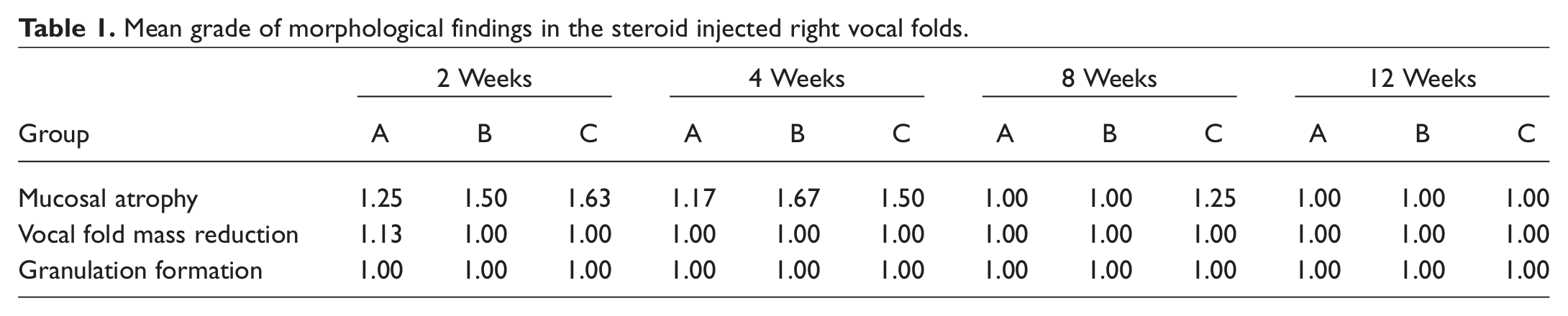
In all groups, mucosal atrophy was mild at 2 weeks. But, mucosal atrophy had recovered, and there was no difference compared to control vocal folds at 12 weeks ( Figure 1 ). In group C, mild mucosal atrophy was observed until 8 weeks. In group A, a mild degree of vocal fold mass reduction was observed at 2 weeks. After 2 weeks, in all groups, there was no difference in vocal fold mass reduction compared to control vocal folds. In all groups, there was no granulation formation throughout the experimental period ( Figure 2 ).
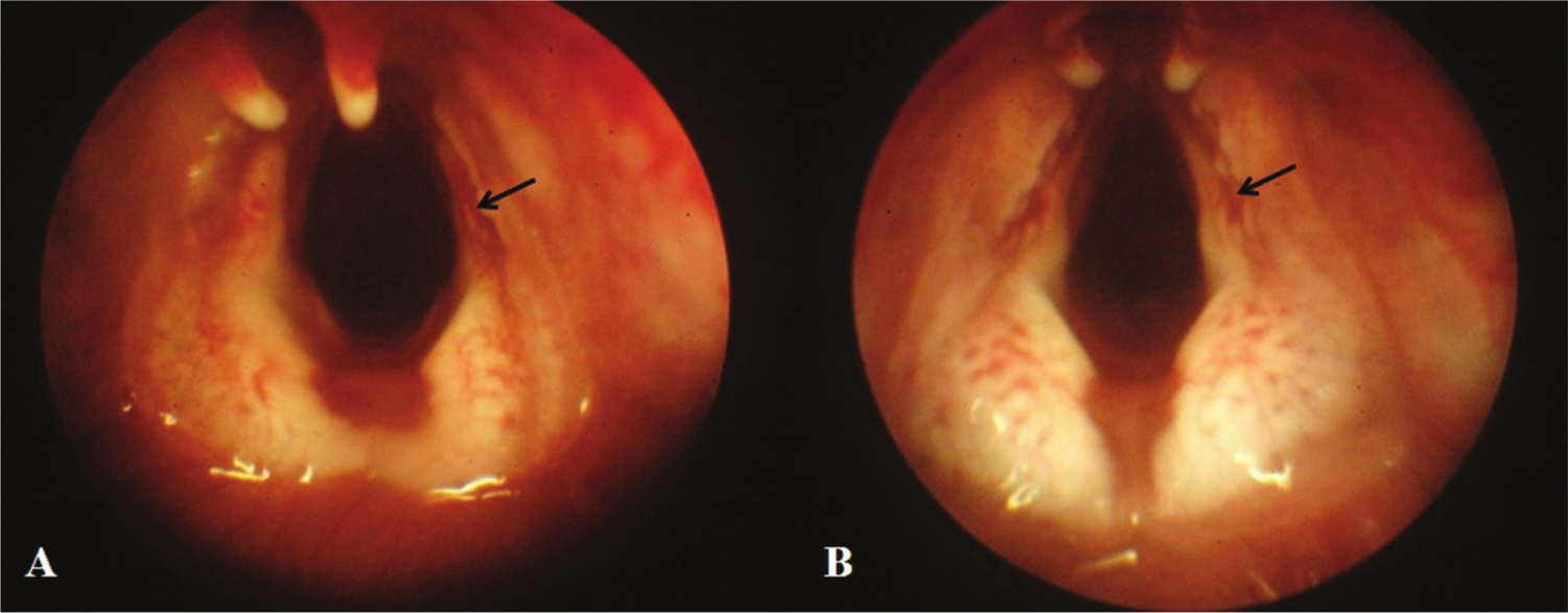
A morphological assessment of vocal folds in group A. (A) There was slightly more mucosal atrophic change in steroid injected right vocal folds at 2 weeks (arrow). (B) There was no difference in mucosal atrophy in both vocal folds at 12 weeks.
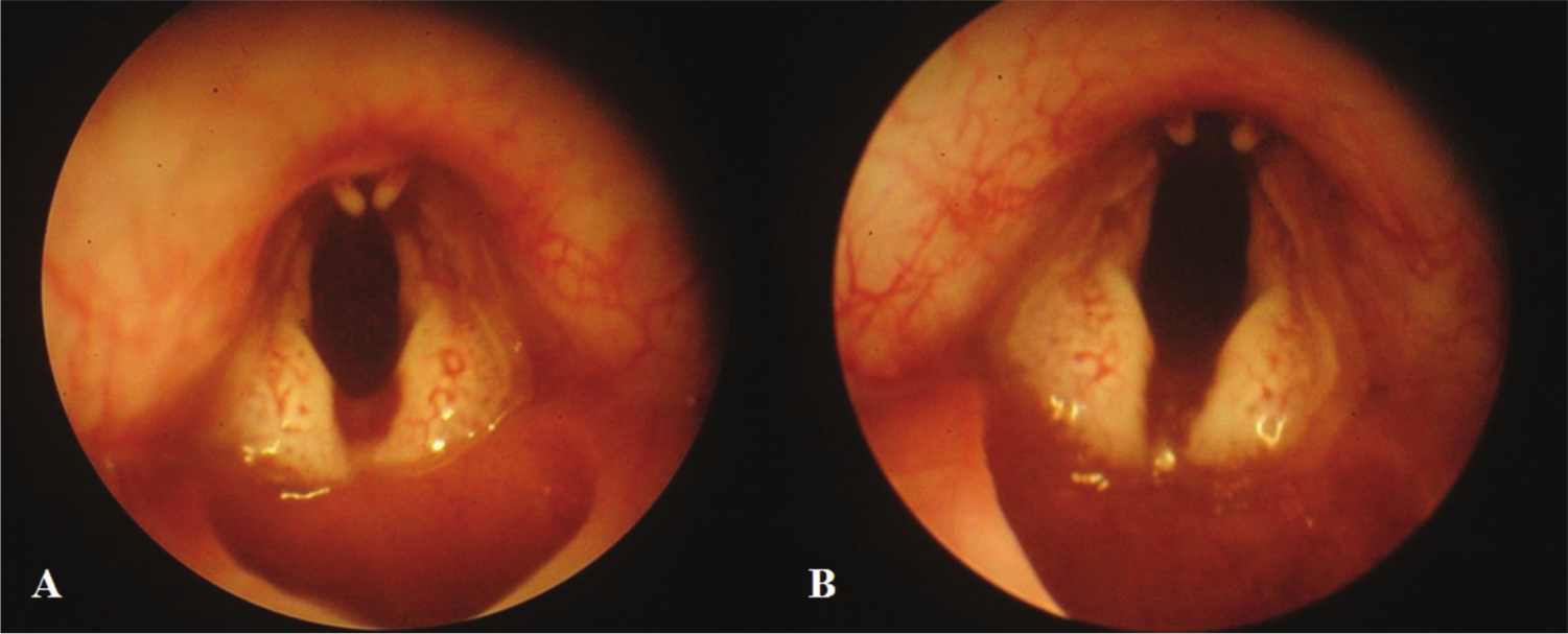
A morphological assessment of vocal folds in group B. There was no difference in vocal fold mass reduction and granulation formation at (A) 2 weeks and (B) 12 weeks.
By each group, in group A (0.4 mg/mL), little change of vocal fold mass reduction and granulation formation were found in steroid-injected right vocal fold. Mild mucosal atrophy was found until 4 weeks, but it was not found at 8 and 12 weeks. In group B (0.8 mg/mL), no change in vocal fold mass reduction and granulation formation was found in steroid-injected right vocal fold. Mild mucosal atrophy was found until 4 weeks, but it was not found at 8 and 12 weeks. In group C (0.8 mg/mL), there was no change of vocal fold mass reduction and granulation formation in steroid-injected right vocal fold. Various rates of mild mucosal atrophy were found until 8 weeks, but they were not found at 12 weeks ( Table 1 ).
Mean grade of morphological findings in the steroid injected right vocal folds.
Histological Analysis
In all groups, there was no sign of inflammatory response and glandular atrophy in both vocal folds throughout the experimental period (P > .05) ( Table 2 , Figure 3 ). Various degrees of muscular atrophy were observed in all groups ( Figure 4 ). In group A, muscular atrophy was initially observed at 4 weeks, and it had recovered after 8 weeks. In group B, muscular atrophy was observed at 2 weeks, and it was recovered after 4 weeks. In group C, muscular atrophy was observed from 2 to 8 weeks. In all groups, there was no difference in muscular atrophy compared to control vocal folds at 12 weeks (P > .05).
Mean grade of histological findings in the steroid injected right vocal folds.
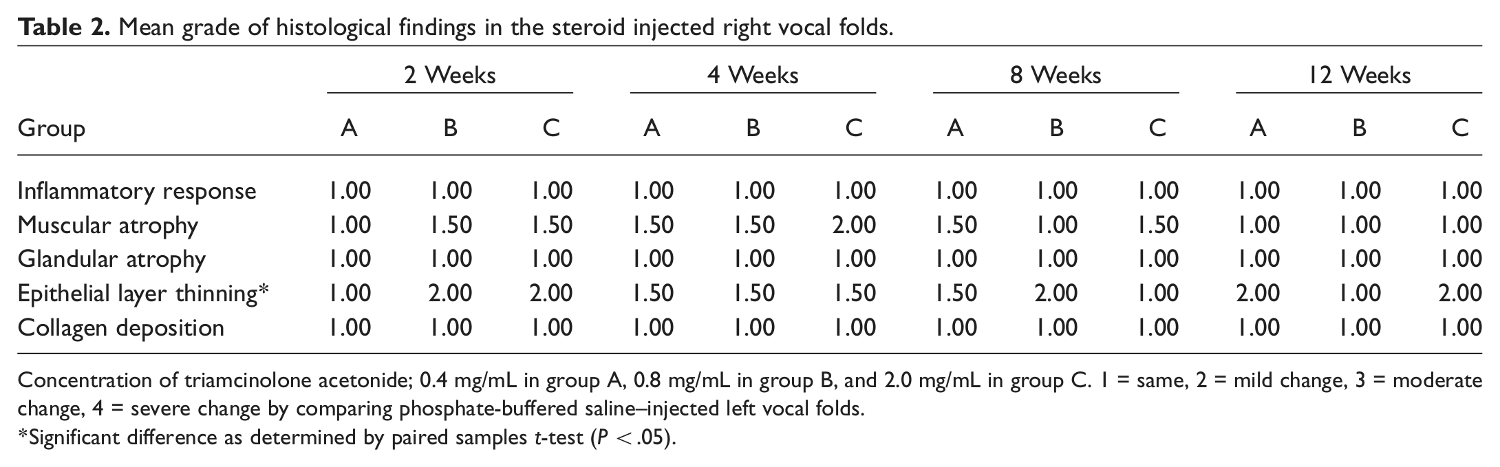
Concentration of triamcinolone acetonide; 0.4 mg/mL in group A, 0.8 mg/mL in group B, and 2.0 mg/mL in group C. 1 = same, 2 = mild change, 3 = moderate change, 4 = severe change by comparing phosphate-buffered saline–injected left vocal folds.
Significant difference as determined by paired samples t-test (P < .05).
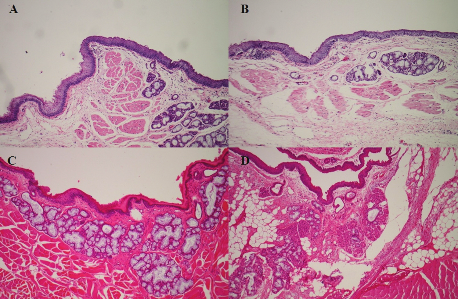
Histological findings in the vocal folds in group A at 4 weeks (A, B) and in group C at 8 weeks (C, D). A phosphate-buffered saline–injected left vocal fold (A, C) and steroid-injected right vocal fold (B, D) show no difference in inflammatory response and glandular atrophy (H&E stain, ×100).
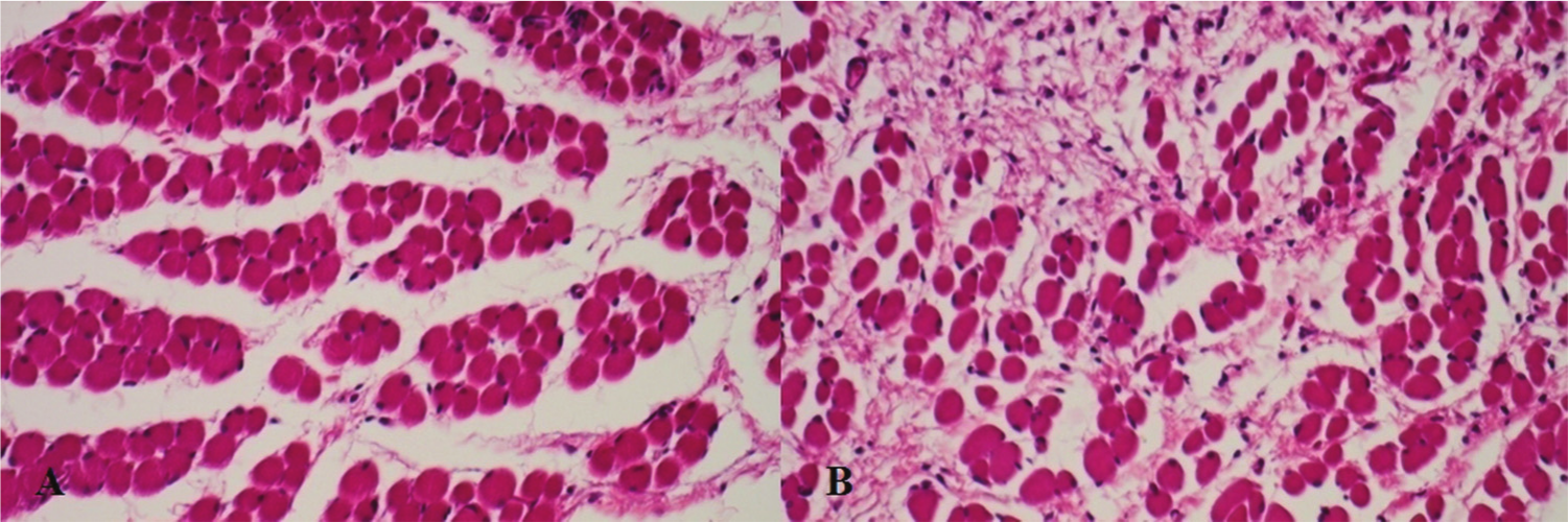
Histological findings in the vocal folds in group C at 4 weeks. A phosphate-buffered saline–injected left vocal fold (A) and steroid-injected right vocal fold (B) show that the right one revealed more muscular atrophy and stromal fibrosis compared to the left one (H&E stain, ×200).
However, various degree of epithelial layer thinning was observed over weeks in all groups ( Figure 5 ). Epithelial layer thinning was initially observed at 2 weeks in groups B and C, and continued until 12 weeks in groups A and C (P < .05). There was no difference in collagen deposition between the both vocal folds throughout experimental period (P > .05) ( Figure 6 , Table 2 ). Depending on the concentration, there was no significant difference in muscular atrophy and epithelial thinning (P > .05) ( Table 3 ).
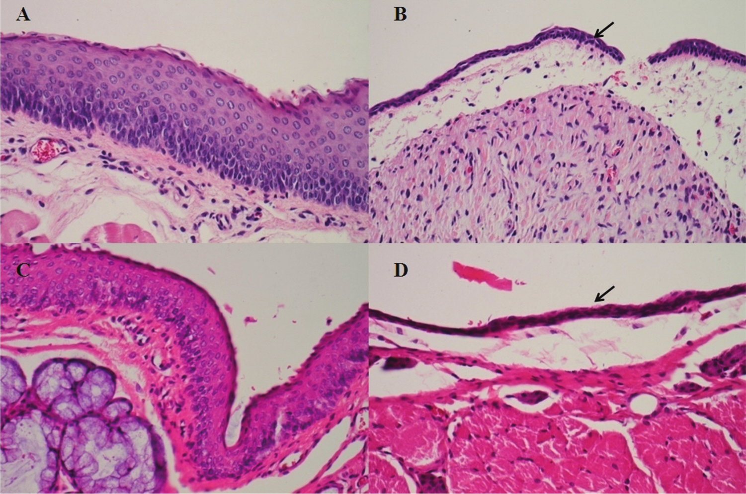
Histological findings in the vocal folds in group A at 4 weeks (A, B) and in group C at 12 weeks (C, D). A steroid-injected right vocal fold (B, D) shows thinner epithelium compared to a phosphate-buffered saline–injected left vocal fold (A, C) (arrow) (H&E stain, ×200).
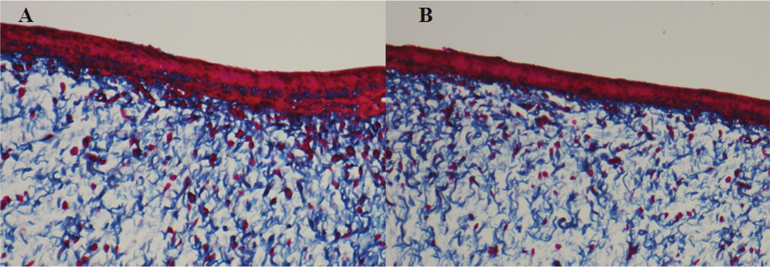
Histological findings in the vocal folds in group C at 4 weeks. A phosphate-buffered saline–injected left vocal fold (A) and steroid-injected right vocal fold (B) show no difference in collagen deposition (Masson’s trichrome staining, ×100).
Average histological grade following dosage of triamcinolone.
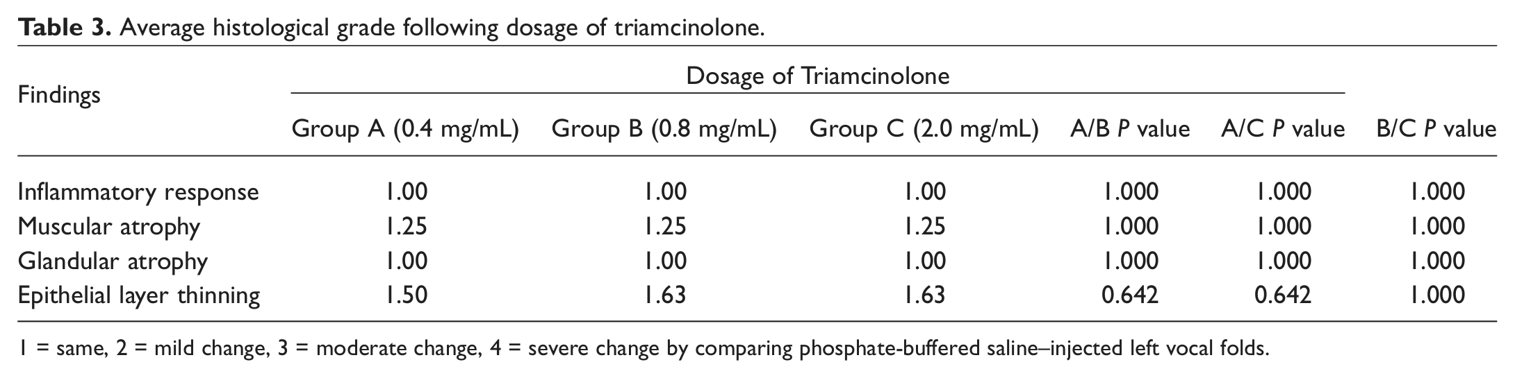
1 = same, 2 = mild change, 3 = moderate change, 4 = severe change by comparing phosphate-buffered saline–injected left vocal folds.
Discussion
Although short-term prescription of oral and systemic steroid is generally safe, prolonged usage of steroid can cause some side effects. A recent clinical practice guideline for hoarseness and dysphonia has recommended that the clinician should not routinely prescribe oral corticosteroids to treat hoarseness. 11 Given this situation, local steroid injection directly into the vocal fold (VF) benign lesions has recently been focused on alternative treatment options, and good results have been reported using different methods. By injecting steroid directly into the VF, it is possible to administer a potent drug locally and avoid the systemic adverse effects of the steroid. There were a few previous histologic studies of VF after injection of steroid or stem cells in animal models, but these studies have focused on wound healing process after injury, and the study of steroid injection was about acute phase results—follow-up period was 3 and 7 days.12,13 However, there have been few studies regarding the histological changes of VF after steroid injection without injury. As such, the aim of this study was to investigate the morphologic and histologic properties of rabbit VFs following injection of steroid.
Histologically, vocal nodules have stromal edema with fibroblast proliferation and dilated vasculature. 14 And Czerwonka et al reported that vocal fold intravascular pressure is likely to rise significantly during vocal fold vibration and may lead to the type of injury seen in benign vocal fold lesions. Resulting edema and increased intravascular pressures may lead to capillary failure and leakage of erythrocytes. 15 This tissue remodeling process is thought to be the pathogenesis of a vocal nodule. Campagnolo et al reported that injected steroid reduced collagen deposition during acute vocal fold wound healing. 12 As the treatment for vocal nodules, steroid may have reduced the formation of vocal nodules while changing the tissue remodeling process. In this present study, there was no significant difference in collagen deposition between the both vocal folds throughout experimental period.
Because of the effects of steroids, some clinicians are concerned about the complications of vocal fold steroid injection. Sulica and Behrman described the possible complication of steroid injection in the management of benign vocal fold lesions; it can delay wound healing and promote scar formation. 9 Also, Tateya et al 6 and Tateya 16 described the possibility of vocal fold atrophy following steroid injection. However, they reported that it was not observed even 5 years after the injection.6,16 In our experience, we had 4 cases of mild vocal fold atrophy. Although the vocal nodules had disappeared by the second week after the steroid injection, all 4 patients complained of a mild breathy voice. In stroboscopic findings, we observed decreased amplitude of mucosal wave and mild vocal fold bowing. 10 In this study, we identified histologically that the vocal fold steroid injection does not induce inflammatory response, glandular atrophy, and collagen deposition in a rabbit model, and mild muscular atrophy recovered at 12 weeks. These results suggest that intralesional steroid injection was well tolerated and safe modality in rabbit vocal fold tissue. However, the vocal fold steroid injection may induce epithelial layer thinning compared to PBS injection. This epithelial layer thinning was not influenced by the dosage of steroid. A recent study reported that the glucocorticoid receptors were observed in the epithelium, and dexamethasone decreased the expression of the glucocorticoid receptors, fibroblast proliferation, and TGF-β–induced collagen synthesis. 17 Epithelial layer thinning shown in this study may be associated with a lot of glucocorticoid receptors in the epithelium.
Our main limitation was the small number of subjects enrolled, and the scoring system used to judge treatment differences was entirely too simple and subjective. Also, this study focused on effects of steroids within 12 weeks, which was relatively an acute stage after injection. Further study with long-term follow-up will be needed to confirm the changes in chronic stages. And functional analysis was not performed. If further studies could be performed comparing steroid injection group and control group, rheological properties of vocal fold following steroid injection could be assessed. Our study showed that there were no inflammatory responses or any significant signs of local side effects. However, a mild grade of epithelial layer thinning was observed in steroid-injected vocal folds. In performing vocal fold steroid injection in humans, these possible changes need to be considered.
Conclusion
We found that vocal fold steroid injection does not induce any significant side effects. However, muscular atrophy was observed in a few vocal folds, and it completely recovered. Epithelial layer thinning induced by steroids was continued until 12 weeks. VF steroid injection may induce several histological changes, but it is generally safe and the morbidity is very low in a rabbit model. Our results seem to correlate with human VF steroid injection data in that aspect, and further studies with long-term follow-up periods are required for investing safety of VF steroid injection.
Author Contributions
Disclosures
Footnotes
Sponsorships or competing interests that may be relevant to content are disclosed at the end of this article.