
Abstract
Objectives
To evaluate clinical and immunohistopathological effects of topical glycyl-histidyl-lysine-copper (GHK-Cu) on in vivo irradiated rat wounds.
Design
Animal model.
Setting
Academic institution.
Subjects and Methods
After dorsal irradiation and a 28-day recovery period, 2 × 8 cm cranially based dorsal flaps were created in Sprague-Dawley rats. Twice daily GHK-Cu gel (test) or aquaphilic ointment (control) was applied for 10 days. Animals were euthanized, digital images of flaps were taken, and harvested tissues were immunohistochemically stained for a vascular endothelium marker, caveolin-1, and vascular endothelial growth factor (VEGF). Digital image analysis was used for outcome measures. Unpaired t-tests were used for statistical analyses; significance of P < .01 accounted for multiple comparisons.
Results
By digital analysis of clinical images, 13 test and 10 control animals showed mean ischemic areas of 5.0 cm2 (SD = 0.9) for tests and 3.8 cm2 (SD = 1.1; P = .011) for controls. Whole slide digitized images allowed quantification of caveolin-1-stained blood vessels and VEGF expression in fibroblasts at the interface of healing flaps. Caveolin-1 analyses showed a mean of 209.0 vessels (SD = 111.1) and a mean vessel luminal area of 525.7 um2 (SD = 191.0) in tests and 207.4 vessels (SD = 109.4; P = .973) and 422.8 um2 (SD = 109.7; P = .118) in controls. VEGF quantified as the percentage of pixels exceeding a colorimetric threshold, with higher fractions of positive pixels indicating more intense staining, showed a mean intensity score of 0.34 (SD = 0.19) in tests and 0.54 (SD = 0.41; P = .169) in controls.
Conclusions
Irradiated dorsal rat flaps treated with topical GHK-Cu gel demonstrated no difference in flap ischemia, blood vessel number or area, or VEGF expression compared to controls.
Introduction
Nonsurgical treatment of head and neck squamous cell carcinoma has increased in the past 3 decades. 1 Salvage surgery following radiotherapy or chemoradiotherapy increases the risk of soft tissue complications. 2 Interventions that improve the healing of radiation-damaged tissues postoperatively might decrease the morbidity and potential mortality of such complications.
Glycyl-histidyl-lysine is a naturally occurring tripeptide first shown in the 1970s to improve hepatocyte growth and survival. 3 In vitro studies of glycyl-histidyl-lysine-copper (GHK-Cu) have shown increased chemo-attraction of repair cells, reduced inflammation, and increased collagen synthesis, fibroblast proliferation, and angiogenesis.4-5 Irradiated and nonirradiated fibroblasts taken from operative sites treated with GHK-Cu have shown faster population-doubling times and significantly more vascular endothelial growth factor (VEGF) and basic fibroblastic growth factor. 6 Thus, GHK-Cu appears to be a beneficial upstream effector to local growth factor and extracellular matrix environments. In vivo rat studies have repeatedly demonstrated improved healing in dorsal soft tissue wounds treated with GHK-Cu,7-9 but this has never been evaluated in an irradiated model. Randomized, evaluator-blinded, placebo-controlled human clinical trials utilizing topical GHK-Cu have shown improved wound closure and infection rates in diabetic neuopathic ulcers 10 and higher patient satisfaction in healing results following circumoral CO2 laser resurfacing. 11
For the patient requiring salvage surgery after radiation-induced tissue injury, the continued study of molecules to improve wound healing is vital. This study investigates the ability of topical GHK-Cu to improve wound healing in an irradiated, ischemic flap model in rats, including the evaluation of flap survival and of immunohistochemical (IHC) analyses of 2 markers of angiogenesis, caveolin-1, and VEGF.
Methods
Power Calculation
Clinical flap survival was used a priori to find the minimum number of animals required for a statistical power of 80% with a type 1 error of .05 based on a previous study. 9 Mean percentage flap survival of control and treatment groups were used to calculate a minimum of 12 animals per group. Assuming a degree of unknown effects due to anesthesia or complications, 15 animals per group were used.
Radiation Protocol
After institutional approval, male Sprague-Dawley rats matched by species, age, weight, and gender were obtained. Animals were anesthetized with 0.15 mg/kg dexmedetomidine and 0.2 mg/kg fentanyl and reversed with 0.5 mg/kg atipamazole, then placed in lateral decubitus position ( Figure 1A ) in a linear accelerator. After plain radiography confirmed avoidance of the spinal canal ( Figure 1B ), a 12 cm length, 0.75 cm width, and 2.5 cm depth radiation field (posterior axillary line to anterior iliac crest) was administered in four 10-Gray (Gy) fractions at 3-day intervals. The effects of this dose are comparable to humans after radiotherapy 12 and correspond to 66.67 Gy. A 4-week healing period was provided to avoid acute radiation effects. The protocol was based on a previous study. 13
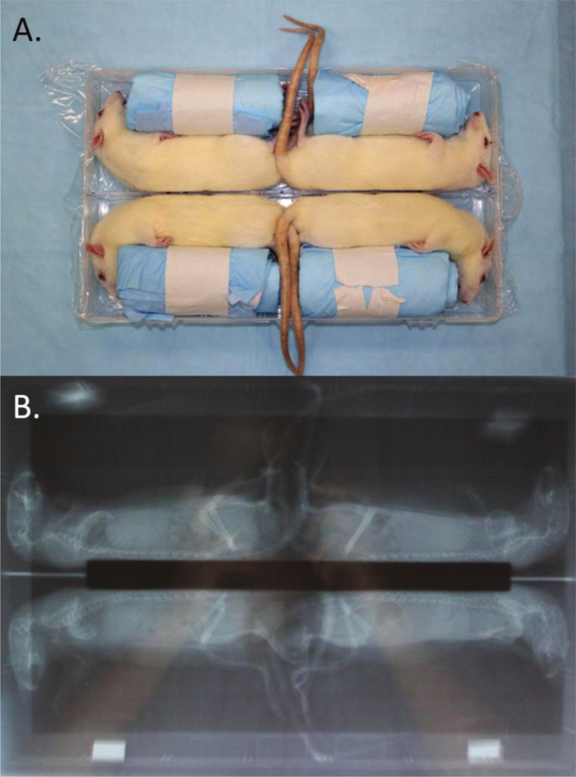
Radiation positioning (A) and x-ray confirmation of spine alignment and radiation exposure confined to dorsal soft tissues (B).
Surgical Procedure
The same anesthetics were used. Preoperative cefazolin was administered. Dorsal skin was shaved and cleansed with povidine-iodine. A centrally located, cranially based 2 cm by 8 cm flap was elevated deep to the panniculus carnosus ( Figure 2 ) based on an ischemic flap model, 14 then closed with nonabsorbable, monocryl suture. Postoperative pain control included 0.05 to 0.1 mg/kg SQ bupernorphine injections for 24 hours, then per os ibuprofen as needed.
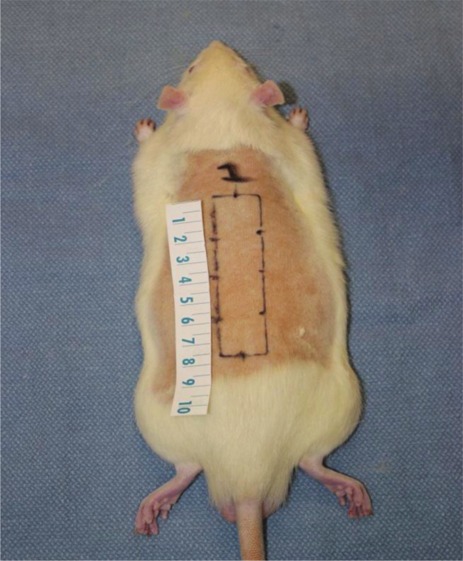
Animal procedure: premarked 2 × 8 cm incision.
Treatment and Control Groups
The treatment group received twice daily topical 0.5% prezatide copper acetate gel (PhotoMedex, Montgomeryville, Pennsylvania), while the control group received twice daily topical aquaphilic ointment (Eucerin, Beirsdorf, Wilton, Connecticut). The gel formulation in test animals is based on previous animal 8 and human studies, 10 while the ointment formulation in controls was chosen due to its routine use postoperatively in our patients.
Clinical Evaluation
Digital photographs taken on postoperative day 10 were converted to 8-bit images, and pixel -measurements were scaled according to rulers included in photographs using ImageJ software (Rasband, W.S., ImageJ, US National Institutes of Health, Bethesda, Maryland, http://imagej.nih.gov/ij/, 1997-2011). Conversion to binary images ( Figure 3 ) allowed for objective calculation of total area of necrosis.
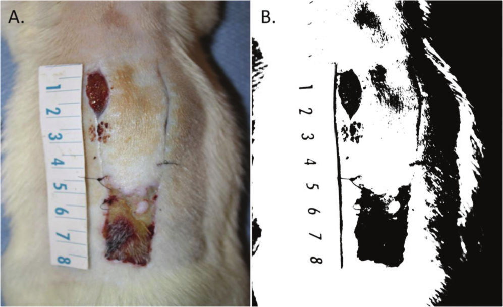
Clinical flap survival: control in vivo (A) converted to binary image for analysis (B).
Histological and Immunohistochemical Preparation
Animals were euthanized on postoperative day 10 with carbon dioxide. Dorsal soft tissues were excised, placed in formalin, processed, and paraffin imbedded. Hematoxylin and eosin (H&E) and IHC staining was performed. IHC staining was performed for caveolin-1 and VEGF to evaluate angiogenesis. Caveolae are structures within endothelial cells that coalesce to generate trans-endothelial channels acting as molecular conduits. Expression of caveolin-1 within endothelial cells is sufficient to drive the formation of caveolae, and targeted disruption of the caveolin-1 gene results in their complete loss. Caveolin-1 identifies vascular endothelium and is considered a marker of angiogenesis. 15 VEGF has been shown to be a strong angiogenic factor in nonirradiated 14 and irradiated 16 models.
IHC staining was performed in a standard fashion. Pertinent agents and steps included antigen retrieval (ReVeal Decloaking reagent, Biocare Medical, Concord, California), antibody incubation (dilute polyclonal anti-caveolin-1, Cell Signaling Technology, Danvers, Massachusetts; dilute monoclonal anti-VEGF, Santa Cruz Biotechnology, Santa Cruz, California), polymer detection (Rabbit on Rodent 2-step polymer detection, Biocare [caveolin-1]; biotinylated mouse secondary, Covance, Dedham, Massachusetts [VEGF]), and avidin biotin complex application (Vector ABC Elite) and detection (Covance). The protocols were based on previous studies in rats.17,18
Histological and Immunohistochemical Evaluation
Digital images of slides were created at 20× magnification using a scanner (ScanScope XT, Aperio Technologies, Vista, California). Using digital software (ImageScope 11.1, Aperio), 4 representative tissue areas (100 um × 100 um) located at the tissue-flap interface were annotated, including the distal end of the flap, 0.5 cm from the distal end, 1.0 cm from the distal end, and 1.5 cm from the distal end. Using image analysis software (Color Deconvolution Algorithm, Aperio) the color vectors for the individual staining components of the Hematoxylin counterstain (blue) and DAB chromagen (brown) were measured. For caveolin-1, analysis software (Microvessel Algorithm, Aperio) identified pixels exceeding intensity threshold limits, then adjusted them to remove background staining and preserve only endothelial-stained cells ( Figure 4A ), which allowed for isolation of pixels arranged in a continuous line, indicating the geometry of blood vessels ( Figure 4B ), and calculation of blood vessel number and area. For VEGF, pixels exceeding threshold limits were quantified as a fraction of total pixels; then the average number of positive pixels expressed as a fraction total was calculated. These methods have been shown to accurately analyze vascular endothelium in animal tissues. 18
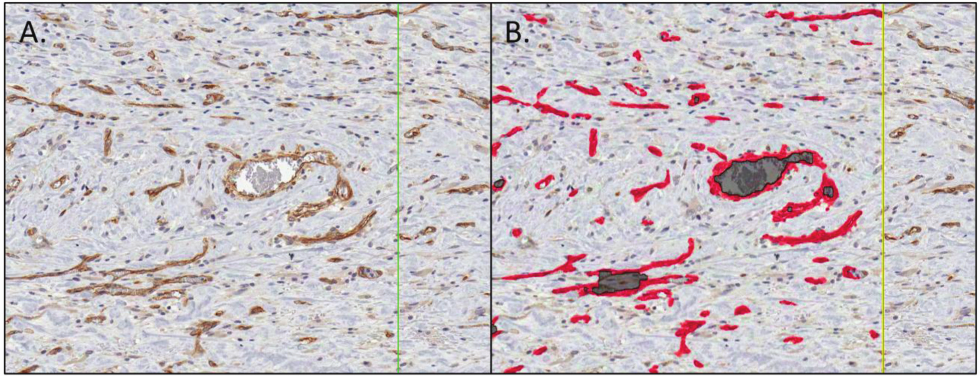
Caveolin-1 staining: magnified view of blood vessels before (A) and after (B) vessel analysis algorithm.
Treatment Group Versus Control Group Analyses
Means and standard deviations of ischemic areas were calculated and compared using unpaired t tests using QuickCalcs (GraphPad Software, La Jolla, California). Means and standard deviations of blood vessel number, vessel area, and VEGF intensity score were calculated and compared using unpaired t tests. To account for multiple comparisons within the same data set, the Bonferroni procedure was performed, and P < .01 was considered statistically significant.
Results
One animal suffered seizure-like activity following reversal of anesthesia during radiotherapy and recovered with conservative measures. One animal expired following anesthesia administration for radiotherapy, while 6 expired following anesthesia administration for surgery. Ten control group and 13 test group animals survived to the study endpoint. Clinical and immunohistochemical results are listed in Table 1 . Differences in mean ischemic area, vessels number, vessel area, and VEGF intensity scores were not statistically different.
Clinical and immunohistochemical results.
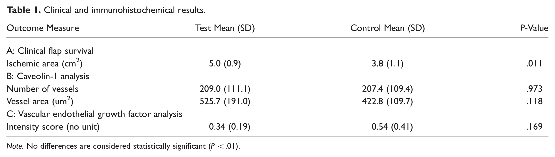
Note. No differences are considered statistically significant (P < .01).
Discussion
While much attention has been paid to local growth factors and their effects on wound healing, little attention has been paid to upstream molecules that affect the local molecular environment. We sought to perform the first study evaluating the effects of topical GHK-Cu on irradiated soft tissue wound healing and did so using objective analysis of flap survival in an ischemic model and of angiogenesis through caveolin-1 and VEGF analyses. This is the first in vivo study evaluating GHK-Cu as applied topically to closed wounds; prior models used either subcutaneous impregnation in closed wounds7-9 or topical application to open wounds.10-12 The topical application was chosen because ultimately, patients at high risk for wound healing complications, such as those treated with radiotherapy or chemoradiotherapy, have closed wounds after salvage surgery.
In the current study, ischemia trended toward being greater in the GHK-Cu group as compared to the test group, but this was not statistically significant. This is contrary to prior animal and human studies reporting improved wound healing.7-12 We sought to explain the clinical effects at a molecular level through analysis of angiogenesis via caveolin-1 and VEGF expression. We found no difference in caveolin-1 analyses and VEGF expression at the flap-tissue interface, which indicated no improvement in blood vessel formation or in a primary growth factor related to angiogenesis.
Although animal 8 and human 19 studies used GHK-Cu gel topically, it is unclear whether this formulation had a negative impact on the healing of flaps compared to the petroleum-based, hydrophilic ointment used as a control. It is unclear how the topical treatment of such a formulation was affected by tissue absorption. There are also no data reporting the effects of differential frequency of application on the healing of wounds. The study is limited by the survival of animals related to anesthesia during irradiation and the surgical procedures. Perhaps a larger sample size would have allowed for more definitive statistical analysis, while a smaller sample may have led to biased outcomes.
Conclusions
A safe radiation protocol and reproducible animal model was used to evaluate the effect of topical GHK-Cu on wound healing. Objective quantification of flap survival, blood vessel formation, and growth factor expression was performed. Dorsal soft tissue flaps in irradiated rats treated with topical GHK-Cu gel showed no difference in area of clinical ischemia, blood vessel number or vessel area, or VEGF expression compared to controls.
Author Contributions
Disclosures
Footnotes
Acknowledgements
This project utilized BioNet core facilities which are supported by the University of Minnesota Academic Health Center. Specifically, Colleen Forster from the BioNet histology core provided expertise in histology and immunohistochemistry, and Jonathan Henriksen of the BioNet digital imaging core provided expertise in digital image analysis. We are indebted to Kim Janisch, Susan Jenson, and Dr Dan Feeney, DVM, from the University of Minnesota Veterinary Medical Center for their assistance with animal irradiation.
Sponsorships or competing interests that may be relevant to content are disclosed at the end of this article.
This article was presented at the 2012 AAO-HNSF Annual Meeting & OTO EXPO; September 9-12, 2012; Washington, DC.